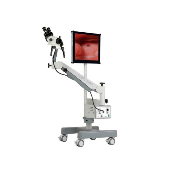

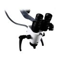
Ecleris C100-FID Binocular Colposcope
$5,042.00
Shipped from Abroad
Content Includes:
Forearm,
Pantographic Arm,
Floor Stand (H Shaped Base and Column),
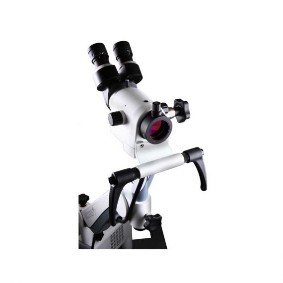
5 Magnifications Head,
Green Filter and Inclined Binocular,
LED Light Source,
Fiber Optic Cable,
110/220V Power Cable and User`s Guide,
Stand for LCD Monitor and Printer,
Digital Capturing System for Images, Videos and Sounds,
USB 2.0. Includes Main Unit Processor with three Camera Inputs,
USB Cable,
Software,
Hands Free Microphone and Footswitch.
Delivery & Availability:
Typically 10 working days – excluding furniture and heavy/bulky equipment. Please contact us for further information.

Description
Ecleris Colposcope Series C-100 was designed to cover all the diagnosis and therapeutic needs of modern gynecology. All models can be transformed into video colposcopes with our high resolution video camera. Digital handling of patients and image filing is achieved through the endoDIGI software which is easily adaptable to a portable PC or desktop computer. Great and accurate quality images, improved clearness, resolution and focal range can be obtained through our new C-100 Colposcope optic system. Floor and wall-mounted models include 5 magnifications (4, 6, 10, 16 and 25x).
The C-100 model, with its pantographic arm has enhanced maneuverability, as the arms are mounted on bearings and guarantee smooth movements and greater stability (WBS, weight balance system). Its state-of-the-art designed base allows for easy transport. We provide accessories that allow using the microscope light source and video camera also to carry out endoscopic studies, without need to have two light sources and two video cameras at the doctor’s office.
A new dimension in microsurgery
Ecleris 3D Splitter
- Is a new device that integrates a beam-splitter with an HD 3D video system that expands the borders of surgical visualization as it allows the surgeon to share the stereoscopic images that he sees through the binocular; and in high definition.
- It is an ideal method for education since 3D visualization improves understanding, motivation and retention of knowledge.
- Compatible with Microscopes and Colposcopes of multiple brands.
Ecleris HD Splitter
- This beam splitter integrates a full HD video camera in an ergonomic and compact way.
- Both devices are installed between the optical head and the binocular. This configuration results in great balance since it avoids the use of photographic and/or video equipment located on the side of the optical head which usually alter the stability of the movements of it.
- Both products are complemented by sophisticated image capture systems from the ECLERIS endoDIGI family.
TECHNICAL SPECIFICATION
Colposcope
- Binocular – 45º Inclined (straight optional)
- Objective Lens – Standard 300 mm included. Optional: 200 / 250 mm.
- Magnifications – Manual changer 5 positions: Factor 4 / 6 / 10 / 16 / 25 X
- Fine Focus – Manual integrated in focal lens.
- Eyepieces – 10 X Wide angle. Dioptric setting: + / – 5.
- Field of View (10 X) – Ø 24 mm / 0,95“ (for f: 200 mm) Ø 31 mm / 1,22” (for f: 250 mm). Ø 36 mm / 1,42” (for f: 300 mm) Ø 50 mm
- Interpupilar Distance – 2,16”- 2,95”. 55 – 75 mm
- Filter – Green
Illumination
- Type of Illumination – Coaxial Illumination through 7 mm fiber optic light guide cable
- Light Source – LED (80 W 50.000 hours life)
- Illuminated Field – Ø 70 mm / 2,75” (for f: 200 mm) Ø 90 mm / 3,54” (for f: 250 mm). Ø 107 mm / 4,2” (for f: 300 mm) Ø 145 mm
- Illumination Control – Electronic dimmer with continuous adjustment. Constant light color
- Power Supply – 100 – 240 VAC, 50 / 60 Hz
Video
- Video Camera – Video camera connection input and video output integrated into light source
Mechanics
- Type – Stand floor unit, 5 wheels.
- Rotation – 360◦
- Height Adjustment – 97,5 / 120 cm, 38 / 47
- Weight – 13,5 kg / 30 lb
Content Includes:
Forearm,
Pantographic Arm,
Floor Stand (H Shaped Base and Column),
5 Magnifications Head,
Green Filter and Inclined Binocular,
LED Light Source,
Fiber Optic Cable,
110/220V Power Cable and User`s Guide,
Stand for LCD Monitor and Printer,
Digital Capturing System for Images, Videos and Sounds,
USB 2.0. Includes Main Unit Processor with three Camera Inputs,
USB Cable,
Software,
Hands Free Microphone and Footswitch.
Click Here To Download Catalogue
Quick Comparison
| Ecleris C100-FID Binocular Colposcope remove | Sonoscape S11 Ultrasound Machine remove | Bistos BT-770-12.1" Touchscreen Patient Monitor remove | ASPEL Stress ECG with Ergometer and Software remove | Sonoscape P10 Ultrasound Machine remove | Sonoscape S8 Exp Portable Ultrasound remove | |
|---|---|---|---|---|---|---|
| Name | Ecleris C100-FID Binocular Colposcope remove | Sonoscape S11 Ultrasound Machine remove | Bistos BT-770-12.1" Touchscreen Patient Monitor remove | ASPEL Stress ECG with Ergometer and Software remove | Sonoscape P10 Ultrasound Machine remove | Sonoscape S8 Exp Portable Ultrasound remove |
| Image | 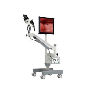 |  | 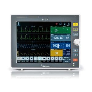 | 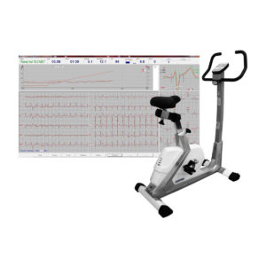 |  |  |
| SKU | SF1033560087-1 | SF1033560012-1 | SF1033560059-1 | SF1033560075-1 | SF1033560012-7 | SF1033560012-15 |
| Rating | ||||||
| Price | $5,042.00 | $6,380.00 | $902.00 | $4,202.00 | $9,350.00 | $9,350.00 |
| Stock | ||||||
| Availability | ||||||
| Add to cart | ||||||
| Description | Shipped from Abroad Content Includes: Forearm, Pantographic Arm, Floor Stand (H Shaped Base and Column), 5 Magnifications Head, Green Filter and Inclined Binocular, LED Light Source, Fiber Optic Cable, 110/220V Power Cable and User`s Guide, Stand for LCD Monitor and Printer, Digital Capturing System for Images, Videos and Sounds, USB 2.0. Includes Main Unit Processor with three Camera Inputs, USB Cable, Software, Hands Free Microphone and Footswitch. Delivery & Availability: Typically 10 working days – excluding furniture and heavy/bulky equipment. Please contact us for further information. | In Stock A Value Choice beyond Your Expectation. SonoScape’s trolley color Doppler system S11 redefines price and performance with practical design. The S11 will go beyond your expectations but not your budget. Delivery & Availability: Typically 2 working days – excluding furniture and heavy/bulky equipment. Please contact us for further information. | Shipped from Abroad The Bistos BT-770 patient monitor is equipped with a 12.1" touchscreen display, which allows for an easy operation and readability with a powerful rechargeable battery guaranteeing a continuous operation of 5 hours to monitor ECG, SpO2, NIBP, temperature and respiration Delivery & Availability: Typically 14 working days – excluding furniture and heavy/bulky equipment. Please contact us for further information. | Shipped from Abroad Ergometer CRG 200 is dedicated for Exercise Stress Tests System CardioTEST. The Ergometer has been designed according to modern technologies. It is controlled from PC equipped in CardioTEST software. Load level is controlled by a microprocessor, therefore it does not depend on speed, which in turn can be adjusted according to patient’s individual needs. Ergometer is equipped with ECG mode recording 12 standard leads. Delivery & Availability: Typically 21 working days – excluding furniture and heavy/bulky equipment. Please contact us for further information. | Shipped from Abroad The P10 color Doppler ultrasound system is a new generation product from SonoScape. It is designed to give high quality images, rich probe configurations, various clinical tools and automatic analysis software to provide you with comprehensive solutions for your growing demand for clinical applications. Delivery & Availability: Typically 5-7 working days – excluding furniture and heavy/bulky equipment. Please contact us for further information. | Shipped from Abroad With ultra-modern innovative design and the clinically-proven technologies, S8 Exp is portable ultrasound scanner well equipped as a low-physical-effort and enhanced-image-quality ultrasound scanner, which not only provides optimized solutions for versatile applications, but does help to improve the user-experience for both routine and non-traditional challenges. Delivery & Availability: Typically 5-7 working days – excluding furniture and heavy/bulky equipment. Please contact us for further information. |
| Content | Ecleris Colposcope Series C-100 was designed to cover all the diagnosis and therapeutic needs of modern gynecology. All models can be transformed into video colposcopes with our high resolution video camera. Digital handling of patients and image filing is achieved through the endoDIGI software which is easily adaptable to a portable PC or desktop computer. Great and accurate quality images, improved clearness, resolution and focal range can be obtained through our new C-100 Colposcope optic system. Floor and wall-mounted models include 5 magnifications (4, 6, 10, 16 and 25x).
The C-100 model, with its pantographic arm has enhanced maneuverability, as the arms are mounted on bearings and guarantee smooth movements and greater stability (WBS, weight balance system). Its state-of-the-art designed base allows for easy transport. We provide accessories that allow using the microscope light source and video camera also to carry out endoscopic studies, without need to have two light sources and two video cameras at the doctor’s office.
A new dimension in microsurgery
Ecleris 3D Splitter
Click Here To Download Catalogue | DETAILS
SonoScape’s trolley colour Doppler system S11 redefines price and performance with practical design. The S11 will go beyond your expectations but not your budget. As an easy-to-use ultrasound system, the S11 is integrated with a new software platform, especially optimized for a smooth workflow and convenient operation. The system speeds up the exam process and makes file management easier.
SPECIFICATION
- 15-inch high definition LCD monitor with articulating arm
- Compact and agile trolley design
- 3 active transducer sockets available for a wide range of applications
- Duplex, Color Doppler, DPI, PW Doppler, tissue harmonic imaging, μ-scan speckle reduction imaging, compound imaging, trapezoidal imaging
- Customized settings based on your own working style
- Full patient database and image management solutions
Click Here To Download Catalogue |
Bistos BT-770 is a 12.1" touchscreen patient monitor designed for easy operations.
SPECIFICATIONS
Click Here To Download Catalogue | Ergometer CRG 200 is dedicated for Exercise Stress Tests System CardioTEST. The Ergometer has been designed according to modern technologies. It is controlled from PC equipped in CardioTEST software. Load level is controlled by a microprocessor, therefore it does not depend on speed, which in turn can be adjusted according to patient’s individual needs. Ergometer is equipped with ECG mode recording 12 standard leads.
Technical Specifications:
Click Here To Download Catalogue | DETAILS
B + Compound
B + Compound utilizes several lines of sight for optimal contrast resolution, speckle reduction and border detection, with which P10 is ideal for superficial and abdominal imaging with better clarity and improved continuity of structures.
μ-Scan
The new generation μ-Scan imaging technology gives you better image quality by reducing noise, improving signal strength and improving visualization.
P10 offers a comprehensive selection of electronic probes to maximize its capabilities to meet a wide range of applications including abdomen, pediatric, OB/GYN, cardiovascular, musculoskeletal, etc. The advanced probe technologies also effectively enhance the image quality and confidence in reaching clinical diagnoses, even in difficult patients.
Convex Probe 3C-A
Ideal for an abundant of application such as abdomen, gynecology, obstetrics, urology and even abdomen biopsy.
Linear Probe L741
This linear probe is designed to satisfy vascular, breast, thyroid, and other small parts diagnosis, and its adjustable parameters could also present users a clear view of MSK and deep vessels.
Phase Array Probe 3P-A
For the purpose of adult and pediatric cardiology and emergency, the phase array probe provides elaborate presets for different exam modes, even for difficult patients.
Intracavitary Probe 6V1
Intracavitary probe could face application of gynecology, urology, prostate, and its temperature detection technology not only protects the patient but also extends the service life.
Click Here To Download Catalogue | Sonoscape S8 Exp Portable Ultrasound scannerDETAILS Agile and Versatile With ultra-modern innovative design and the clinically-proven technologies, S8 Exp Portable Ultrasound scanner is well equipped as a low-physical-effort and enhanced-image-quality ultrasound scanner, which not only provides optimized solutions for versatile applications but does help to improve the user experience for both routine and non-traditional challenges. Working with S8 Exp, it will trigger your unlimited reverie and endow you with endless charm. Carrying forward the classical design of SonoScape's portable ultrasound products, S8 Exp successfully combines the best ergonomics, attractive design and ease of use. This charismatic identity is also enhanced by a sophisticated color palette—with sedate grey as its interior paint color and pearl white as exterior cover, S8 Exp reveals a style of aristocrat and strong character among SonoScape's ultrasound systems. Workflow The S8 Exp is a portable ultrasound scanner that adapts to your workflow, whether you are in the consulting room, at the bedside, or at a remote location. With easy-to-use new platform designed for sonographers' needs and full connection interfaces for easy connectivity and data sharing, S8 Exp leads to improved user comfort and clinical outcome as well as patient throughput and working efficiency. Powerful Platform Embedded with SonoScape's core imaging technologies such as μ-scan, PHI and Spatial Compound, S8 Exp boasts exceptional 2D image, sensitive spectral, Color and Power Doppler, displaying well-defined anatomy and pathology and facilitating a highly optimized diagnostic user environment for conclusive diagnoses. Besides, S8 Exp offers a comprehensive selection of electronic probes to maximally extend its capabilities to meet a wide range of applications including the abdomen, pediatric, OB/GYN, cardiovascular, musculoskeletal, etc. The advanced probe technologies also effectively enhance the image quality and confidence in reaching clinical diagnoses even in difficult patients.Click Here To Download Catalogue |
| Weight | N/A | N/A | N/A | N/A | N/A | N/A |
| Dimensions | N/A | N/A | N/A | N/A | N/A | N/A |
| Additional information |


Reviews
There are no reviews yet.