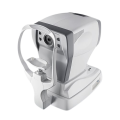
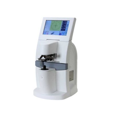
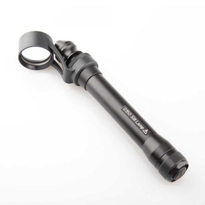
FA-6500 Automatic Computerized Refractor
$0.00
Shipped From Abroad
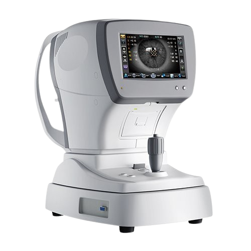
The FA-6500 Automatic Computerized Refractor provides fast and accurate refraction measurements for precise vision correction. With advanced optics, ergonomic design, and user-friendly operation, it enhances efficiency in eye clinics. Its compact design, high-resolution display, and reliable automation make it ideal for professional ophthalmic and optometry practices.
Typically 10-21 working days – excluding furniture and heavy/bulky equipment. Please contact us for further information.
Description
The FA-6500 Automatic Computerized Refractor is a next-generation ophthalmic diagnostic device engineered to deliver rapid, precise, and reliable refraction measurements. Designed for modern eye care practices, it simplifies the examination process by automating refraction testing with advanced optical technology. The device provides accurate readings for myopia, hyperopia, astigmatism, and other refractive errors, ensuring superior accuracy for eyeglass and contact lens prescriptions. Its ergonomic design enhances patient comfort, while the intuitive interface allows practitioners to operate the system efficiently with minimal training. The FA-6500 is equipped with a high-resolution LCD screen, easy data transfer options, and built-in memory for seamless workflow integration. With automatic measurement, smooth data recording, and connectivity to external systems, it improves both productivity and patient satisfaction. Compact, reliable, and user-friendly, the FA-6500 is an essential tool for ophthalmologists, optometrists, and optical clinics seeking consistent and accurate vision assessments.
Features
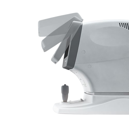
● Auto focus function
Just move, it will automatically focus the eye and get the value.
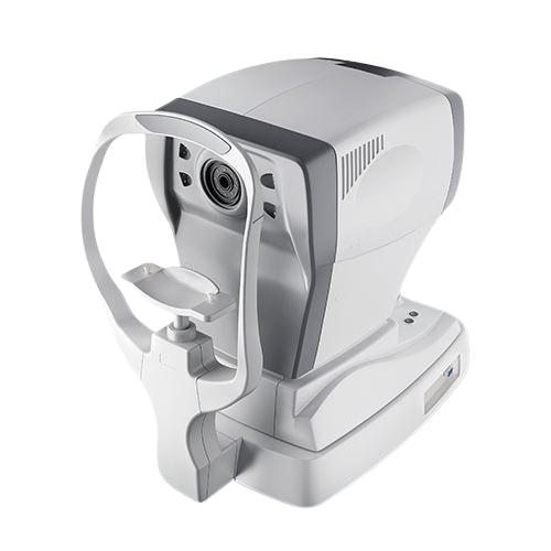
● Motorized joystick
A motorized joystick with auto vertical tracking mode is adopted, so the operation is easier.
Specifications
|
Refractometer |
|
| Vertex distance | 0mm, 12mm, 13.75mm |
| Spherical | -20~+20m-1 (VD=12) 0.12/0.25m-1Step |
| Cylinder | -8~+8m-1 0.12/0.25m-1 Step |
| Axis | 0~180° 1°Step |
| Cylinder form | -, + , ± |
| Pupil distance | 45~88mm, 1mm Step |
| Min. pupil size | 2.0mm |
| Keratometry | |
| Radius of curvature | 5.0~10mm(increment:1mm) |
| Corneal power | 33.75~67.50m-1 (when corner equivalent refractive index is 1.337) |
| (increment selectable from 0.12, 0.25m-1) | |
| Corneal astigmatism | 0.0~8.00m-1 (increment selectable from 0.12,0.25m-1 ) |
| Axis | 1~180°(increment:1°) |
| Corneal diameter | 2.0~14.0mm(increment: 0.1mm) |
| Others | |
| Chart | Follow-up on the colorful fogging chart system |
| Memory of data | 10 measure value for each right and left eye |
| Display | 7″LCD/TFT |
| Thermal printer | |
| Power supply | 220V±10% 50Hz 60VA |
| Dimension | 478mm×268mm×472mm, |
| Weight | ~15.5kg |
Quick Comparison
| FA-6500 Automatic Computerized Refractor remove | Ophthalmic AB Scan Machine remove | Slit Lamp with Workstation remove | View Tester (Manual Phoropter) remove | ENT/Neurosurgery Operating Microscope remove | Timesco Ophthalmoscope remove | ||||||||||||||||||||||||||||||||||||||||||||||||||||||||||||||||||||||||||||||||||||||||||||||||||||||||||||||||||||||||||||||||
|---|---|---|---|---|---|---|---|---|---|---|---|---|---|---|---|---|---|---|---|---|---|---|---|---|---|---|---|---|---|---|---|---|---|---|---|---|---|---|---|---|---|---|---|---|---|---|---|---|---|---|---|---|---|---|---|---|---|---|---|---|---|---|---|---|---|---|---|---|---|---|---|---|---|---|---|---|---|---|---|---|---|---|---|---|---|---|---|---|---|---|---|---|---|---|---|---|---|---|---|---|---|---|---|---|---|---|---|---|---|---|---|---|---|---|---|---|---|---|---|---|---|---|---|---|---|---|---|---|---|---|---|---|---|
| Name | FA-6500 Automatic Computerized Refractor remove | Ophthalmic AB Scan Machine remove | Slit Lamp with Workstation remove | View Tester (Manual Phoropter) remove | ENT/Neurosurgery Operating Microscope remove | Timesco Ophthalmoscope remove | |||||||||||||||||||||||||||||||||||||||||||||||||||||||||||||||||||||||||||||||||||||||||||||||||||||||||||||||||||||||||||||||
| Image |  |  |  | 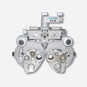 |  | 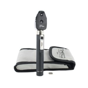 | |||||||||||||||||||||||||||||||||||||||||||||||||||||||||||||||||||||||||||||||||||||||||||||||||||||||||||||||||||||||||||||||
| SKU | SF1033560107-8 | SF1033560107-7 | SF1033560107-26 | SF1033560109-1 | SF1033560084-282 | ||||||||||||||||||||||||||||||||||||||||||||||||||||||||||||||||||||||||||||||||||||||||||||||||||||||||||||||||||||||||||||||||
| Rating | |||||||||||||||||||||||||||||||||||||||||||||||||||||||||||||||||||||||||||||||||||||||||||||||||||||||||||||||||||||||||||||||||||||
| Price |
| $4,895.00 | $3,740.00 | $858.00 |
| $140.00 | |||||||||||||||||||||||||||||||||||||||||||||||||||||||||||||||||||||||||||||||||||||||||||||||||||||||||||||||||||||||||||||||
| Stock | |||||||||||||||||||||||||||||||||||||||||||||||||||||||||||||||||||||||||||||||||||||||||||||||||||||||||||||||||||||||||||||||||||||
| Availability | |||||||||||||||||||||||||||||||||||||||||||||||||||||||||||||||||||||||||||||||||||||||||||||||||||||||||||||||||||||||||||||||||||||
| Add to cart | |||||||||||||||||||||||||||||||||||||||||||||||||||||||||||||||||||||||||||||||||||||||||||||||||||||||||||||||||||||||||||||||||||||
| Description | Shipped From Abroad
The FA-6500 Automatic Computerized Refractor provides fast and accurate refraction measurements for precise vision correction. With advanced optics, ergonomic design, and user-friendly operation, it enhances efficiency in eye clinics. Its compact design, high-resolution display, and reliable automation make it ideal for professional ophthalmic and optometry practices.
Delivery & Availability:
Typically 10-21 working days – excluding furniture and heavy/bulky equipment. Please contact us for further information.
| Shipped from abroad
| Shipped from abroad
| Ship from abroad
| Shipped from abroad
Corder Microscope has Fluid, Responsive and Accurate.Fluid. Responsive. Accurate. These were a few of the principles guiding every phase in the design of the Corder Microscope. With the choicest mechanical machined components, the Corder Microscope has the grace and agility to adjust to every desired position on command. Well designed Apochromatic optics treated with Corder's Mcoatings produce true-to life sharp images with high depth, definition and contrast. | In Stock
| |||||||||||||||||||||||||||||||||||||||||||||||||||||||||||||||||||||||||||||||||||||||||||||||||||||||||||||||||||||||||||||||
| Content | The FA-6500 Automatic Computerized Refractor is a next-generation ophthalmic diagnostic device engineered to deliver rapid, precise, and reliable refraction measurements. Designed for modern eye care practices, it simplifies the examination process by automating refraction testing with advanced optical technology. The device provides accurate readings for myopia, hyperopia, astigmatism, and other refractive errors, ensuring superior accuracy for eyeglass and contact lens prescriptions. Its ergonomic design enhances patient comfort, while the intuitive interface allows practitioners to operate the system efficiently with minimal training. The FA-6500 is equipped with a high-resolution LCD screen, easy data transfer options, and built-in memory for seamless workflow integration. With automatic measurement, smooth data recording, and connectivity to external systems, it improves both productivity and patient satisfaction. Compact, reliable, and user-friendly, the FA-6500 is an essential tool for ophthalmologists, optometrists, and optical clinics seeking consistent and accurate vision assessments. FeaturesSpecifications
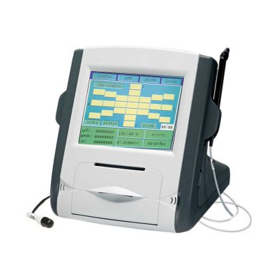
| Functions of Ophthalmic AB Scan Machine:
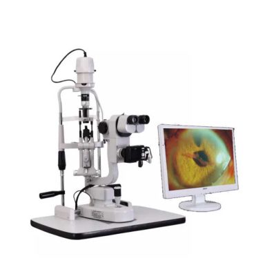
| Slit Lamp with Workstation Features:
| Features:
| Features:Corder Microscope has Fluid, Responsive and Accurate.Fluid. Responsive. Accurate. These were a few of the principles guiding every phase in the design of the Corder Microscope. With the choicest mechanical machined components, the Corder Microscope has the grace and agility to adjust to every desired position on command. Well designed Apochromatic optics treated with Corder's Mcoatings produce true-to life sharp images with high depth, definition and contrast. More comfortable operation Tiltable binocular tubes available, which can incline more than 60° depending on the posture and physique of the operating surgeon. Movable range: 30° (straight) to 90° (inclined) Corder microscope configured with XYZ motorized movement operated through a comfortable foot /Handle control, a veryeffective co-axial illumnation and 50W halogen light source makes it ideal for Neuro surgeries.Doctor-patient communication is easierTo address digital documentation needs, a host of digital SLR, video camera, and CCD adapters are made available with the ProLine in addition to Corder's proprietary iVu multi-functional imaging solution. 1080P full hd image quality, efficient image management during the operation. Integrate your digital workflow to facilitate case management and facilitate more intuitive patient communication. Technical Permeants: Magnification: motorized zoom system, 1:6 zoom ratio, magnification 3x~16x Focusing range: 50mm Binocular tube: 30°~90° tiltable tube ,(0° ~200° optional) Eyepiece: 12.5x / 10x Objective lens: F 300mm(175mm, 250mm, 350mm optional) pupil distance: 55mm~75mm diopter adjustment: +6D ~ -6D Field of view: Φ74~Φ12mm X-Y translator: Motorized by foot switch or handle controller, ±30mm Assistant tube: 360° Rotating assistant tube Reset functions: YES Illumination System: Coaxial illumination Light source: Halogen lamp Light intensity adjustment: Continuous brightness adjustment 0-100000lux Fiber optic illumination: Dual fiber Field of illumination: Φ50mm Filter: Red free filter, small spot Accessories CCD Camera system: Beam splitter, CCD adapter, CCD, Display XENON LAMP: 150000lux Integrated Video Adapter: SONY / CANON CameraClick Here To Download Catalogue | Timesco Ophthalmoscope features a head made from lightweight hermetically sealed durable plastic, precision optics and a latex free rubber eyebrow rest. A bright white light from long life standard bulbs provides crystal clear illumination in ophthalmic diagnostic procedures.
Click Here To Download Catalogue | |||||||||||||||||||||||||||||||||||||||||||||||||||||||||||||||||||||||||||||||||||||||||||||||||||||||||||||||||||||||||||||||
| Weight | N/A | N/A | N/A | N/A | N/A | N/A | |||||||||||||||||||||||||||||||||||||||||||||||||||||||||||||||||||||||||||||||||||||||||||||||||||||||||||||||||||||||||||||||
| Dimensions | N/A | N/A | N/A | N/A | N/A | N/A | |||||||||||||||||||||||||||||||||||||||||||||||||||||||||||||||||||||||||||||||||||||||||||||||||||||||||||||||||||||||||||||||
| Additional information | |||||||||||||||||||||||||||||||||||||||||||||||||||||||||||||||||||||||||||||||||||||||||||||||||||||||||||||||||||||||||||||||||||||


Reviews
There are no reviews yet.