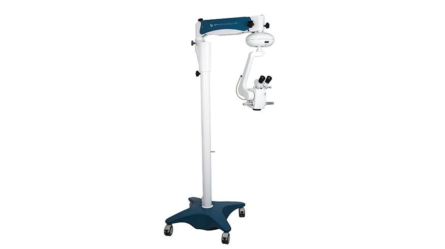
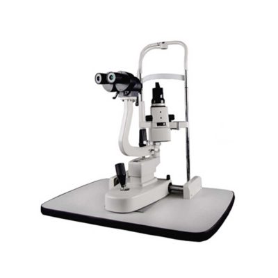
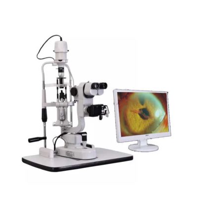
FOCUS ADVANCED MICROSCOPE
$0.00
Shipped From Abroad
The FOCUS series is a line of equipment specially designed in detail to meet the most demanding standards of anterior and posterior segment surgical procedures.
The optics developed for FOCUS line microscopes enable external transmission of LED light, generating excellent clarity and image depth of the observed field.
Ideal for Cataract and Retina procedures.
The optics developed for FOCUS ADVANCED line microscopes enable external transmission of LED light (oblique), generating excellent clarity and image depth of the observed field. The equipment has an independent coaxial lighting system, activated via a multifunction pedal, ensuring total image clarity and excellent red reflection quality, providing total safety and visibility during cataract procedures.
Having functions such as zoom, microfocusing, intensity control activated via pedal and being compatible with the best non-contact lens adapters for vitreectomy on the market, it enables all retinal procedures with total quality.
Typically 10-21 working days – excluding furniture and heavy/bulky equipment. Please contact us for further information.
Description
Description
Versatility in multiple configuration possibilities
Focus Microscopes stand out in the market for presenting multidisciplinary characteristics and high optical quality. Additionally, they allow you to add options and accessories according to the need for use, including binoculars, double iris, image inverter, among others.
The Focus* line of microscopes has a pedal-operated motorized microfocusing and zooming system, aiming for greater precision and comfort for the user during surgical procedures.
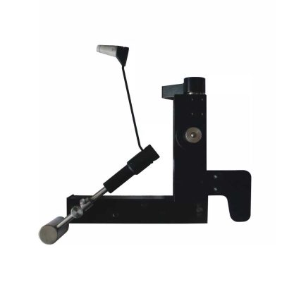
Pedal controlled XY positioner
The DFVasconcellos XY Positioner allows precise alignment between the user’s vision and the surgical field. This becomes an increasingly necessary resource as microscopy techniques become more widespread.
Quick Comparison
| Settings | FOCUS ADVANCED MICROSCOPE remove | Retinoscope remove | Portable Fundus Camera remove | Portable Rebound Tonometer remove | View Tester (Manual Phoropter) remove | Ophthalmic AB Scan Machine remove | |||||||||||||||||||||||||||||||||||||||||||||||||||||||||||||||||||||||||||||||||||||||||||||||||||||||||||||||||||||
|---|---|---|---|---|---|---|---|---|---|---|---|---|---|---|---|---|---|---|---|---|---|---|---|---|---|---|---|---|---|---|---|---|---|---|---|---|---|---|---|---|---|---|---|---|---|---|---|---|---|---|---|---|---|---|---|---|---|---|---|---|---|---|---|---|---|---|---|---|---|---|---|---|---|---|---|---|---|---|---|---|---|---|---|---|---|---|---|---|---|---|---|---|---|---|---|---|---|---|---|---|---|---|---|---|---|---|---|---|---|---|---|---|---|---|---|---|---|---|---|---|---|---|---|
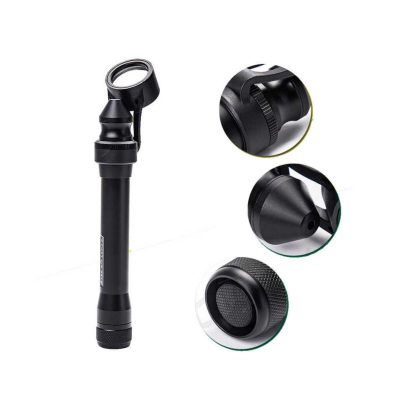
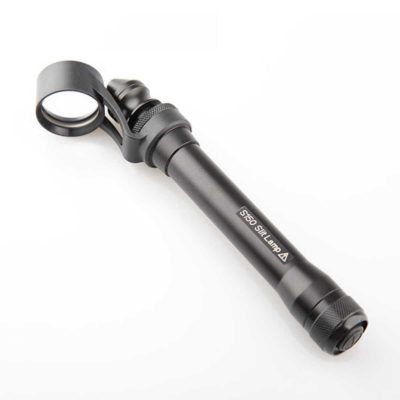
| Name | FOCUS ADVANCED MICROSCOPE remove | Retinoscope remove | Portable Fundus Camera remove | Portable Rebound Tonometer remove | View Tester (Manual Phoropter) remove | Ophthalmic AB Scan Machine remove | |||||||||||||||||||||||||||||||||||||||||||||||||||||||||||||||||||||||||||||||||||||||||||||||||||||||||||||||||||||
| Image |  | 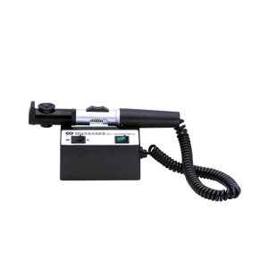 | 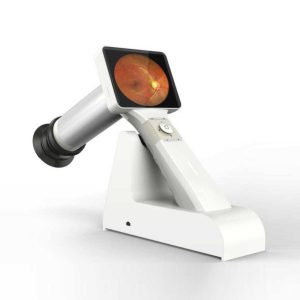 | 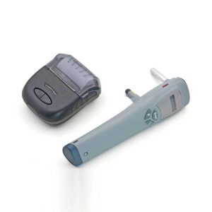 | 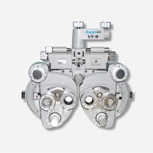 | 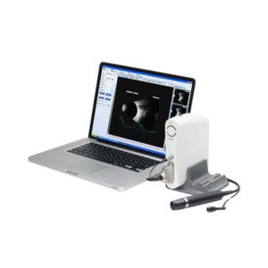 | |||||||||||||||||||||||||||||||||||||||||||||||||||||||||||||||||||||||||||||||||||||||||||||||||||||||||||||||||||||
| SKU | SF103356013091-8 | SF1033560107-12 | SF1033560107-23 | SF1033560107-17 | SF1033560107-26 | SF1033560107-8 | |||||||||||||||||||||||||||||||||||||||||||||||||||||||||||||||||||||||||||||||||||||||||||||||||||||||||||||||||||||
| Rating | |||||||||||||||||||||||||||||||||||||||||||||||||||||||||||||||||||||||||||||||||||||||||||||||||||||||||||||||||||||||||||
| Price |
| $165.00 | $2,310.00 | $1,815.00 | $858.00 | $4,895.00 | |||||||||||||||||||||||||||||||||||||||||||||||||||||||||||||||||||||||||||||||||||||||||||||||||||||||||||||||||||||
| Stock | |||||||||||||||||||||||||||||||||||||||||||||||||||||||||||||||||||||||||||||||||||||||||||||||||||||||||||||||||||||||||||
| Availability | |||||||||||||||||||||||||||||||||||||||||||||||||||||||||||||||||||||||||||||||||||||||||||||||||||||||||||||||||||||||||||
| Add to cart | |||||||||||||||||||||||||||||||||||||||||||||||||||||||||||||||||||||||||||||||||||||||||||||||||||||||||||||||||||||||||||
| Description | Shipped From Abroad
The FOCUS series is a line of equipment specially designed in detail to meet the most demanding standards of anterior and posterior segment surgical procedures.
The optics developed for FOCUS line microscopes enable external transmission of LED light, generating excellent clarity and image depth of the observed field.
Ideal for Cataract and Retina procedures.
The optics developed for FOCUS ADVANCED line microscopes enable external transmission of LED light (oblique), generating excellent clarity and image depth of the observed field. The equipment has an independent coaxial lighting system, activated via a multifunction pedal, ensuring total image clarity and excellent red reflection quality, providing total safety and visibility during cataract procedures.
Having functions such as zoom, microfocusing, intensity control activated via pedal and being compatible with the best non-contact lens adapters for vitreectomy on the market, it enables all retinal procedures with total quality.
Delivery & Availability:
Typically 10-21 working days – excluding furniture and heavy/bulky equipment. Please contact us for further information. | Shipped from abroad
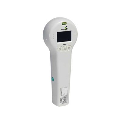
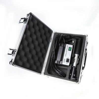
The product can quickly and precisely measure the astigmatism axis and is one of the necessary instruments in optometry inspection.
| 83Shipped from abroad
This is a portable medical camera for fundus imaging, diagnosis, and especially for fundus disease screening. It's compact, easy to obtain high definition fundus image. It can be conveniently applied to rapid screening, out diagnosis, bedside diagnosis and remote medical treatment, etc.
| Shipped from abroad
Tonometer SW-500 with vertical and horizontal two working modes, wireless output print data.
| Ship from abroad
| Shipped from abroad
| |||||||||||||||||||||||||||||||||||||||||||||||||||||||||||||||||||||||||||||||||||||||||||||||||||||||||||||||||||||
| Content | Description
Versatility in multiple configuration possibilities
Focus Microscopes stand out in the market for presenting multidisciplinary characteristics and high optical quality. Additionally, they allow you to add options and accessories according to the need for use, including binoculars, double iris, image inverter, among others.
The Focus* line of microscopes has a pedal-operated motorized microfocusing and zooming system, aiming for greater precision and comfort for the user during surgical procedures.
Pedal controlled XY positioner
The DFVasconcellos XY Positioner allows precise alignment between the user's vision and the surgical field. This becomes an increasingly necessary resource as microscopy techniques become more widespread.
| The product can quickly and precisely measure the astigmatism axis and is one of the necessary instruments in optometry inspection.
Features:
| Portable Fundus Camera is a portable medical camera for fundus imaging, diagnosis, and especially for fundus disease screening. It's compact, easy to obtain high definition fundus image. It can be conveniently applied to rapid screening, out diagnosis, bedside diagnosis and remote medical treatment, etc.
Features of Portable Fundus Camera:
| Tonometer SW-500 with vertical and horizontal two working modes, wireless output print data. The equipment is used to measure intraocular pressure, using the principle of: the probe hits the surfaces of different hardness at a certain speed, has a different reaction when the probe rebounds. Be of advantages of high accuracy, portable, without anesthesia, without the cross-infection, etc.
Features:
| Features:
| Functions of Ophthalmic AB Scan Machine:
| |||||||||||||||||||||||||||||||||||||||||||||||||||||||||||||||||||||||||||||||||||||||||||||||||||||||||||||||||||||
| Weight | N/A | N/A | N/A | N/A | N/A | N/A | |||||||||||||||||||||||||||||||||||||||||||||||||||||||||||||||||||||||||||||||||||||||||||||||||||||||||||||||||||||
| Dimensions | N/A | N/A | N/A | N/A | N/A | N/A | |||||||||||||||||||||||||||||||||||||||||||||||||||||||||||||||||||||||||||||||||||||||||||||||||||||||||||||||||||||
| Additional information | |||||||||||||||||||||||||||||||||||||||||||||||||||||||||||||||||||||||||||||||||||||||||||||||||||||||||||||||||||||||||||


Reviews
There are no reviews yet.