


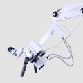
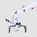
ENT/Neurosurgery Operating Microscope
$0.00
Shipped from abroad
Delivery & Availability:
Typically 14-21 working days – excluding furniture and heavy/bulky equipment. Please contact us for further information.
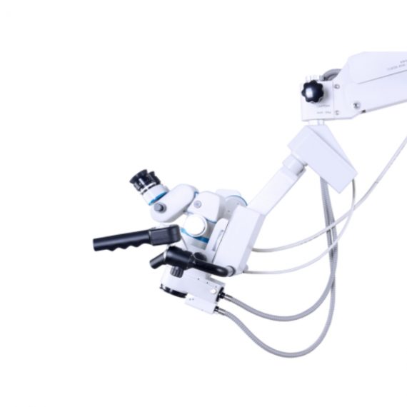
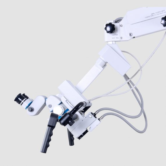
Corder Microscope has Fluid, Responsive and Accurate.
Fluid. Responsive. Accurate. These were a few of the principles guiding every phase in the design of the Corder Microscope. With the choicest mechanical machined components, the Corder Microscope has the grace and agility to adjust to every desired position on command. Well designed Apochromatic optics treated with Corder’s Mcoatings produce true-to life sharp images with high depth, definition and contrast.
Description
Features:
Corder Microscope has Fluid, Responsive and Accurate.
Fluid. Responsive. Accurate. These were a few of the principles guiding every phase in the design of the Corder Microscope. With the choicest mechanical machined components, the Corder Microscope has the grace and agility to adjust to every desired position on command. Well designed Apochromatic optics treated with Corder’s Mcoatings produce true-to life sharp images with high depth, definition and contrast.
More comfortable operation
Tiltable binocular tubes available, which can incline more than 60° depending on the posture and physique of the operating surgeon.
Movable range: 30° (straight) to 90° (inclined) Corder microscope configured with XYZ motorized movement operated through a comfortable foot /Handle control, a veryeffective co-axial illumnation and 50W halogen light source makes it ideal for Neuro surgeries.
Doctor-patient communication is easier
To address digital documentation needs, a host of digital SLR, video camera, and CCD adapters are made available with the ProLine in addition to Corder’s proprietary iVu multi-functional imaging solution. 1080P full hd image quality, efficient image management during the operation. Integrate your digital workflow to facilitate case management and facilitate more intuitive patient communication.
Technical Permeants:
Magnification: motorized zoom system, 1:6 zoom ratio, magnification 3x~16x
Focusing range: 50mm
Binocular tube: 30°~90° tiltable tube ,(0° ~200° optional)
Eyepiece: 12.5x / 10x
Objective lens: F 300mm(175mm, 250mm, 350mm optional)
pupil distance: 55mm~75mm
diopter adjustment: +6D ~ -6D
Field of view: Φ74~Φ12mm
X-Y translator: Motorized by foot switch or handle controller, ±30mm
Assistant tube: 360° Rotating assistant tube
Reset functions: YES
Illumination
System: Coaxial illumination
Light source: Halogen lamp
Light intensity adjustment: Continuous brightness adjustment 0-100000lux
Fiber optic illumination: Dual fiber
Field of illumination: Φ50mm
Filter: Red free filter, small spot
Accessories
CCD Camera system: Beam splitter, CCD adapter, CCD, Display
XENON LAMP: 150000lux
Integrated Video Adapter: SONY / CANON Camera
Click Here To Download Catalogue
Quick Comparison
| ENT/Neurosurgery Operating Microscope remove | ASPEL Ambulatory BP Machine remove | Sonoscape P50 Ultrasound Machine remove | Sonoscape E2 Ultrasound Machine remove | DrGem Ceiling Analogue X-ray Machine remove | Sonoscape P20 Ultrasound Machine remove | |
|---|---|---|---|---|---|---|
| Name | ENT/Neurosurgery Operating Microscope remove | ASPEL Ambulatory BP Machine remove | Sonoscape P50 Ultrasound Machine remove | Sonoscape E2 Ultrasound Machine remove | DrGem Ceiling Analogue X-ray Machine remove | Sonoscape P20 Ultrasound Machine remove |
| Image | 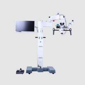 | 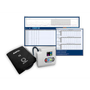 |  |  |  |  |
| SKU | SF1033560109-1 | SF1033560075-13 | SF1033560012-11 | SF1033560012-17 | SF1033560074-7 | SF1033560012-9 |
| Rating | ||||||
| Price |
| $920.00 |
| $5,500.00 |
|
|
| Stock | ||||||
| Availability | ||||||
| Add to cart | ||||||
| Description | Shipped from abroad
Corder Microscope has Fluid, Responsive and Accurate.Fluid. Responsive. Accurate. These were a few of the principles guiding every phase in the design of the Corder Microscope. With the choicest mechanical machined components, the Corder Microscope has the grace and agility to adjust to every desired position on command. Well designed Apochromatic optics treated with Corder's Mcoatings produce true-to life sharp images with high depth, definition and contrast. | Shipped from Abroad ASPEL Ambulatory BP Machine - is a recorder of long-term records of non-invasive measurement of blood pressure intended for use in clinics, hospitals, outpatient centers and specialist surgeries. The recorder enables the assessment of blood pressure by the oscillometric method in adult patients, pregnant women, including preeclampsia and pediatric patients (from 3 years of age). Blood pressure is assessed by using an inflatable cuff, an accurate pressure transducer, and a deflation valve. Delivery & Availability: Typically 10 working days – excluding furniture and heavy/bulky equipment. Please contact us for further information. | Shipped from Abroad Easily accomplish more with SonoScape’s new P50 ultrasound system. Incorporating single crystal clarity, automatic corrections and calculation, and user defined flexibility promises a confident diagnostic experience as well as opening new doors of opportunity for ultrasound use. Delivery & Availability: Typically 7-14 working days – excluding furniture and heavy/bulky equipment. Please contact us for further information. | Shipped from Abroad Sonoscape E2 portable ultrasound machine is a color Doppler ultrasound system that reaches beyond your expectations due to its compact and fashionable appearance. It fulfills GI, OB/GYN, Cardiac and POC applications to fit your routine scanning needs while its color mode will help you for more accurate and efficient diagnosis of lesions. E2 provides a wide range of applications to assist users with routine scanning. E2 provides automatic calculations to enhance your diagnostic confidence and save you time for patient communication. Delivery & Availability: Typically 14 working days – excluding furniture and heavy/bulky equipment. Please contact us for further information. | Shipped from abroad The DrGem Ceiling Analogue X-ray Machine is a diagnostic radiography system that provides reliable high quality radiographic images with a reduced dose. The reliable high-frequency x-ray generators that are known worldwide for their excellent performance, lifetime and stability. Patient tables and wall stands are also offered. Delivery & Availability: Typically 21 working days – excluding furniture and heavy/bulky equipment. Please contact us for further information. | Shipped from Abroad Incorporating innovative technologies, P20’s user-friendly design with a simple operation panel, intuitive user interface and a variety of intelligent auxiliary scanning tools, will significantly improve your daily examination experience. Besides general imaging applications, P20 has entitled with diagnostic 4D technology which has an extraordinary performance in obstetrics and gynecology applications. Delivery & Availability: Typically 5-7 working days – excluding furniture and heavy/bulky equipment. Please contact us for further information. |
| Content | Features:Corder Microscope has Fluid, Responsive and Accurate.Fluid. Responsive. Accurate. These were a few of the principles guiding every phase in the design of the Corder Microscope. With the choicest mechanical machined components, the Corder Microscope has the grace and agility to adjust to every desired position on command. Well designed Apochromatic optics treated with Corder's Mcoatings produce true-to life sharp images with high depth, definition and contrast. More comfortable operation Tiltable binocular tubes available, which can incline more than 60° depending on the posture and physique of the operating surgeon. Movable range: 30° (straight) to 90° (inclined) Corder microscope configured with XYZ motorized movement operated through a comfortable foot /Handle control, a veryeffective co-axial illumnation and 50W halogen light source makes it ideal for Neuro surgeries.Doctor-patient communication is easierTo address digital documentation needs, a host of digital SLR, video camera, and CCD adapters are made available with the ProLine in addition to Corder's proprietary iVu multi-functional imaging solution. 1080P full hd image quality, efficient image management during the operation. Integrate your digital workflow to facilitate case management and facilitate more intuitive patient communication. Technical Permeants: Magnification: motorized zoom system, 1:6 zoom ratio, magnification 3x~16x Focusing range: 50mm Binocular tube: 30°~90° tiltable tube ,(0° ~200° optional) Eyepiece: 12.5x / 10x Objective lens: F 300mm(175mm, 250mm, 350mm optional) pupil distance: 55mm~75mm diopter adjustment: +6D ~ -6D Field of view: Φ74~Φ12mm X-Y translator: Motorized by foot switch or handle controller, ±30mm Assistant tube: 360° Rotating assistant tube Reset functions: YES Illumination System: Coaxial illumination Light source: Halogen lamp Light intensity adjustment: Continuous brightness adjustment 0-100000lux Fiber optic illumination: Dual fiber Field of illumination: Φ50mm Filter: Red free filter, small spot Accessories CCD Camera system: Beam splitter, CCD adapter, CCD, Display XENON LAMP: 150000lux Integrated Video Adapter: SONY / CANON CameraClick Here To Download Catalogue | ASPEL Ambulatory BP Machine - is a recorder of long-term records of non-invasive measurement of blood pressure intended for use in clinics, hospitals, outpatient centers and specialist surgeries. The recorder enables the assessment of blood pressure by the oscillometric method in adult patients, pregnant women, including preeclampsia and pediatric patients (from 3 years of age). Blood pressure is assessed by using an inflatable cuff, an accurate pressure transducer, and a deflation valve.
Features:
Save-2-Safe: Double security system
Thanks to the use of two independent measuring systems with an additional valve, it meets the highest standards and takes care of patient safety even better.
Start-Easy: Quick start in two moves
The quick launch function allows you to use the device instantly, easily allows you to start recording in holter mode.
Memo-Care: Cuff pressure memory
Recorder remembers the pressure in the cuff. Thanks to the use of Intelligent Solutions, it adapts individually to the patient.
Power-Usb: USB connection
The device can work without batteries: by connecting to a computer via a USB cable.
Technical Specification:
Click Here To Download Catalogue | DETAILS
Powerful Compact Precision
Taking into consideration the evolving expectations and needs for ultrasound, the P50 is a slim and unobtrusive trolley system that is comfortable in tight, congested spaces with little room to work in. Providing everything you need for a comfortable examination in a small space for both you and your patient.
Single Crystal Transducer
Wideband single crystal probes greatly improve the signal ratio, acquire stunning images and provide superior sensitivity and resolution for both the near and far-fields.
μ-Scan+
The new generation μ-Scan imaging technologies give you better image quality by reducing noise, improving signal strength and improving visualization.
Dynamic Color
Dynamic colour improves upon already existing colour Doppler technologies for clear capture of colour flow and detail visualization of even tiny veins with lower velocities.
Solution for Radiology
P50, is a leading-edge ultrasound system that can meet the demands of any clinical setting. You can experience a superior performance in multi-dimensional imaging for a full range of clinical applications – abdominal, breast and cardiovascular.
C-xlasto Imaging
By understanding that tissue stiffness varies depending on the type of tissue, we can use C-xlasto Imaging to easily find abnormalities and tumours within soft tissue. The differences in tissue responses are detected and visualized in real-time by the elastography algorithms through different representations, which can be particularly helpful in analyzing breast, thyroid and musculoskeletal structures. Predominately used only in linear probes, SonoScape’s new transvaginal and bi-plane probe for gynaecology and urology are breaking the mould and expanding elastography applications.
Real-time Color Panoramic
With the combination of colour flow and real-time panoramic, visualizing the blood flow of an entire vein or artery is now an easy task. Accomplished in real-time for the convenience of the sonographers, any mistakes can also be easily backtracked and corrected without interrupting the scan.
Contrast Imaging
Contrast Imaging on P50 makes full use of the infra harmonic signal and second harmonic signal to improve the image resolution and deep penetration. What’s more, the Dynamic Acoustic Control technology effectively controls the acoustic pressure for the contrast agent, decreasing the required agent dose and assures uniform image quality, guaranteeing longer contrast agent duration and better lesion perfusion of delayed phase observation.
Solution for OB/GYN
P50 has superior image quality, automated measurement tools, and a variety of volume technologies to provide ideal solutions for clinical examinations such as pregnancy examinations, and gynecologic disease diagnosis. With a new 4D transvaginal probe, P50 helps you to see and detect fetal abnormalities and significantly improves your diagnostic confidence during your examinations.
S-Live Silhouette
A unique transparent 3D anatomical image of the fetus for improved initial anatomical review. By using this new application, the system can create completely different fetal images from conventional ultrasound images, which can depict the fetal's intracorporeal anatomical structure.
Pelvic Floor 4D
Working in conjunction with SonoScape’s latest transvaginal probes, trans-perineal 4D pelvic floor ultrasound provides a useful clinical assessment of the impact of vaginal delivery on the female anterior compartment. Allowing doctors to judge whether the pelvic organs prolapsed or not, the extent of prolapse, and determining whether the pelvic muscles tore correctly.
S-Guide
S-Guide gives the user an extensive list of example obstetric ultrasound images as reference guides and a convenient checklist system to keep track of their progress during their obstetrics examination.
Auto Face
Automatically removes masking layers in front of the fetus’s face for a clearer vision of the fetus’s face.
AVC Follicle
AVC Follicle automatically identifies how many follicles are present and calculates their individual volumes.
Solution for Cardiology
P50 provides clear 2D clinical images and Doppler sensitivity to assess critical cardiac performance. Compatible with SonoScape’s single crystal probes, the P50 can provide images with better resolution and penetration in Cardiac diagnosis.
Tissue Doppler Imaging
Tissue Doppler Imaging allows clinical doctors to quantitatively evaluate local myocardial movements and functions, facilitating them with the ability to analyze and compare the motions of the different parts of the patient’s heart.
Stress Echo
Stress echocardiography is the combination of 2D echocardiography with physical, pharmacological or electrical stress of the patient. It also then provides users with report management tools such as configurable template editor, multiple loops to select one for storage, wall motion scoring, stress echo report, etc
Auto IMT
Auto IMT is used when determining the level of vascular sclerosis present in the patient by automatically tracing and calculating the thickness of the carotid vessels. What distinguishes the P50 is that it provides an instant and accurate Mean and Max index at the touch of a single button.
Auto EF
Automated 2D Cardiac Quantification is a fully intelligent trace function for endocardium with 19 easily-adjustable points providing rapid access to proven 2D EF and volumes.
Click Here To Download Catalogue | SONOSCAPE E2 DETAILS
Auto Image Optimization
A portable ultrasound machine with the press of a button, the image is automatically adjusted and optimized, saving you time with parameter adjustments. Additionally, with Auto Focus on, the focus area follows the depth of the ROI box as it is moved in the scanning field, providing users with excellent image quality in the desired area of interest.
Automated Calculation
Auto IMT is used when determining the level of vascular sclerosis present in the patient by automatically tracing the thickness of the carotid vessels.
Auto trace provides users sensitive and accurate wave tracing, avoiding the error of manual trace and giving out calculation result in no time
In-Build Battery pack
This portable ultrasound machine was equipped with an in-build battery pack which enable the user to perform image scanning when AC power is not available.
Click Here To Download Catalogue | DrGem Ceiling Analogue X-ray Machine is a diagnostic radiography system X-ray Machine that provides reliable high quality radiographic images with a reduced dose. The reliable high-frequency x-ray generators that are known worldwide for their excellent performance, lifetime and stability. Patient tables and wall stands are also offered.
Features of DrGem Ceiling Analogue X-ray Machine
Click Here To Download Catalogue | DETAILS
Upgraded Images with More Clarity
SonoScape never stops making progress in improving the image quality of its ultrasound products to enhance the confidence of diagnosis for doctors. With extraordinary images provided by P20, the anatomy structures are clearer than ever.
C-Xlasto Imaging
With C-xlasto Imaging, P20 enables comprehensive quantitative elastic analysis. Meanwhile, C-xlasto on P20 is supported by linear, convex and transvaginal probes, to ensure good reproducibility and highly consistent quantitative elastic results.
S-Live
S-Live allows for detailed visualization of subtle anatomical features, thereby enabling intuitive diagnosis with real-time 3D images and enriching patient communication.
Pelvic Floor 4D
Transperineal 4D pelvic floor ultrasound can provide useful clinical values in assessing the vaginal delivery impact on the female anterior compartment, judging whether the pelvic organs are prolapsed or not and the extent, determining if the pelvic muscles were torn accurately.
Anatomic M Mode
Anatomic M Mode helps you observe the myocardial motion at different phases by freely placing sample lines. It accurately measures the myocardial thickness and the heart size of even difficult patients and supports the myocardial function and LV wall-motion assessment.
Tissue Doppler Imaging
P20 is endowed with Tissue Doppler Imaging which provides velocities and other clinical information on myocardial functions, facilitating clinical doctors with the ability to analyze and compare the motions of different parts of the patient's heart.
Click Here To Download Catalogue |
| Weight | N/A | N/A | N/A | N/A | N/A | N/A |
| Dimensions | N/A | N/A | N/A | N/A | N/A | N/A |
| Additional information |


Reviews
There are no reviews yet.