Nio Gray 5.8MP (MDNG‑6221)
$0.00
Shipped From Abroad
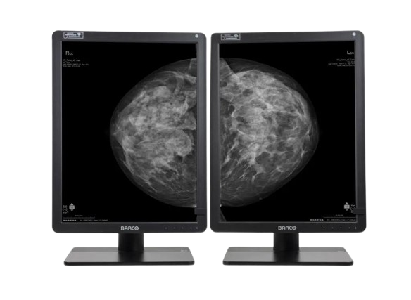
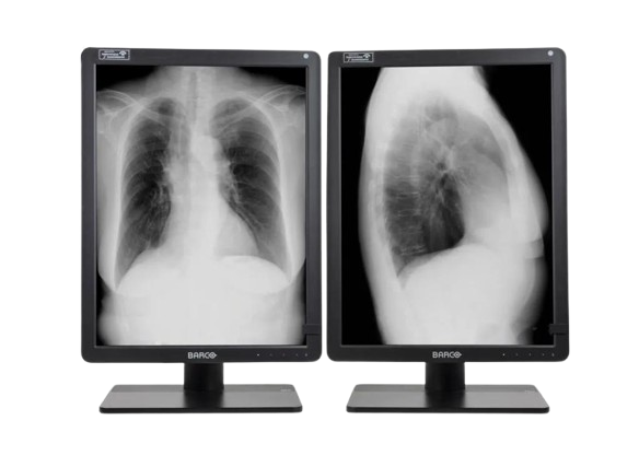
The Barco Nio Gray 5.8MP (MDNG-6221) is a high-performance grayscale medical display designed for mammography and diagnostic imaging. It ensures superior image clarity, consistent luminance, and advanced calibration for precise and reliable clinical interpretation in breast imaging and radiology.
Typically 10-21 working days – excluding furniture and heavy/bulky equipment. Please contact us for further information.
Description
The Barco Nio Gray 5.8MP (MDNG-6221) is a premium grayscale diagnostic display optimized for mammography and breast imaging. With a 5.8-megapixel resolution, exceptional luminance, and DICOM compliance, it delivers outstanding image quality for accurate detection and interpretation. Equipped with I-Guard for continuous quality assurance and QAWeb for remote management, this display ensures long-term consistency and reliability. Its ergonomic design and advanced grayscale rendering make it the ideal solution for radiologists requiring precise visualization in demanding diagnostic environments.
Bigger image, more details
Why 5.8MP? Well, in contrast to conventional 5.2MP display systems, you get 12% more pixels on your screen, which means that you can see more details at any given moment. Combine this with the tall 4:3 aspect ratio, which offers more room to view images in their entirety.
Reliable reading
The Nio Gray 5.8MP offers you more Just Noticeable Differences, thanks to its high brightness and contrast ratio. Our integrated stability, calibration, and uniformity technologies make sure that image quality, light output and DICOM compliance remain consistent over the years. The ambient light measurement sensor helps you stay in control of the environment you work in.
Efficient workflow
The Nio Gray 5.8MP is more than a grayscale display alone. It offers many ways to personalize settings to your liking, such as preferred tints of white or viewing angle. The KVM option helps you switch between workstations at the touch of a button.
On top of that, the display can help you improve your efficiency and speed, thanks to the set of Intuitive Workflow Tools included with our MXRT medical display controllers.
Did you know that SpotView, for example, makes it possible to make an area you choose twice as bright as it was originally? It’s been proven to help radiologists reduce their reading time by as much as 15.5%.
Long lifetime, clear view
Install our free and secure QAWeb Enterprise application, and rely on intervention-free, remote quality assurance.
To summarize, your Nio Gray 5.8MP is a functional, easy-to-use diagnostic display system, fully up to date with today’s innovations in general grayscale radiology, as well as 2D and 3D mammography. It comes with a 5-year warranty on all its components.
Ensuring diagnostic confidence with MDR Class IIa
Our radiology displays are MDR-certified as Class IIa. Their product information has been reviewed and cleared by independent medical and technical experts, and is audited yearly. In other words, we ensure diagnostic confidence and peace of mind for our users.
Technologies that enhance image quality:
- More detail on your screen, with 5.8MP resolution
- Designed to show breast images entirely, with a 3:4 aspect ratio
- Increased contrast, with a 1400:1 contrast ratio and 600 to 1000 cd/m² calibrated luminance
- Consistent brightness and grays, with Uniform Luminance Technology and SteadyGray
- Always stable DICOM images and auto QA, with I-Guard front sensor and optionally, QAWeb Enterprise
- Possibility to boost luminance, with I-Luminate and SpotView
- Optional settings and tools to adjust the display to your workflow, with Intuitive Workflow Tools
Ecolabel A for Nio Gray 5.8MP
The Nio Gray 5.8MP has been subjected to Barco’s ecoscoring protocol and has received an A rating. Some key factors that contributed to this rating are:
- Energy-efficient power supply, energy-efficient standby, and off modes
- Possibility to automatically switch to standby mode when the device is not in use
- Halogen-free cables and plastics
- Use of recycled cardboard in packaging (>85% recycled content)
- Product design optimized for disassembly with common tools
Features
-
5.8MP high-resolution grayscale medical display
-
Optimized for mammography and diagnostic imaging
-
High luminance and contrast for clinical accuracy
-
I-Guard sensor for real-time quality assurance
-
DICOM calibration for consistent grayscale performance
-
QAWeb for remote quality and compliance management
-
Ergonomic design for comfortable, extended use
Specifications
| Category | Specification |
|---|---|
| Screen Technology | LCD |
| Active Screen Size | 21.3″ (541 mm); 324 × 433 mm (12.77″ × 17″) |
| Aspect Ratio | 3:4 per display (portrait), 3:2 overall |
| Resolution | 5.8 MP (2100 × 2800 pixels) |
| Pixel Pitch | 0.1545 mm |
| Color Imaging | No |
| Gray Imaging | Yes |
| Bit Depth | 10-bit |
| Viewing Angle | 178° (H/V) |
| Uniformity Correction | ULT |
| SteadyGray | Yes |
| SteadyColor | N/A |
| I-Luminate | Yes |
| Ambient Light Presets | Yes, reading room selection |
| Ambient Light Sensor | Yes |
| Front Sensor | Yes (I-Guard) |
| Presence Sensor | N/A |
| Maximum Luminance | 1560 cd/m² |
| DICOM Calibrated Luminance | Factory default: 600 cd/m²; Warrantied max: 1000 cd/m² |
| Contrast Ratio | 1400:1 |
| Response Time | 12.5 ms ((Tr + Tf)/2) |
| Housing Color | Black (RAL 9004) / White (RAL 9003) |
| Video Input Signals | 2 × DisplayPort 1.4 |
| Video Output Signals | N/A |
| USB Ports | 2 × USB-B 2.0 upstream; 5 × USB-A 2.0 downstream (1 charging) |
| KVM Switch | Yes |
| Power Rating | 24 VDC, 5 A |
| Power Requirements | AdapterTech ATM160T-P240 (100–240 Vac, 50–60 Hz, 1.8–0.9 A; Output: 24 VDC, 6.6 A) |
| Power Consumption | 60 W (nominal); 0.4 W (hibernate/off) |
| Dimensions with Stand | Portrait: 378 × 528~628 × 235 mm; Landscape: 491 × 472~572 × 235 mm |
| Dimensions without Stand | Portrait: 378 × 491 × 84 mm; Landscape: 491 × 378 × 84 mm |
| Packaged Dimensions | 500 × 280 × 670 mm |
| Net Weight with Stand | With cover: 11.9 kg; Without cover: 10.6 kg |
| Net Weight without Stand | With cover: 6.9 kg; Without cover: 5.6 kg |
| Packaged Weight | With cover: 16.9 kg; Without cover: 15.6 kg |
| Tilt / Swivel / Pivot | Tilt: -10° to +30°; Swivel: ±45°; Pivot: 90° |
| Height Adjustment Range | 100 mm |
| Mounting Standard | VESA (100 mm) |
| Screen Protection | Optional protective, anti-reflective glass |
| Recommended Modalities | All digital images, including digital mammography |
| Certifications | FDA 510(K) K170476, CE0123, CCC, KC, INMETRO, BIS |
| Safety Standards | IEC/EN/AAMI/CSA 62368-1, 60601-1 |
| EMI Compliance | IEC/EN 60601-1-2, FCC Part 15 Class B, ICES-001 Level B, VCCI |
| Environmental Compliance | EU RoHS, China RoHS, REACH, Canada Health, WEEE, Packaging Directive |
| Supplied Accessories | User Guide, Documentation Disc, System Sheet, DisplayPort Cable, USB Cable, Mains Cable(s), External Power Supply |
| Optional Accessories | Graphics Board, QA Software, QAWeb Enterprise |
| Warranty | 5 years, including 40,000 hours backlight warranty |
| Operating Temperature | 0–40 °C (specs: 15–30 °C) |
| Storage Temperature | -20–60 °C |
| Operating Humidity | 8–80% (non-condensing) |
| Storage Humidity | 5–85% (non-condensing) |
| Operating Pressure | 70 kPa |
| Storage Pressure | 50–106 kPa |
Quick Comparison
| Nio Gray 5.8MP (MDNG‑6221) remove | DrGem Ceiling Analogue X-ray Machine remove | Anke Supermark 1.5T MRI Machine remove | Anke MRI Openmark 5000 Permanent System remove | Sonoscape S8 Exp Portable Ultrasound remove | IBIS Neeo R9 Digital Surgical C-Arm remove | |||||||||||||||||||||||||||||||||||||||||||||||||||||||||||||||||||||||||||||||||||||||||||||||||||||||||||||||||||||||||||||||||||||||||||||||||||||||||||||||||||||||||||||||||||||||||||||||||||||||||||||||||||||||||||||||||||||||||||||||||||||||||||||||||||||||||||||||||||||||||||||||||||||||||||||||||||||||||||||||||||||||||||||||||||||||||||||||||||||||||||||||||||||||||||||||||||||||||||||||||||||||||||||||||||||||||||||||||||||||||||||||||||||||||||||||||||||||||||||||||||||||||||||||||||||||||||||||||||||||||||||||||||||||||||||||||||||||||||||||||||||||||||||||||||||||||||||||||||||||||||||||||||||||||||||||||||||||||||||||||||||||||||||||||||||||||||||||||||||||||||||||||||||||||||||||||||||||||||
|---|---|---|---|---|---|---|---|---|---|---|---|---|---|---|---|---|---|---|---|---|---|---|---|---|---|---|---|---|---|---|---|---|---|---|---|---|---|---|---|---|---|---|---|---|---|---|---|---|---|---|---|---|---|---|---|---|---|---|---|---|---|---|---|---|---|---|---|---|---|---|---|---|---|---|---|---|---|---|---|---|---|---|---|---|---|---|---|---|---|---|---|---|---|---|---|---|---|---|---|---|---|---|---|---|---|---|---|---|---|---|---|---|---|---|---|---|---|---|---|---|---|---|---|---|---|---|---|---|---|---|---|---|---|---|---|---|---|---|---|---|---|---|---|---|---|---|---|---|---|---|---|---|---|---|---|---|---|---|---|---|---|---|---|---|---|---|---|---|---|---|---|---|---|---|---|---|---|---|---|---|---|---|---|---|---|---|---|---|---|---|---|---|---|---|---|---|---|---|---|---|---|---|---|---|---|---|---|---|---|---|---|---|---|---|---|---|---|---|---|---|---|---|---|---|---|---|---|---|---|---|---|---|---|---|---|---|---|---|---|---|---|---|---|---|---|---|---|---|---|---|---|---|---|---|---|---|---|---|---|---|---|---|---|---|---|---|---|---|---|---|---|---|---|---|---|---|---|---|---|---|---|---|---|---|---|---|---|---|---|---|---|---|---|---|---|---|---|---|---|---|---|---|---|---|---|---|---|---|---|---|---|---|---|---|---|---|---|---|---|---|---|---|---|---|---|---|---|---|---|---|---|---|---|---|---|---|---|---|---|---|---|---|---|---|---|---|---|---|---|---|---|---|---|---|---|---|---|---|---|---|---|---|---|---|---|---|---|---|---|---|---|---|---|---|---|---|---|---|---|---|---|---|---|---|---|---|---|---|---|---|---|---|---|---|---|---|---|---|---|---|---|---|---|---|---|---|---|---|---|---|---|---|---|---|---|---|---|---|---|---|---|---|---|---|---|---|---|---|---|---|---|---|---|---|---|---|---|---|---|---|---|---|---|---|---|---|---|---|---|---|---|---|---|---|---|---|---|---|---|---|---|---|---|---|---|---|---|---|---|---|---|---|---|---|---|---|---|---|---|---|---|---|---|---|---|---|---|---|---|---|---|---|---|---|---|---|---|---|---|---|---|---|---|---|---|---|---|---|---|---|---|---|---|---|---|---|---|---|---|---|---|---|---|---|---|---|---|---|---|---|---|---|---|---|---|---|---|---|---|---|---|---|---|---|---|---|---|---|---|---|---|---|---|---|---|---|---|---|---|---|---|---|---|---|---|---|---|---|---|---|---|---|---|---|---|---|---|---|---|---|---|---|---|---|---|---|---|---|---|---|---|---|---|---|---|---|---|---|---|---|---|---|---|---|---|---|---|---|---|---|---|---|---|---|---|---|---|---|---|---|---|---|---|---|---|---|---|---|---|---|---|---|---|---|---|---|---|---|---|---|---|---|---|---|---|---|---|---|---|---|---|---|---|---|---|---|---|---|---|---|---|---|---|---|---|---|---|---|---|---|---|---|---|---|---|---|---|---|---|---|---|---|---|---|---|---|---|---|---|---|---|---|---|---|---|---|---|---|---|---|---|---|---|---|---|---|---|---|---|---|---|---|---|---|---|---|---|---|---|---|
| Name | Nio Gray 5.8MP (MDNG‑6221) remove | DrGem Ceiling Analogue X-ray Machine remove | Anke Supermark 1.5T MRI Machine remove | Anke MRI Openmark 5000 Permanent System remove | Sonoscape S8 Exp Portable Ultrasound remove | IBIS Neeo R9 Digital Surgical C-Arm remove | ||||||||||||||||||||||||||||||||||||||||||||||||||||||||||||||||||||||||||||||||||||||||||||||||||||||||||||||||||||||||||||||||||||||||||||||||||||||||||||||||||||||||||||||||||||||||||||||||||||||||||||||||||||||||||||||||||||||||||||||||||||||||||||||||||||||||||||||||||||||||||||||||||||||||||||||||||||||||||||||||||||||||||||||||||||||||||||||||||||||||||||||||||||||||||||||||||||||||||||||||||||||||||||||||||||||||||||||||||||||||||||||||||||||||||||||||||||||||||||||||||||||||||||||||||||||||||||||||||||||||||||||||||||||||||||||||||||||||||||||||||||||||||||||||||||||||||||||||||||||||||||||||||||||||||||||||||||||||||||||||||||||||||||||||||||||||||||||||||||||||||||||||||||||||||||||||||||||||||
| Image | 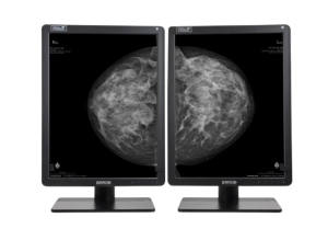 |  |  |  |  |  | ||||||||||||||||||||||||||||||||||||||||||||||||||||||||||||||||||||||||||||||||||||||||||||||||||||||||||||||||||||||||||||||||||||||||||||||||||||||||||||||||||||||||||||||||||||||||||||||||||||||||||||||||||||||||||||||||||||||||||||||||||||||||||||||||||||||||||||||||||||||||||||||||||||||||||||||||||||||||||||||||||||||||||||||||||||||||||||||||||||||||||||||||||||||||||||||||||||||||||||||||||||||||||||||||||||||||||||||||||||||||||||||||||||||||||||||||||||||||||||||||||||||||||||||||||||||||||||||||||||||||||||||||||||||||||||||||||||||||||||||||||||||||||||||||||||||||||||||||||||||||||||||||||||||||||||||||||||||||||||||||||||||||||||||||||||||||||||||||||||||||||||||||||||||||||||||||||||||||||
| SKU | SF1033560074-7 | SF1033560092-4 | SF1033560092-3 | SF1033560012-15 | SF1033560011-1 | |||||||||||||||||||||||||||||||||||||||||||||||||||||||||||||||||||||||||||||||||||||||||||||||||||||||||||||||||||||||||||||||||||||||||||||||||||||||||||||||||||||||||||||||||||||||||||||||||||||||||||||||||||||||||||||||||||||||||||||||||||||||||||||||||||||||||||||||||||||||||||||||||||||||||||||||||||||||||||||||||||||||||||||||||||||||||||||||||||||||||||||||||||||||||||||||||||||||||||||||||||||||||||||||||||||||||||||||||||||||||||||||||||||||||||||||||||||||||||||||||||||||||||||||||||||||||||||||||||||||||||||||||||||||||||||||||||||||||||||||||||||||||||||||||||||||||||||||||||||||||||||||||||||||||||||||||||||||||||||||||||||||||||||||||||||||||||||||||||||||||||||||||||||||||||||||||||||||||||
| Rating | ||||||||||||||||||||||||||||||||||||||||||||||||||||||||||||||||||||||||||||||||||||||||||||||||||||||||||||||||||||||||||||||||||||||||||||||||||||||||||||||||||||||||||||||||||||||||||||||||||||||||||||||||||||||||||||||||||||||||||||||||||||||||||||||||||||||||||||||||||||||||||||||||||||||||||||||||||||||||||||||||||||||||||||||||||||||||||||||||||||||||||||||||||||||||||||||||||||||||||||||||||||||||||||||||||||||||||||||||||||||||||||||||||||||||||||||||||||||||||||||||||||||||||||||||||||||||||||||||||||||||||||||||||||||||||||||||||||||||||||||||||||||||||||||||||||||||||||||||||||||||||||||||||||||||||||||||||||||||||||||||||||||||||||||||||||||||||||||||||||||||||||||||||||||||||||||||||||||||||||||||
| Price |
|
|
|
| $9,350.00 |
| ||||||||||||||||||||||||||||||||||||||||||||||||||||||||||||||||||||||||||||||||||||||||||||||||||||||||||||||||||||||||||||||||||||||||||||||||||||||||||||||||||||||||||||||||||||||||||||||||||||||||||||||||||||||||||||||||||||||||||||||||||||||||||||||||||||||||||||||||||||||||||||||||||||||||||||||||||||||||||||||||||||||||||||||||||||||||||||||||||||||||||||||||||||||||||||||||||||||||||||||||||||||||||||||||||||||||||||||||||||||||||||||||||||||||||||||||||||||||||||||||||||||||||||||||||||||||||||||||||||||||||||||||||||||||||||||||||||||||||||||||||||||||||||||||||||||||||||||||||||||||||||||||||||||||||||||||||||||||||||||||||||||||||||||||||||||||||||||||||||||||||||||||||||||||||||||||||||||||||
| Stock | ||||||||||||||||||||||||||||||||||||||||||||||||||||||||||||||||||||||||||||||||||||||||||||||||||||||||||||||||||||||||||||||||||||||||||||||||||||||||||||||||||||||||||||||||||||||||||||||||||||||||||||||||||||||||||||||||||||||||||||||||||||||||||||||||||||||||||||||||||||||||||||||||||||||||||||||||||||||||||||||||||||||||||||||||||||||||||||||||||||||||||||||||||||||||||||||||||||||||||||||||||||||||||||||||||||||||||||||||||||||||||||||||||||||||||||||||||||||||||||||||||||||||||||||||||||||||||||||||||||||||||||||||||||||||||||||||||||||||||||||||||||||||||||||||||||||||||||||||||||||||||||||||||||||||||||||||||||||||||||||||||||||||||||||||||||||||||||||||||||||||||||||||||||||||||||||||||||||||||||||||
| Availability | ||||||||||||||||||||||||||||||||||||||||||||||||||||||||||||||||||||||||||||||||||||||||||||||||||||||||||||||||||||||||||||||||||||||||||||||||||||||||||||||||||||||||||||||||||||||||||||||||||||||||||||||||||||||||||||||||||||||||||||||||||||||||||||||||||||||||||||||||||||||||||||||||||||||||||||||||||||||||||||||||||||||||||||||||||||||||||||||||||||||||||||||||||||||||||||||||||||||||||||||||||||||||||||||||||||||||||||||||||||||||||||||||||||||||||||||||||||||||||||||||||||||||||||||||||||||||||||||||||||||||||||||||||||||||||||||||||||||||||||||||||||||||||||||||||||||||||||||||||||||||||||||||||||||||||||||||||||||||||||||||||||||||||||||||||||||||||||||||||||||||||||||||||||||||||||||||||||||||||||||||
| Add to cart | ||||||||||||||||||||||||||||||||||||||||||||||||||||||||||||||||||||||||||||||||||||||||||||||||||||||||||||||||||||||||||||||||||||||||||||||||||||||||||||||||||||||||||||||||||||||||||||||||||||||||||||||||||||||||||||||||||||||||||||||||||||||||||||||||||||||||||||||||||||||||||||||||||||||||||||||||||||||||||||||||||||||||||||||||||||||||||||||||||||||||||||||||||||||||||||||||||||||||||||||||||||||||||||||||||||||||||||||||||||||||||||||||||||||||||||||||||||||||||||||||||||||||||||||||||||||||||||||||||||||||||||||||||||||||||||||||||||||||||||||||||||||||||||||||||||||||||||||||||||||||||||||||||||||||||||||||||||||||||||||||||||||||||||||||||||||||||||||||||||||||||||||||||||||||||||||||||||||||||||||||
| Description | Shipped From Abroad
The Barco Nio Gray 5.8MP (MDNG-6221) is a high-performance grayscale medical display designed for mammography and diagnostic imaging. It ensures superior image clarity, consistent luminance, and advanced calibration for precise and reliable clinical interpretation in breast imaging and radiology.
Delivery & Availability:
Typically 10-21 working days – excluding furniture and heavy/bulky equipment. Please contact us for further information.
| Shipped from abroad The DrGem Ceiling Analogue X-ray Machine is a diagnostic radiography system that provides reliable high quality radiographic images with a reduced dose. The reliable high-frequency x-ray generators that are known worldwide for their excellent performance, lifetime and stability. Patient tables and wall stands are also offered. Delivery & Availability: Typically 21 working days – excluding furniture and heavy/bulky equipment. Please contact us for further information. | Shipped from Abroad
SuperMark 1.5T is a new generation superconducting MRI system based on years of experience in production and research. It's applicable to whole body scan, such as, nervous system, spine, joint soft tissue, pelvic and abdominal cavity, etc
Delivery & Availability: Typically 90 working days – excluding furniture and heavy/bulky equipment. Please contact us for further information. | Shipped from Abroad
OPENMARK 5000 is 0.51T MRI. It's approved by FDA and has CE mark. It adopts two-pillar magnet design with 280 degree openness and equipped with powerful
RF and gradient system, together with advanced imaging technology, making it as a high-end system which is comparable to high-field MRI.
Delivery & Availability: Typically 90 working days – excluding furniture and heavy/bulky equipment. Please contact us for further information. | Shipped from Abroad With ultra-modern innovative design and the clinically-proven technologies, S8 Exp is portable ultrasound scanner well equipped as a low-physical-effort and enhanced-image-quality ultrasound scanner, which not only provides optimized solutions for versatile applications, but does help to improve the user-experience for both routine and non-traditional challenges. Delivery & Availability: Typically 5-7 working days – excluding furniture and heavy/bulky equipment. Please contact us for further information. | Shipped from Abroad Our Neeo “C” arms are easy to place, use and are specifically designed to be used in orthopedics, traumatology, abdominal surgery, urology, cardiology and operating rooms. Delivery & Availability: Typically 21 working days – excluding furniture and heavy/bulky equipment. Please contact us for further information. | ||||||||||||||||||||||||||||||||||||||||||||||||||||||||||||||||||||||||||||||||||||||||||||||||||||||||||||||||||||||||||||||||||||||||||||||||||||||||||||||||||||||||||||||||||||||||||||||||||||||||||||||||||||||||||||||||||||||||||||||||||||||||||||||||||||||||||||||||||||||||||||||||||||||||||||||||||||||||||||||||||||||||||||||||||||||||||||||||||||||||||||||||||||||||||||||||||||||||||||||||||||||||||||||||||||||||||||||||||||||||||||||||||||||||||||||||||||||||||||||||||||||||||||||||||||||||||||||||||||||||||||||||||||||||||||||||||||||||||||||||||||||||||||||||||||||||||||||||||||||||||||||||||||||||||||||||||||||||||||||||||||||||||||||||||||||||||||||||||||||||||||||||||||||||||||||||||||||||||
| Content | The Barco Nio Gray 5.8MP (MDNG-6221) is a premium grayscale diagnostic display optimized for mammography and breast imaging. With a 5.8-megapixel resolution, exceptional luminance, and DICOM compliance, it delivers outstanding image quality for accurate detection and interpretation. Equipped with I-Guard for continuous quality assurance and QAWeb for remote management, this display ensures long-term consistency and reliability. Its ergonomic design and advanced grayscale rendering make it the ideal solution for radiologists requiring precise visualization in demanding diagnostic environments.
Bigger image, more details Why 5.8MP? Well, in contrast to conventional 5.2MP display systems, you get 12% more pixels on your screen, which means that you can see more details at any given moment. Combine this with the tall 4:3 aspect ratio, which offers more room to view images in their entirety. Reliable reading The Nio Gray 5.8MP offers you more Just Noticeable Differences, thanks to its high brightness and contrast ratio. Our integrated stability, calibration, and uniformity technologies make sure that image quality, light output and DICOM compliance remain consistent over the years. The ambient light measurement sensor helps you stay in control of the environment you work in. Efficient workflow The Nio Gray 5.8MP is more than a grayscale display alone. It offers many ways to personalize settings to your liking, such as preferred tints of white or viewing angle. The KVM option helps you switch between workstations at the touch of a button. On top of that, the display can help you improve your efficiency and speed, thanks to the set of Intuitive Workflow Tools included with our MXRT medical display controllers. Did you know that SpotView, for example, makes it possible to make an area you choose twice as bright as it was originally? It’s been proven to help radiologists reduce their reading time by as much as 15.5%. Long lifetime, clear view Install our free and secure QAWeb Enterprise application, and rely on intervention-free, remote quality assurance. To summarize, your Nio Gray 5.8MP is a functional, easy-to-use diagnostic display system, fully up to date with today’s innovations in general grayscale radiology, as well as 2D and 3D mammography. It comes with a 5-year warranty on all its components. Ensuring diagnostic confidence with MDR Class IIa Our radiology displays are MDR-certified as Class IIa. Their product information has been reviewed and cleared by independent medical and technical experts, and is audited yearly. In other words, we ensure diagnostic confidence and peace of mind for our users.Technologies that enhance image quality:
Ecolabel A for Nio Gray 5.8MPThe Nio Gray 5.8MP has been subjected to Barco’s ecoscoring protocol and has received an A rating. Some key factors that contributed to this rating are:
Features
Specifications
| DrGem Ceiling Analogue X-ray Machine is a diagnostic radiography system X-ray Machine that provides reliable high quality radiographic images with a reduced dose. The reliable high-frequency x-ray generators that are known worldwide for their excellent performance, lifetime and stability. Patient tables and wall stands are also offered.
Features of DrGem Ceiling Analogue X-ray Machine
Click Here To Download Catalogue | SuperMark 1.5T is a new generation superconducting MRI system based on years of experience in production and research. It's applicable to whole body scan, such as, nervous system, spine, joint soft tissue, pelvic and abdominal cavity, etc. SuperMark 1.5T provides not only conventional pulse sequences and clinical diagnosis functions, but also provides advanced functional applications, for instance, 3D angiography and water imaging. It adopts brand new ANKE APEX operating system which ensures easy operation and fast diagnosis.
Technical Advantages:
Click Here To Download Catalogue | OPENMARK 5000 is 0.51T MRI. It's approved by FDA and has CE mark. It adopts two-pillar magnet design with 280 degree openness and equipped with powerful
RF and gradient system, together with advanced imaging technology, making it as a high-end system which is comparable to high-field MRI.
Features:
Click Here To Download Catalogue | Sonoscape S8 Exp Portable Ultrasound scannerDETAILS Agile and Versatile With ultra-modern innovative design and the clinically-proven technologies, S8 Exp Portable Ultrasound scanner is well equipped as a low-physical-effort and enhanced-image-quality ultrasound scanner, which not only provides optimized solutions for versatile applications but does help to improve the user experience for both routine and non-traditional challenges. Working with S8 Exp, it will trigger your unlimited reverie and endow you with endless charm. Carrying forward the classical design of SonoScape's portable ultrasound products, S8 Exp successfully combines the best ergonomics, attractive design and ease of use. This charismatic identity is also enhanced by a sophisticated color palette—with sedate grey as its interior paint color and pearl white as exterior cover, S8 Exp reveals a style of aristocrat and strong character among SonoScape's ultrasound systems. Workflow The S8 Exp is a portable ultrasound scanner that adapts to your workflow, whether you are in the consulting room, at the bedside, or at a remote location. With easy-to-use new platform designed for sonographers' needs and full connection interfaces for easy connectivity and data sharing, S8 Exp leads to improved user comfort and clinical outcome as well as patient throughput and working efficiency. Powerful Platform Embedded with SonoScape's core imaging technologies such as μ-scan, PHI and Spatial Compound, S8 Exp boasts exceptional 2D image, sensitive spectral, Color and Power Doppler, displaying well-defined anatomy and pathology and facilitating a highly optimized diagnostic user environment for conclusive diagnoses. Besides, S8 Exp offers a comprehensive selection of electronic probes to maximally extend its capabilities to meet a wide range of applications including the abdomen, pediatric, OB/GYN, cardiovascular, musculoskeletal, etc. The advanced probe technologies also effectively enhance the image quality and confidence in reaching clinical diagnoses even in difficult patients.Click Here To Download Catalogue | Our Neeo “C” arms are easy to place, use and are specifically designed to be used in orthopedics, traumatology, abdominal surgery, urology, cardiology and operating rooms.
Using Neeo with the RTP (Real Time Processing) option it is possible to perform vascular, urological and cardiological diagnostics. One of the main functions, digital image subtraction, allows to see, as an example, the passage of contrast liquids in a tissue or in a venous or arterial duct; thanks to the possibility of looping, the acquired video can be reproduced several times to monitor more accurately the passage of the fluid within the area in question. Angiographic measurement is another useful function in the vascular field (QA Quantitative Angiography) that allows the measurement of stenoses. Finally, fluoroscopy allows the correct positioning of stents or expanders.
Neeo is used in various interventional and diagnostic procedures in traumatology and orthopedics wards and operating rooms as well. Thanks to low-dose fluoroscopy, it is possible to use the device for positioning bone or subcutaneous grafts, inserting K-wire (Kirschner wire) for stabilization of bone fragments or for the correct positioning of prostheses. The low dose emitted ensures safe use for both the patient and the surgeon or doctor on the operating field.
On the control panel there is a large touch screen display that allows to adjust the basic functions of the equipment. From this display it is possible to select and adjust the fluoroscopic data for the examination, activate or deactivate the laser pointer, select between pulsed, one shot or standard fluoroscopy, rotate the image and perform all operations on collimator. The four side buttons on the display offer the possibility to move the bow vertically thanks to an extremely silent motor.
Neeo has two 19 “medical grade monitors that can be positioned according to the needs of the medical practitioner. Work monitors and feedback monitors are separated to be managed independently. The possible movements are: rotation, revolution, tilting and possibility of height adjustment.
Features:
Click Here To Download Catalogue | ||||||||||||||||||||||||||||||||||||||||||||||||||||||||||||||||||||||||||||||||||||||||||||||||||||||||||||||||||||||||||||||||||||||||||||||||||||||||||||||||||||||||||||||||||||||||||||||||||||||||||||||||||||||||||||||||||||||||||||||||||||||||||||||||||||||||||||||||||||||||||||||||||||||||||||||||||||||||||||||||||||||||||||||||||||||||||||||||||||||||||||||||||||||||||||||||||||||||||||||||||||||||||||||||||||||||||||||||||||||||||||||||||||||||||||||||||||||||||||||||||||||||||||||||||||||||||||||||||||||||||||||||||||||||||||||||||||||||||||||||||||||||||||||||||||||||||||||||||||||||||||||||||||||||||||||||||||||||||||||||||||||||||||||||||||||||||||||||||||||||||||||||||||||||||||||||||||||||||
| Weight | N/A | N/A | N/A | N/A | N/A | N/A | ||||||||||||||||||||||||||||||||||||||||||||||||||||||||||||||||||||||||||||||||||||||||||||||||||||||||||||||||||||||||||||||||||||||||||||||||||||||||||||||||||||||||||||||||||||||||||||||||||||||||||||||||||||||||||||||||||||||||||||||||||||||||||||||||||||||||||||||||||||||||||||||||||||||||||||||||||||||||||||||||||||||||||||||||||||||||||||||||||||||||||||||||||||||||||||||||||||||||||||||||||||||||||||||||||||||||||||||||||||||||||||||||||||||||||||||||||||||||||||||||||||||||||||||||||||||||||||||||||||||||||||||||||||||||||||||||||||||||||||||||||||||||||||||||||||||||||||||||||||||||||||||||||||||||||||||||||||||||||||||||||||||||||||||||||||||||||||||||||||||||||||||||||||||||||||||||||||||||||
| Dimensions | N/A | N/A | N/A | N/A | N/A | N/A | ||||||||||||||||||||||||||||||||||||||||||||||||||||||||||||||||||||||||||||||||||||||||||||||||||||||||||||||||||||||||||||||||||||||||||||||||||||||||||||||||||||||||||||||||||||||||||||||||||||||||||||||||||||||||||||||||||||||||||||||||||||||||||||||||||||||||||||||||||||||||||||||||||||||||||||||||||||||||||||||||||||||||||||||||||||||||||||||||||||||||||||||||||||||||||||||||||||||||||||||||||||||||||||||||||||||||||||||||||||||||||||||||||||||||||||||||||||||||||||||||||||||||||||||||||||||||||||||||||||||||||||||||||||||||||||||||||||||||||||||||||||||||||||||||||||||||||||||||||||||||||||||||||||||||||||||||||||||||||||||||||||||||||||||||||||||||||||||||||||||||||||||||||||||||||||||||||||||||||
| Additional information |
|
|


Reviews
There are no reviews yet.