
Optika B-293LD1.50 Trinocular LED Fluorescence Microscope
$0.00
Shipped from Abroad
Fluorescence binocular and trinocular microscopes especially designed for tubercolosis and malaria analysis.
Delivery & Availability:
Typically 21 working days – excluding furniture and heavy/bulky equipment. Please contact us for further information.

Description
Fluorescence binocular and trinocular microscopes especially designed for tubercolosis and malaria analysis.
Features:
- Observation mode: Brightfield.
- Head: Trinocular, 360° rotating and 30° inclined. Interpupillary distance 48-75mm.
- Dioptric adjustement: On the left eyepiece tube.
- Eyepieces: WF10x/20 mm, high eye-point and secured by a screw.
- Nosepiece: Quadruple revolving nosepiece, rotation on ball bearings.
- Objectives:
Equipped with binocular head and following objectives:
IOS N-PLAN 10x/0.25 (Cover/No Cover), with anti-fungus treatment
IOS N-PLAN 20x/0.40 (Cover/No Cover), with anti-fungus treatment
IOS N-PLAN 40x/0.65 (Cover/No Cover), with anti-fungus treatment
IOS W-PLAN MET 50x/0.75 (No Cover), with anti-fungus treatment - Specimen stage: Double layer rackless mechanical sliding stage, 150×139 mm, 75×33 mm X-Y movement range. Vernier scale on the two axes, accuracy: 0.1 mm.
- Focusing: Coaxial coarse and fine focusing mechanism with limit stop to prevent the contact between objective and specimen. Adjustable tension of coarse focusing knob.
- Condenser: Abbe N.A. 1.25, with objective-coded iris diaphragm, focusable and centerable.
- Brightfield Illumination (Fixed Koehler type): X-LED3 with white 3.6 W LED (6,300 K) and light intensity control. Multi-plug 100-240Vac/6Vdc external power supply.
- Fluorescence Illumination: Extra efficiency LED, with light intensity control. Peak wavelength: 465 nm, Power: 3.6W.
- Epi Fluorescence Attachment: Slider with 3 positions (2 fluorescence, 1 brightfield), with 1 included filterset: Fluorescence B: EX 460-490, DM 505, EM 515LP: Acridine Yellow, Acridine Orange, Auramine, DiO, DTAF, FITC, GFP, YFP, etc.
Technical Specifications:
| Observation Method –
Transmitted Light |
Brightfield | Yes |
| Phase contrast (Positive type) | As optional | |
| Darkfield | As optional | |
| Simple polarized light | As optional | |
| Observation Method –
Incident Light |
Fluorescence | Yes |
| Main Body | Type | Upright |
| Construction material | Aluminum die-cast | |
| Trasportation handle | Yes | |
| Head | Type | Trinocular (Siedentopf) |
| Split ratio | 50/50 | |
| Inclination | 30° | |
| 360° rotating | Yes | |
| Interpupillary distance (mm) | 48-75 | |
| Dioptric adjustement | On left tube | |
| Fixing screw for eyepieces | Yes | |
| Tube inner diameter (mm) | 23 | |
| Eyepieces | Field number (mm) | 20 |
| Magnification | 10x | |
| Pointer | As optional | |
| Micrometric scale | As optional | |
| Diameter of micrometer glass (mm) | 21 | |
| High eyepoint (for glass wearers) | Yes | |
| Rubber cup | Yes | |
| Nosepiece | Positions | Quadruple |
| Bi-directional | Yes | |
| Rotation on ball bearings | Yes | |
| Objective thread | RMS | |
| Objectives | Optical system | ∞ |
| Anti-fungus treatment | Yes | |
| Parfocal distance (mm) | 45 | |
| Standard magnifications | 40x-400x | |
| Type | IOS | |
| Objectives included | OS N-PLAN
10x/0.25, W.D. 5.8 mm IOS N-PLAN 20x/0.40, W.D. 5.1 mm IOS N-PLAN 40x/0.65, W.D. 0.43 mm IOS W-PLAN MET 50x/0.75, W.D. 0.32 mm |
|
| Stage | Type | Double layer |
| Dimensions (mm) | 150×139 | |
| Moving mechanism | Rackless | |
| Moving range (mm) | 75×33 | |
| Material | Anti-scratch painting | |
| Specimen holder | Yes | |
| Slide number | 1 | |
| X-Y Vernier scale | Yes | |
| Vernier scale accuracy (mm) | 0.1 | |
| Condenser – Single
Position |
Type | Abbe |
| Removable | Yes | |
| Numerical aperture (N.A.) | 1.25 | |
| Magnification scale for simplified positioning | Yes | |
| Diaphragms | Iris | |
| Centrable | Yes | |
| Focusable | By rack and pinion | |
| Focusing System | Type | Coaxial coarse & fine |
| Coarse total travel (mm) | 18 | |
| Fine total travel (per single rotation) (mm) | 0.4 | |
| Fine graduations | 100 | |
| Fine resolution (µm) | 4 | |
| Upper stop to prevent contact | Yes | |
| Adjustable tension | Yes | |
| Transmitted
Illumination |
Kohler illumination | Fixed |
| Type | X-LED | |
| X-LED type | X-LED3 | |
| Light source power (W) | 3.6 | |
| Brightness control | Manual | |
| Lifetime (hours) | > 65,000 | |
| Temperature (K) | 6,300 | |
| Max. required power (W) | 6 | |
| Power Supply for
Transmitted Illumination |
Type | External |
| Microscope connector | Jack, 2.1 mm | |
| Power plug type | Multi-plug (EU, UK, US) | |
| Input voltage | 100/240 Vac, 50/60 Hz | |
| Output voltage | 6 Vdc 2.5A | |
| Accessories Included | Dust cover | Yes |
| Immersion oil (10ml) | Yes | |
| Tension adjustment tool | Yes | |
| User Manual | Digital version (downloadable) | |
| Additional Information | Mirror for transmitted light (as optional).
External rechargeable battery pack (as optional) |
|
| Product Weight | Height (mm) | 515 |
| Width (mm) | 235 | |
| Depth (mm) | 340 | |
| Product Weight | (kg) | 7.5 |
| Fluorescence
Attachment |
Number of positions | 3 |
| Blue filter set (included) specs | Excitation: 460 – 490 nm; Dichroic: 505 nm;
Emission: 515LP nm |
|
| Filter dimensions | Excitation: 18 mm diam.;
Dichroic: 26.5 mm x 19 mm; Emission: 18 mm diam. |
|
| Filter set selection | Manual | |
| Fluorescence Light
Source |
Light source | Blue LED |
| Watt (W) | 3.6 | |
| LED wavelength | Blue LED: 465 nm | |
| Lifetime (hours) | > 65,000 | |
| Brightness control | Yes |
Quick Comparison
| Optika B-293LD1.50 Trinocular LED Fluorescence Microscope remove | Eppendorf Minispin Centrifuge remove | Motic BA210 Series Biomedical Microscope remove | 8 Bucket Centrifuge remove | Eppendorf Pipette Tip 20ul Per Pack remove | Hawksley 1400 Haematocrit Centrifuge remove | ||||||||||||||||||||||||||||||||||||||||||||||||||||||||||||||||||||||||||||||||||||||||||||||||||||||||||||||||||||||||||||||||||||||||||||||||||||||||||||||||||||||||||||||||||||||||||||||||||||||||||||||||||||||||||||||||||||||||||||||||||||||||||||||||||||||||||||||||||||||||||||||
|---|---|---|---|---|---|---|---|---|---|---|---|---|---|---|---|---|---|---|---|---|---|---|---|---|---|---|---|---|---|---|---|---|---|---|---|---|---|---|---|---|---|---|---|---|---|---|---|---|---|---|---|---|---|---|---|---|---|---|---|---|---|---|---|---|---|---|---|---|---|---|---|---|---|---|---|---|---|---|---|---|---|---|---|---|---|---|---|---|---|---|---|---|---|---|---|---|---|---|---|---|---|---|---|---|---|---|---|---|---|---|---|---|---|---|---|---|---|---|---|---|---|---|---|---|---|---|---|---|---|---|---|---|---|---|---|---|---|---|---|---|---|---|---|---|---|---|---|---|---|---|---|---|---|---|---|---|---|---|---|---|---|---|---|---|---|---|---|---|---|---|---|---|---|---|---|---|---|---|---|---|---|---|---|---|---|---|---|---|---|---|---|---|---|---|---|---|---|---|---|---|---|---|---|---|---|---|---|---|---|---|---|---|---|---|---|---|---|---|---|---|---|---|---|---|---|---|---|---|---|---|---|---|---|---|---|---|---|---|---|---|---|---|---|---|---|---|---|---|---|---|---|---|---|---|---|---|---|---|---|---|---|---|---|---|---|---|---|---|---|---|---|---|---|---|---|---|---|---|---|---|---|---|---|---|---|---|---|---|---|---|---|
| Name | Optika B-293LD1.50 Trinocular LED Fluorescence Microscope remove | Eppendorf Minispin Centrifuge remove | Motic BA210 Series Biomedical Microscope remove | 8 Bucket Centrifuge remove | Eppendorf Pipette Tip 20ul Per Pack remove | Hawksley 1400 Haematocrit Centrifuge remove | |||||||||||||||||||||||||||||||||||||||||||||||||||||||||||||||||||||||||||||||||||||||||||||||||||||||||||||||||||||||||||||||||||||||||||||||||||||||||||||||||||||||||||||||||||||||||||||||||||||||||||||||||||||||||||||||||||||||||||||||||||||||||||||||||||||||||||||||||||||||||||||
| Image |  | 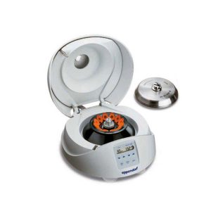 | 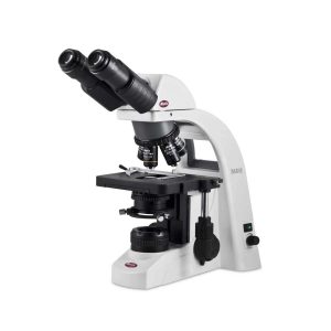 | 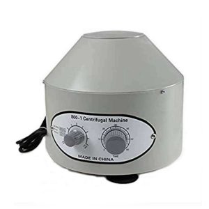 |  |  | |||||||||||||||||||||||||||||||||||||||||||||||||||||||||||||||||||||||||||||||||||||||||||||||||||||||||||||||||||||||||||||||||||||||||||||||||||||||||||||||||||||||||||||||||||||||||||||||||||||||||||||||||||||||||||||||||||||||||||||||||||||||||||||||||||||||||||||||||||||||||||||
| SKU | SF1033560098-7 | SF1033560084-116 | SF1033560084-78 | SF1033560084-114 | SF1033560084-136 | SF1033560084-118 | |||||||||||||||||||||||||||||||||||||||||||||||||||||||||||||||||||||||||||||||||||||||||||||||||||||||||||||||||||||||||||||||||||||||||||||||||||||||||||||||||||||||||||||||||||||||||||||||||||||||||||||||||||||||||||||||||||||||||||||||||||||||||||||||||||||||||||||||||||||||||||||
| Rating | |||||||||||||||||||||||||||||||||||||||||||||||||||||||||||||||||||||||||||||||||||||||||||||||||||||||||||||||||||||||||||||||||||||||||||||||||||||||||||||||||||||||||||||||||||||||||||||||||||||||||||||||||||||||||||||||||||||||||||||||||||||||||||||||||||||||||||||||||||||||||||||||||||
| Price |
|
|
| $72.50 |
| $690.00 | |||||||||||||||||||||||||||||||||||||||||||||||||||||||||||||||||||||||||||||||||||||||||||||||||||||||||||||||||||||||||||||||||||||||||||||||||||||||||||||||||||||||||||||||||||||||||||||||||||||||||||||||||||||||||||||||||||||||||||||||||||||||||||||||||||||||||||||||||||||||||||||
| Stock | |||||||||||||||||||||||||||||||||||||||||||||||||||||||||||||||||||||||||||||||||||||||||||||||||||||||||||||||||||||||||||||||||||||||||||||||||||||||||||||||||||||||||||||||||||||||||||||||||||||||||||||||||||||||||||||||||||||||||||||||||||||||||||||||||||||||||||||||||||||||||||||||||||
| Availability | |||||||||||||||||||||||||||||||||||||||||||||||||||||||||||||||||||||||||||||||||||||||||||||||||||||||||||||||||||||||||||||||||||||||||||||||||||||||||||||||||||||||||||||||||||||||||||||||||||||||||||||||||||||||||||||||||||||||||||||||||||||||||||||||||||||||||||||||||||||||||||||||||||
| Add to cart | |||||||||||||||||||||||||||||||||||||||||||||||||||||||||||||||||||||||||||||||||||||||||||||||||||||||||||||||||||||||||||||||||||||||||||||||||||||||||||||||||||||||||||||||||||||||||||||||||||||||||||||||||||||||||||||||||||||||||||||||||||||||||||||||||||||||||||||||||||||||||||||||||||
| Description | Shipped from Abroad
Fluorescence binocular and trinocular microscopes especially designed for tubercolosis and malaria analysis.
| In stock
| In stock The BA210 series is a line of basic biological microscopes manufactured by Motic. The unit is equipped with Motic Infinity Optics (CCIS®, offering quality performance in the fields of education and training. The device features a coaxial coarse and fine housing with a quadruple reverse-mounted nose piece. Its contrast methods, such as phase contrast, polarization and dark field, are easily performed through optional accessories. Delivery & Availability: Typically 5-7 working days – excluding furniture and heavy/bulky equipment. Please contact us for further information. | In stock
| In stock
| In stock
| |||||||||||||||||||||||||||||||||||||||||||||||||||||||||||||||||||||||||||||||||||||||||||||||||||||||||||||||||||||||||||||||||||||||||||||||||||||||||||||||||||||||||||||||||||||||||||||||||||||||||||||||||||||||||||||||||||||||||||||||||||||||||||||||||||||||||||||||||||||||||||||
| Content | Fluorescence binocular and trinocular microscopes especially designed for tubercolosis and malaria analysis.
Features:
| Powerful and user-friendly MiniSpin Centrifuge are small enough so that each workstation can be equipped with a “personal” centrifuge. The rotor, inner centrifuge lid, and lid latch are made of metal for maximum operation safety.
Features:
Click Here To Download Catalogue | The BA210 series is a line of basic biological microscopes manufactured by Motic. The unit is equipped with Motic Infinity Optics (CCIS®, offering quality performance in the fields of education and training. The device features a coaxial coarse and fine housing with a quadruple reverse-mounted nose piece. Its contrast methods, such as phase contrast, polarization and dark field, are easily performed through optional accessories. Technical applications: Biomedical Type: Optical Observation technique: Dark field Magnification: 4 unit, 10 unit, 40 unit, 100 unit | 800-1 electric centrifuge is widely used in hospitals, chemical and Biological Chemistry Laboratory for serum, plasma, immune qualitative analysis experiment. Has beautiful shape, large capacity, small volume, complete function. Its performance is stable, the speed is adjustable and can automatically adjust the balance, low temperature, high efficiency and wide applicability and advantages.
Technical Specifications:
Click Here To Download Catalogue |
| The Hawksley Haematospin 1400 Centrifuges are built in the UK and are made from durable and high-quality components. These centrifuges are ideal for veterinary laboratories and practices specifically designed to produce reliable and accurate results on blood and urine. One of the main features of this centrifuge is that it takes both 2ml microtubes and 75mm microhaematocrits. The classic design of these centrifuges is used worldwide with guaranteed support for all servicing and parts.
Technical specifications:
Click Here To Download Catalogue | |||||||||||||||||||||||||||||||||||||||||||||||||||||||||||||||||||||||||||||||||||||||||||||||||||||||||||||||||||||||||||||||||||||||||||||||||||||||||||||||||||||||||||||||||||||||||||||||||||||||||||||||||||||||||||||||||||||||||||||||||||||||||||||||||||||||||||||||||||||||||||||
| Weight | N/A | N/A | N/A | N/A | N/A | N/A | |||||||||||||||||||||||||||||||||||||||||||||||||||||||||||||||||||||||||||||||||||||||||||||||||||||||||||||||||||||||||||||||||||||||||||||||||||||||||||||||||||||||||||||||||||||||||||||||||||||||||||||||||||||||||||||||||||||||||||||||||||||||||||||||||||||||||||||||||||||||||||||
| Dimensions | N/A | N/A | N/A | N/A | N/A | N/A | |||||||||||||||||||||||||||||||||||||||||||||||||||||||||||||||||||||||||||||||||||||||||||||||||||||||||||||||||||||||||||||||||||||||||||||||||||||||||||||||||||||||||||||||||||||||||||||||||||||||||||||||||||||||||||||||||||||||||||||||||||||||||||||||||||||||||||||||||||||||||||||
| Additional information |


Reviews
There are no reviews yet.