Description
For gastroenterology
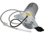
PNEUMOCOLON COMPLETE FOR EXAMINATION IN DOUBLE CONTRAST WALTER KREBS
Bock sealed composed of a tank for the contrast, pear Richardson, a hose and a field for carrying
Out the examinations of the colon in double contrast.
For routine barium irrigation or double contrast enema examination…
Pneumocolon is a lightweigh apparatus which is easy to handle….
Examinations with double contrasts are carried out simply by turning the apparatus….
Constant pressure gives a controlled flow of barium meal…
Quick Comparison
| PNE 1000-Pneumocolon Set remove | Anke Supermark 1.5T MRI Machine remove | Lab/Ward Coat remove | Jade Mobile X-ray machine (Analogue) remove | Sonoscape P15 Ultrasound Machine With Four Probes remove | Sonoscape S11 Ultrasound Machine remove | ||||||||||||||||||||||||||||||||||||||||||||||||||||||||||||||||||||||||||||||||||||||||||||||||||||||||||||||||||||||||||||||||||||||||||||||||||||||||||||||||||||||||||||||||||||||||||||||||||||||||||||||||||||||||||||||||||||||||||||||||||||||||||||||||||||||||||||||||||||||||||||||||||||||||||||||||
|---|---|---|---|---|---|---|---|---|---|---|---|---|---|---|---|---|---|---|---|---|---|---|---|---|---|---|---|---|---|---|---|---|---|---|---|---|---|---|---|---|---|---|---|---|---|---|---|---|---|---|---|---|---|---|---|---|---|---|---|---|---|---|---|---|---|---|---|---|---|---|---|---|---|---|---|---|---|---|---|---|---|---|---|---|---|---|---|---|---|---|---|---|---|---|---|---|---|---|---|---|---|---|---|---|---|---|---|---|---|---|---|---|---|---|---|---|---|---|---|---|---|---|---|---|---|---|---|---|---|---|---|---|---|---|---|---|---|---|---|---|---|---|---|---|---|---|---|---|---|---|---|---|---|---|---|---|---|---|---|---|---|---|---|---|---|---|---|---|---|---|---|---|---|---|---|---|---|---|---|---|---|---|---|---|---|---|---|---|---|---|---|---|---|---|---|---|---|---|---|---|---|---|---|---|---|---|---|---|---|---|---|---|---|---|---|---|---|---|---|---|---|---|---|---|---|---|---|---|---|---|---|---|---|---|---|---|---|---|---|---|---|---|---|---|---|---|---|---|---|---|---|---|---|---|---|---|---|---|---|---|---|---|---|---|---|---|---|---|---|---|---|---|---|---|---|---|---|---|---|---|---|---|---|---|---|---|---|---|---|---|---|---|---|---|---|---|---|---|---|---|---|---|---|---|---|---|---|---|---|
| Name | PNE 1000-Pneumocolon Set remove | Anke Supermark 1.5T MRI Machine remove | Lab/Ward Coat remove | Jade Mobile X-ray machine (Analogue) remove | Sonoscape P15 Ultrasound Machine With Four Probes remove | Sonoscape S11 Ultrasound Machine remove | |||||||||||||||||||||||||||||||||||||||||||||||||||||||||||||||||||||||||||||||||||||||||||||||||||||||||||||||||||||||||||||||||||||||||||||||||||||||||||||||||||||||||||||||||||||||||||||||||||||||||||||||||||||||||||||||||||||||||||||||||||||||||||||||||||||||||||||||||||||||||||||||||||||||||||||||
| Image | |  |  |  |  |  | |||||||||||||||||||||||||||||||||||||||||||||||||||||||||||||||||||||||||||||||||||||||||||||||||||||||||||||||||||||||||||||||||||||||||||||||||||||||||||||||||||||||||||||||||||||||||||||||||||||||||||||||||||||||||||||||||||||||||||||||||||||||||||||||||||||||||||||||||||||||||||||||||||||||||||||||
| SKU | SF103356013086-28 | SF1033560092-4 | SF1033560084-222 | SF1033560074-2 | SF1033560012-8 | SF1033560012-1 | |||||||||||||||||||||||||||||||||||||||||||||||||||||||||||||||||||||||||||||||||||||||||||||||||||||||||||||||||||||||||||||||||||||||||||||||||||||||||||||||||||||||||||||||||||||||||||||||||||||||||||||||||||||||||||||||||||||||||||||||||||||||||||||||||||||||||||||||||||||||||||||||||||||||||||||||
| Rating | |||||||||||||||||||||||||||||||||||||||||||||||||||||||||||||||||||||||||||||||||||||||||||||||||||||||||||||||||||||||||||||||||||||||||||||||||||||||||||||||||||||||||||||||||||||||||||||||||||||||||||||||||||||||||||||||||||||||||||||||||||||||||||||||||||||||||||||||||||||||||||||||||||||||||||||||||||||
| Price |
|
| $11.00 |
| $13,900.00 | $6,380.00 | |||||||||||||||||||||||||||||||||||||||||||||||||||||||||||||||||||||||||||||||||||||||||||||||||||||||||||||||||||||||||||||||||||||||||||||||||||||||||||||||||||||||||||||||||||||||||||||||||||||||||||||||||||||||||||||||||||||||||||||||||||||||||||||||||||||||||||||||||||||||||||||||||||||||||||||||
| Stock | |||||||||||||||||||||||||||||||||||||||||||||||||||||||||||||||||||||||||||||||||||||||||||||||||||||||||||||||||||||||||||||||||||||||||||||||||||||||||||||||||||||||||||||||||||||||||||||||||||||||||||||||||||||||||||||||||||||||||||||||||||||||||||||||||||||||||||||||||||||||||||||||||||||||||||||||||||||
| Availability | |||||||||||||||||||||||||||||||||||||||||||||||||||||||||||||||||||||||||||||||||||||||||||||||||||||||||||||||||||||||||||||||||||||||||||||||||||||||||||||||||||||||||||||||||||||||||||||||||||||||||||||||||||||||||||||||||||||||||||||||||||||||||||||||||||||||||||||||||||||||||||||||||||||||||||||||||||||
| Add to cart | |||||||||||||||||||||||||||||||||||||||||||||||||||||||||||||||||||||||||||||||||||||||||||||||||||||||||||||||||||||||||||||||||||||||||||||||||||||||||||||||||||||||||||||||||||||||||||||||||||||||||||||||||||||||||||||||||||||||||||||||||||||||||||||||||||||||||||||||||||||||||||||||||||||||||||||||||||||
| Description | Shipped from Abroad
SuperMark 1.5T is a new generation superconducting MRI system based on years of experience in production and research. It's applicable to whole body scan, such as, nervous system, spine, joint soft tissue, pelvic and abdominal cavity, etc
Delivery & Availability: Typically 90 working days – excluding furniture and heavy/bulky equipment. Please contact us for further information. | In stock
| In Stock JADE is one of the lightest portable X-ray systems on the market, allowing it to be used in any imaginable way including bedside, operating rooms, intensive care units and in veterinary fields. With a simple, easy-to-use operator console, three-way control, two-step foldable stand and auto lock system, JADE is a user-friendly portable X-ray system. Delivery & Availability: Typically 21 working days – excluding furniture and heavy/bulky equipment. Please contact us for further information. | In Stock A feature-rich system inheriting the Wi-Sono high-end platform, the P15 uses an array of advanced tools to help enhance the image quality. It's a cost-effective, simplified console with an intuitive user interface and multiple intelligent functions. Delivery & Availability: Typically 2 working days – excluding furniture and heavy/bulky equipment. Please contact us for further information. | In Stock A Value Choice beyond Your Expectation. SonoScape’s trolley color Doppler system S11 redefines price and performance with practical design. The S11 will go beyond your expectations but not your budget. Delivery & Availability: Typically 2 working days – excluding furniture and heavy/bulky equipment. Please contact us for further information. | ||||||||||||||||||||||||||||||||||||||||||||||||||||||||||||||||||||||||||||||||||||||||||||||||||||||||||||||||||||||||||||||||||||||||||||||||||||||||||||||||||||||||||||||||||||||||||||||||||||||||||||||||||||||||||||||||||||||||||||||||||||||||||||||||||||||||||||||||||||||||||||||||||||||||||||||||
| Content | For gastroenterology PNEUMOCOLON COMPLETE FOR EXAMINATION IN DOUBLE CONTRAST WALTER KREBS Bock sealed composed of a tank for the contrast, pear Richardson, a hose and a field for carrying Out the examinations of the colon in double contrast. For routine barium irrigation or double contrast enema examination... Pneumocolon is a lightweigh apparatus which is easy to handle.... Examinations with double contrasts are carried out simply by turning the apparatus.... Constant pressure gives a controlled flow of barium meal… | SuperMark 1.5T is a new generation superconducting MRI system based on years of experience in production and research. It's applicable to whole body scan, such as, nervous system, spine, joint soft tissue, pelvic and abdominal cavity, etc. SuperMark 1.5T provides not only conventional pulse sequences and clinical diagnosis functions, but also provides advanced functional applications, for instance, 3D angiography and water imaging. It adopts brand new ANKE APEX operating system which ensures easy operation and fast diagnosis.
Technical Advantages:
Click Here To Download Catalogue |
| JADE Mobile X-ray machine is one of the lightest portable X-ray systems on the market, allowing it to be used in any imaginable way including bedside, operating rooms, intensive care units and veterinary fields. With a simple, easy-to-use operator console, three-way control, two-step foldable stand and auto-lock system, the JADE Mobile X-ray machine is a user-friendly portable X-ray system.
Convenient & Intuitive Operation:
JADE is one of the lightest portable X-ray systems on the market, allowing it to be used in any imaginable way including bedside, operating rooms, intensive care units and in veterinary fields. With a simple, easy-to-use operator console, three-way control, two-step foldable stand and auto-lock system, JADE is a user-friendly portable X-ray system.
Compact & Powerful Design:
JADE Mobile X-ray machine is an innovative, highly versatile portable X-ray system suitable for a variety of clinical uses. Utilizing the unique technology used in DRGEM’s universally recognized X-ray generators, JADE is a compact but powerful unit with a 4kW output and thoughtfully designed components to increase efficiency and maximize workflow. The core part of X-ray source adopts high-quality tube assembly, X-ray collimator and high frequency X-ray generator with excellent performance, lifetime and stability.
Features:
Click Here To Download Catalogue | DETAILS
Super Wide-bandwidth Platform
Inheriting Wi-sono's ultra-wide system platform and with the advanced probe technology, high-resolution and deep penetration images are provided for precision medicine.
Spatial Compound Imaging
Spatial Compound Imaging utilizes several lines of sight for optimal contrast resolution, speckle reduction and border detection, with which P15 is ideal for superficial and abdominal imaging with better clarity and improved continuity of structures.
μ-Scan+
The new generation μ-Scan imaging technology gives you better image quality by reducing noise, improving signal strength and improving visualization.
Dynamic Color
Dynamic color improves upon already existing color Doppler technologies for a clearer capture of color flow and detailed visualization of even tiny veins with lower velocities.
Real-time Panoramic
With real-time panoramic, you can acquire an extended field of view for large organs or long vessels for easy measurement and diagnostic efficiency. Accomplished in real-time for the convenience of the sonographers, any mistake can also be easily back tracked and corrected without interrupting the scan.
3D/4D
Outstanding volume performance with speed and convenience makes P15 outshine others on volume imaging.
Tissue Doppler Imaging
Tissue Doppler Imaging allows clinical doctors to quantitatively evaluate local myocardial movements and functions, facilitating them with the ability to analyze and compare the motions of the different parts of the patient's heart.
Auto IMT
Quick measurement of intra-media vessel thickness ensures good reproducibility and high diagnostic efficiency.
Click Here To Download Catalogue | DETAILS
SonoScape’s trolley colour Doppler system S11 redefines price and performance with practical design. The S11 will go beyond your expectations but not your budget. As an easy-to-use ultrasound system, the S11 is integrated with a new software platform, especially optimized for a smooth workflow and convenient operation. The system speeds up the exam process and makes file management easier.
SPECIFICATION
- 15-inch high definition LCD monitor with articulating arm
- Compact and agile trolley design
- 3 active transducer sockets available for a wide range of applications
- Duplex, Color Doppler, DPI, PW Doppler, tissue harmonic imaging, μ-scan speckle reduction imaging, compound imaging, trapezoidal imaging
- Customized settings based on your own working style
- Full patient database and image management solutions
Click Here To Download Catalogue | |||||||||||||||||||||||||||||||||||||||||||||||||||||||||||||||||||||||||||||||||||||||||||||||||||||||||||||||||||||||||||||||||||||||||||||||||||||||||||||||||||||||||||||||||||||||||||||||||||||||||||||||||||||||||||||||||||||||||||||||||||||||||||||||||||||||||||||||||||||||||||||||||||||||||||||||
| Weight | N/A | N/A | N/A | N/A | N/A | N/A | |||||||||||||||||||||||||||||||||||||||||||||||||||||||||||||||||||||||||||||||||||||||||||||||||||||||||||||||||||||||||||||||||||||||||||||||||||||||||||||||||||||||||||||||||||||||||||||||||||||||||||||||||||||||||||||||||||||||||||||||||||||||||||||||||||||||||||||||||||||||||||||||||||||||||||||||
| Dimensions | N/A | N/A | N/A | N/A | N/A | N/A | |||||||||||||||||||||||||||||||||||||||||||||||||||||||||||||||||||||||||||||||||||||||||||||||||||||||||||||||||||||||||||||||||||||||||||||||||||||||||||||||||||||||||||||||||||||||||||||||||||||||||||||||||||||||||||||||||||||||||||||||||||||||||||||||||||||||||||||||||||||||||||||||||||||||||||||||
| Additional information |
|


Reviews
There are no reviews yet.