Description
For gastroenterology
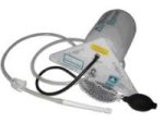
PNEUMOCOLON COMPLETE FOR EXAMINATION IN DOUBLE CONTRAST WALTER KREBS
Bock sealed composed of a tank for the contrast, pear Richardson, a hose and a field for carrying
Out the examinations of the colon in double contrast.
For routine barium irrigation or double contrast enema examination…
Pneumocolon is a lightweigh apparatus which is easy to handle….
Examinations with double contrasts are carried out simply by turning the apparatus….
Constant pressure gives a controlled flow of barium meal…
Quick Comparison
| PNE 1000-Pneumocolon Set remove | Sonoscape E1 Ultrasound Machine With Two Probes remove | Anke Anatom 64 Clarity Multi-Slice Spiral CT Scan remove | Topaz Digital X-ray Machine remove | Single X-Ray Viewing Box remove | DrGem Floor Mounted Analogue X-ray remove | |||||||||||||||||||||||||||||||||||||||||||||||||||||||||||||||||||||||||||||||||||||||||||||||||||||||||||||||||||||||||||||||||||||||||||||||||||||||||||||||||||||||||||||||||||||||||||||||||||||||||||||||||||||||||||||||||||||||||||||||||||||||||||||||||||||||||||||||||||||||||||
|---|---|---|---|---|---|---|---|---|---|---|---|---|---|---|---|---|---|---|---|---|---|---|---|---|---|---|---|---|---|---|---|---|---|---|---|---|---|---|---|---|---|---|---|---|---|---|---|---|---|---|---|---|---|---|---|---|---|---|---|---|---|---|---|---|---|---|---|---|---|---|---|---|---|---|---|---|---|---|---|---|---|---|---|---|---|---|---|---|---|---|---|---|---|---|---|---|---|---|---|---|---|---|---|---|---|---|---|---|---|---|---|---|---|---|---|---|---|---|---|---|---|---|---|---|---|---|---|---|---|---|---|---|---|---|---|---|---|---|---|---|---|---|---|---|---|---|---|---|---|---|---|---|---|---|---|---|---|---|---|---|---|---|---|---|---|---|---|---|---|---|---|---|---|---|---|---|---|---|---|---|---|---|---|---|---|---|---|---|---|---|---|---|---|---|---|---|---|---|---|---|---|---|---|---|---|---|---|---|---|---|---|---|---|---|---|---|---|---|---|---|---|---|---|---|---|---|---|---|---|---|---|---|---|---|---|---|---|---|---|---|---|---|---|---|---|---|---|---|---|---|---|---|---|---|---|---|---|---|---|---|---|---|---|---|---|---|---|---|---|---|---|---|---|---|---|---|---|---|---|---|---|---|---|---|---|---|---|---|
| Name | PNE 1000-Pneumocolon Set remove | Sonoscape E1 Ultrasound Machine With Two Probes remove | Anke Anatom 64 Clarity Multi-Slice Spiral CT Scan remove | Topaz Digital X-ray Machine remove | Single X-Ray Viewing Box remove | DrGem Floor Mounted Analogue X-ray remove | ||||||||||||||||||||||||||||||||||||||||||||||||||||||||||||||||||||||||||||||||||||||||||||||||||||||||||||||||||||||||||||||||||||||||||||||||||||||||||||||||||||||||||||||||||||||||||||||||||||||||||||||||||||||||||||||||||||||||||||||||||||||||||||||||||||||||||||||||||||||||||
| Image | |  |  |  |  |  | ||||||||||||||||||||||||||||||||||||||||||||||||||||||||||||||||||||||||||||||||||||||||||||||||||||||||||||||||||||||||||||||||||||||||||||||||||||||||||||||||||||||||||||||||||||||||||||||||||||||||||||||||||||||||||||||||||||||||||||||||||||||||||||||||||||||||||||||||||||||||||
| SKU | SF103356013086-28 | SF1033560012-20 | SF1033560092-2 | SF1033560074-1 | SF1033560084-203 | SF1033560074-6 | ||||||||||||||||||||||||||||||||||||||||||||||||||||||||||||||||||||||||||||||||||||||||||||||||||||||||||||||||||||||||||||||||||||||||||||||||||||||||||||||||||||||||||||||||||||||||||||||||||||||||||||||||||||||||||||||||||||||||||||||||||||||||||||||||||||||||||||||||||||||||||
| Rating | ||||||||||||||||||||||||||||||||||||||||||||||||||||||||||||||||||||||||||||||||||||||||||||||||||||||||||||||||||||||||||||||||||||||||||||||||||||||||||||||||||||||||||||||||||||||||||||||||||||||||||||||||||||||||||||||||||||||||||||||||||||||||||||||||||||||||||||||||||||||||||||||||
| Price |
| $4,620.00 |
|
| $95.20 |
| ||||||||||||||||||||||||||||||||||||||||||||||||||||||||||||||||||||||||||||||||||||||||||||||||||||||||||||||||||||||||||||||||||||||||||||||||||||||||||||||||||||||||||||||||||||||||||||||||||||||||||||||||||||||||||||||||||||||||||||||||||||||||||||||||||||||||||||||||||||||||||
| Stock | ||||||||||||||||||||||||||||||||||||||||||||||||||||||||||||||||||||||||||||||||||||||||||||||||||||||||||||||||||||||||||||||||||||||||||||||||||||||||||||||||||||||||||||||||||||||||||||||||||||||||||||||||||||||||||||||||||||||||||||||||||||||||||||||||||||||||||||||||||||||||||||||||
| Availability | ||||||||||||||||||||||||||||||||||||||||||||||||||||||||||||||||||||||||||||||||||||||||||||||||||||||||||||||||||||||||||||||||||||||||||||||||||||||||||||||||||||||||||||||||||||||||||||||||||||||||||||||||||||||||||||||||||||||||||||||||||||||||||||||||||||||||||||||||||||||||||||||||
| Add to cart | ||||||||||||||||||||||||||||||||||||||||||||||||||||||||||||||||||||||||||||||||||||||||||||||||||||||||||||||||||||||||||||||||||||||||||||||||||||||||||||||||||||||||||||||||||||||||||||||||||||||||||||||||||||||||||||||||||||||||||||||||||||||||||||||||||||||||||||||||||||||||||||||||
| Description | Shipped from Abroad SonoScape has developed a new probe and function for the E1 Exp. With these additions the E1 Exp will bring users a more efficient examination experience with satisfying image quality and a smooth workflow. Delivery & Availability: Typically 5-7 working days – excluding furniture and heavy/bulky equipment. Please contact us for further information. | Shipped from Abroad
The ANATOM 64 CT scanner is the latest innovation for cardiac imaging based on Precision Platform system. The excellent design of Ahart technology which innovatively combined single spiral scan + gated imaging + mA modulation for easy heart imaging at extremely low radiation dose. We provide you ANATOM 64 Clarity/Precision of two models which are low/high configurations for preferences. It also offers you conventional clinical applications of low dose, better image quality and faster exams.
Delivery & Availability: Typically 90 working days – excluding furniture and heavy/bulky equipment. Please contact us for further information. | In Stock DRGEM’s TOPAZ X-ray machine is a state-of-the-art mobile digital radiography system, designed with maximum comfort for patients and users in mind. From its user-friendly software to smooth movements, TOPAZ is made to improve your workflow and provide you with high-quality images. Delivery & Availability: Typically 21 working days – excluding furniture and heavy/bulky equipment. Please contact us for further information. | In stock
| In Stock GXR Analogue X-ray system matches with a radiographic room which perfectly fits your workow and can be easily upgraded to DR system with the help of DR interface and PC interface in GXR generator as well as Bucky suitable to Flat Panel Detector. GXR X-ray system is equipped with a high frequency X-ray generator which consistently produces high quality radiograph in favor of high quality X-ray output with a very small kV ripple and accurate mA and mAs. GXR X-ray system is designed to provide convenience to operator and comfort to patient. Delivery & Availability: Typically 21 working days – excluding furniture and heavy/bulky equipment. Please contact us for further information. | |||||||||||||||||||||||||||||||||||||||||||||||||||||||||||||||||||||||||||||||||||||||||||||||||||||||||||||||||||||||||||||||||||||||||||||||||||||||||||||||||||||||||||||||||||||||||||||||||||||||||||||||||||||||||||||||||||||||||||||||||||||||||||||||||||||||||||||||||||||||||||
| Content | For gastroenterology PNEUMOCOLON COMPLETE FOR EXAMINATION IN DOUBLE CONTRAST WALTER KREBS Bock sealed composed of a tank for the contrast, pear Richardson, a hose and a field for carrying Out the examinations of the colon in double contrast. For routine barium irrigation or double contrast enema examination... Pneumocolon is a lightweigh apparatus which is easy to handle.... Examinations with double contrasts are carried out simply by turning the apparatus.... Constant pressure gives a controlled flow of barium meal… | DETAILS
Efficient Diagnosis
μ-Scan, Speckle Reduction & Edge Enhancement
Spatial Compound Imaging
PIH - Pure Inversion Harmonic
Wide Scan - Enlarged Image Area
Tissue-Specific Imaging
SR Flow
Ergonomic Designs
Up to 2 Transducer Ports
Light Weight and Compact
15.6 inch Anti-flickering HD LED Screen
Tilting Monitor Angle Adjustment
Backlit Keyboard and Intelligent Panel
Long-lasting Battery for 90 mins
Ease of Use
Quick Boot Up
Auto-Brightness Adjustment
Auto Image Optimization
Auto IMT
Auto Trace
Equipped Accessories
Wi-Fi and Bluetooth Available
DICOM
500GB Hard Disk
Height Adjustable Trolley
Durable, Carry-on Site Suitcase
Click Here To Download Catalogue | The ANATOM 64 CT scanner is the latest innovation for cardiac imaging based on Precision Platform system. The excellent design of Ahart technology which innovatively combined single spiral scan + gated imaging + mA modulation for easy heart imaging at extremely low radiation dose. We provide you ANATOM 64 Clarity/Precision of two models which are low/high configurations for preferences. It also offers you conventional clinical applications of low dose, better image quality and faster exams.
Features:
Click Here To Download Catalogue | TOPAZ X-ray machine is among the high end X-ray machine manufactured by DRGEM, a digital X-ray system that provides quality images with little or no effort.
It begins with Advanced Technology
Integrating high technology and over a decade of experience in conventional and digital radiography systems, DRGEM’s TOPAZ X-ray machine is a state-of-the-art mobile digital radiography system, designed with maximum comfort for patients and users. From its user-friendly software to smooth movements, TOPAZ X-ray machine is made to improve your workflow and provide you with high-quality images.
Full Featured Imaging Software & Excellent Digital Image Processing
With a high-performance, built-in touchscreen, TOPAZ X-ray machine offers a user-friendly interface and powerful software for easy operation and increased workflow. The anatomical view-based digital image processing, automatically optimizes and enhances the quality of the image. it also comes with automatic image storage and print with DICOM 3.0 networking capability. additionally, the system offers increasing exam throughput while decreasing examination time.
Click Here To Download Catalogue |
| DrGem GXR Floor Mounted Analogue X-ray system matches with a radiographic room which perfectly fits your workflow and can be easily upgraded to DR system with the help of DR interface and PC interface in GXR generator as well as Bucky suitable to Flat Panel Detector. GXR (Analogue X-ray)system is equipped with a high frequency X-ray generator which consistently produces high quality radiograph in favor of high quality X-ray output with a very small kV ripple and accurate mA and mAs. GXR (Analogue X-ray) system is designed to provide convenience to operator and comfort to patient.
Features of DrGem GXR Floor Mounted Analogue X-ray:
Click Here To Download Catalogue | ||||||||||||||||||||||||||||||||||||||||||||||||||||||||||||||||||||||||||||||||||||||||||||||||||||||||||||||||||||||||||||||||||||||||||||||||||||||||||||||||||||||||||||||||||||||||||||||||||||||||||||||||||||||||||||||||||||||||||||||||||||||||||||||||||||||||||||||||||||||||||
| Weight | N/A | N/A | N/A | N/A | N/A | N/A | ||||||||||||||||||||||||||||||||||||||||||||||||||||||||||||||||||||||||||||||||||||||||||||||||||||||||||||||||||||||||||||||||||||||||||||||||||||||||||||||||||||||||||||||||||||||||||||||||||||||||||||||||||||||||||||||||||||||||||||||||||||||||||||||||||||||||||||||||||||||||||
| Dimensions | N/A | N/A | N/A | N/A | N/A | N/A | ||||||||||||||||||||||||||||||||||||||||||||||||||||||||||||||||||||||||||||||||||||||||||||||||||||||||||||||||||||||||||||||||||||||||||||||||||||||||||||||||||||||||||||||||||||||||||||||||||||||||||||||||||||||||||||||||||||||||||||||||||||||||||||||||||||||||||||||||||||||||||
| Additional information |
|


Reviews
There are no reviews yet.