
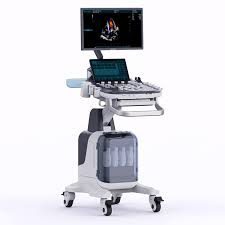
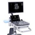
Premium Diagnostic Ultrasound System PU-MT241A
$0.00
Shipped From Abroad
Delivery & Availability:
Typically 10-21 working days – excluding furniture and heavy/bulky equipment. Please contact us for further information.
Typically 10-21 working days – excluding furniture and heavy/bulky equipment. Please contact us for further information.
Description
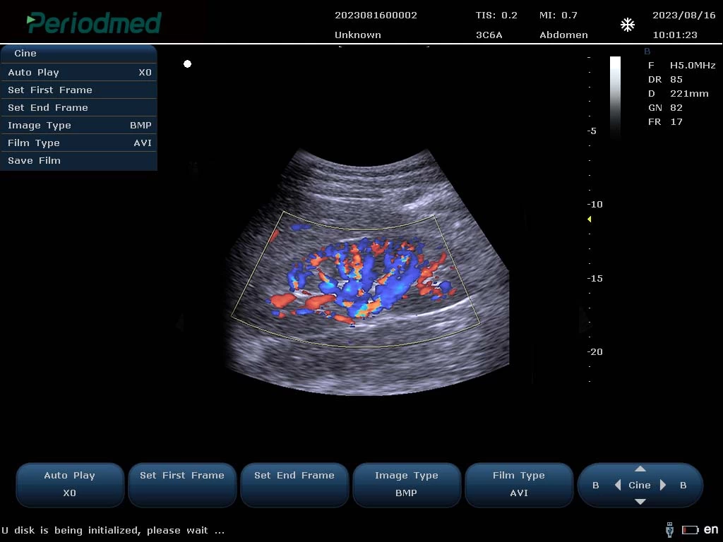
Ultrasound General imaging Images
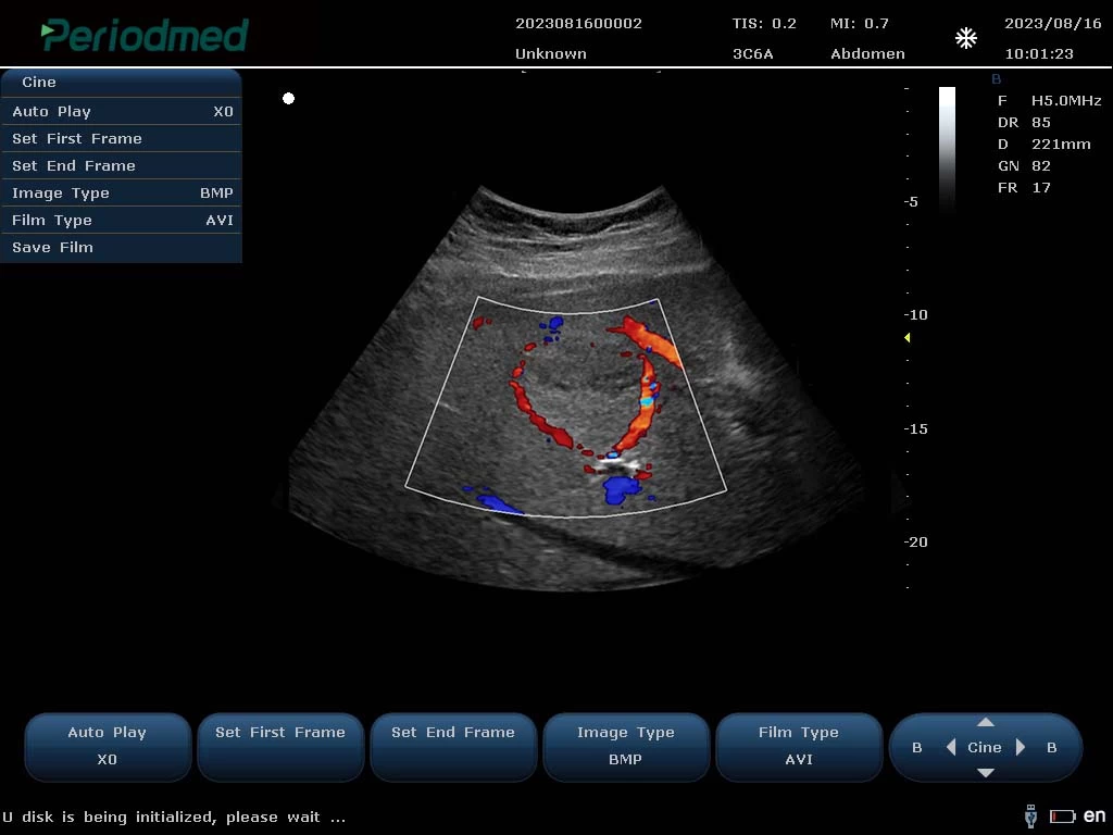
Convex Probe-Color Mode-Liver6
Convex Probe-Color Mode-Kidney 1
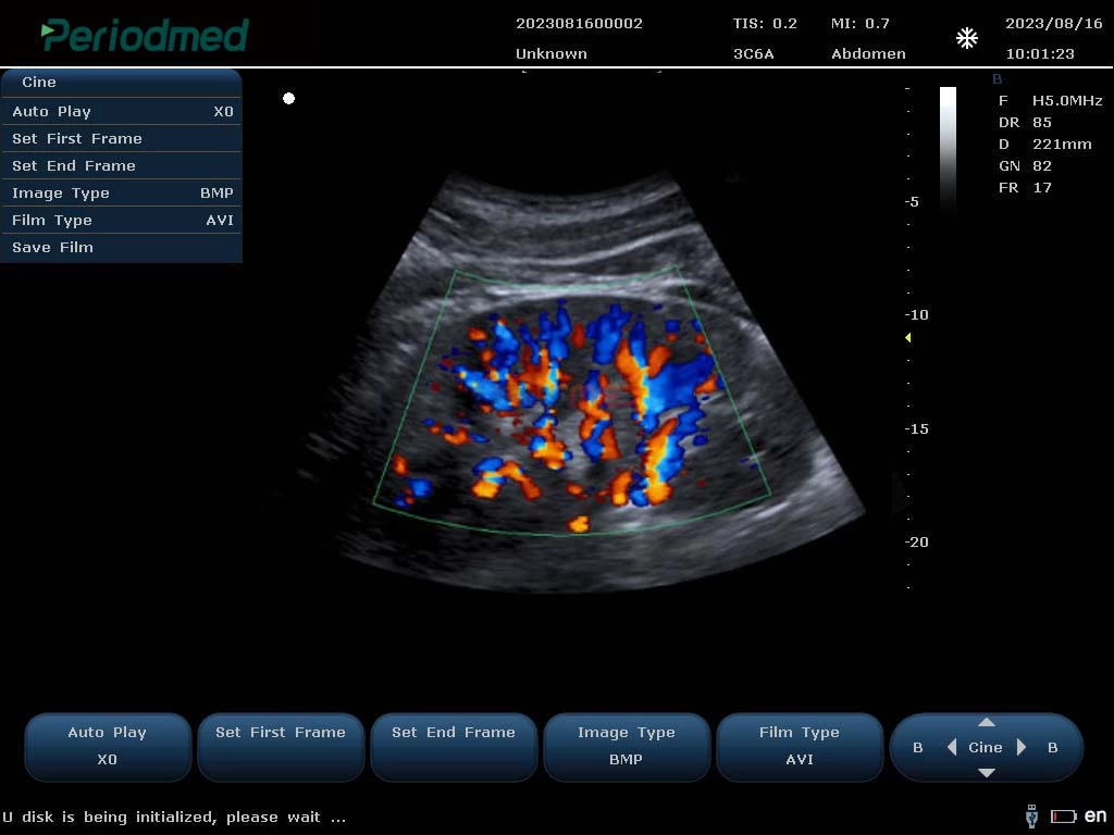
Convex Probe-Color Mode-Kidney 2
Ultrasound Carotid Images
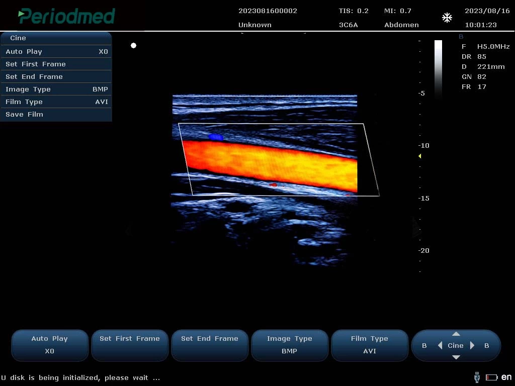
Linear Probe-Color Mode-Carotid1
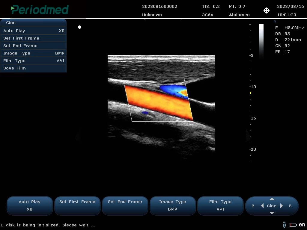
Linear Probe-Color Mode-Carotid2
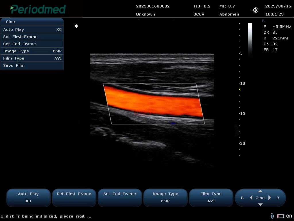
Linear Probe-Color Mode-Carotid3
Ultrasound Cardiovascular Images
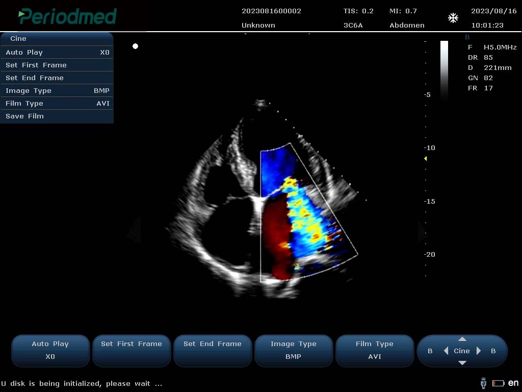
Phased Array Probe-Color Mode-Cardiac1
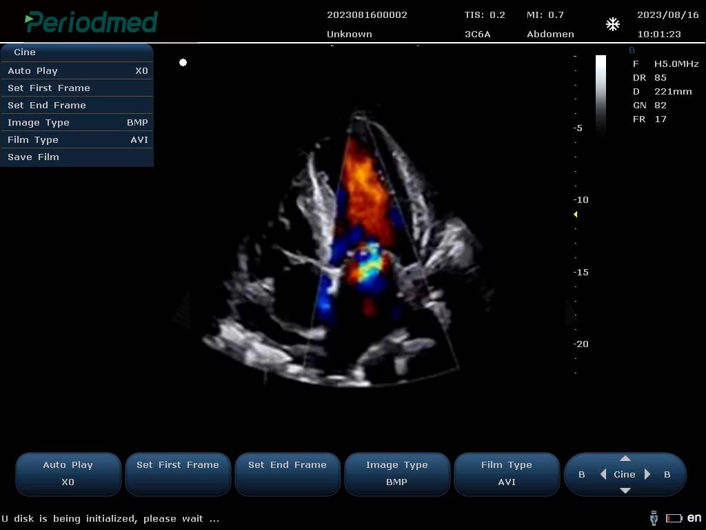
Phased Array Probe-Color Mode-Cardiac2
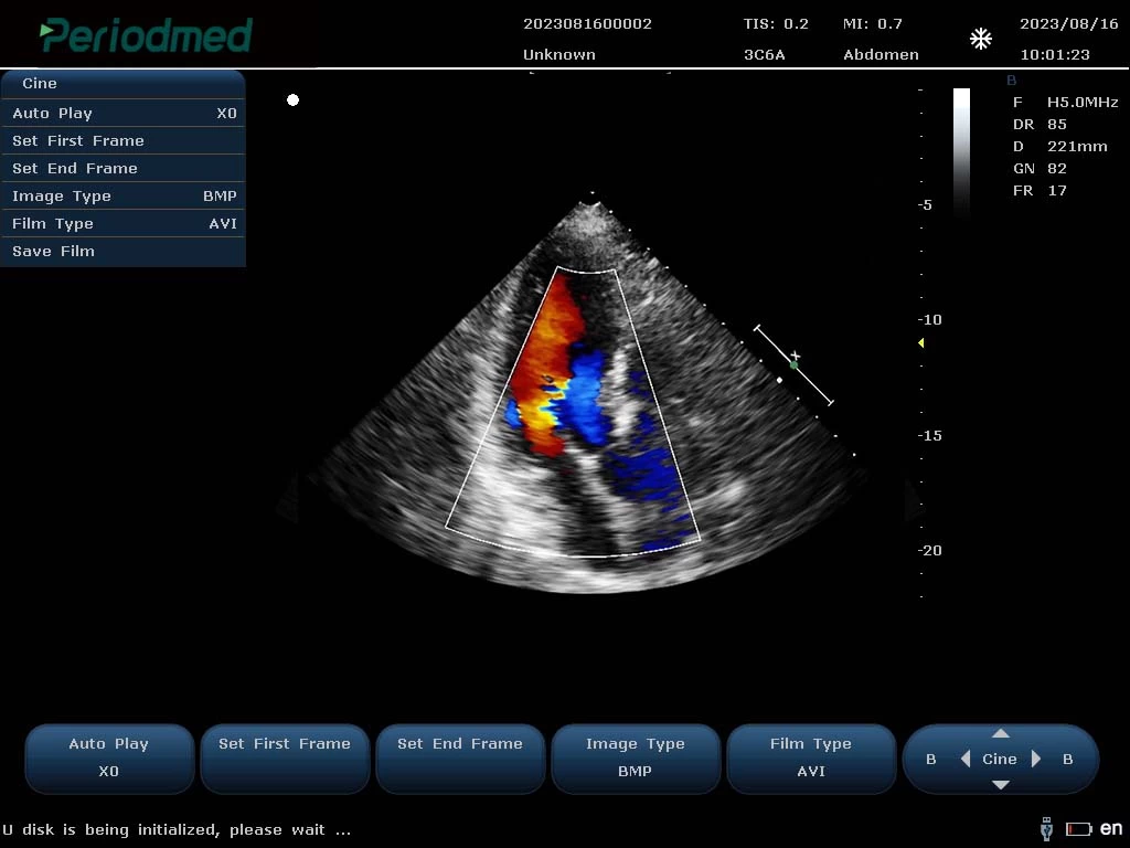
Phased Array Probe-Color Mode-Cardiac3
PU-MT241A Specification
| Multiple Display Modes: | B/2B/4B/BMM/PDI/DPDICF/CW/B+CF/DPDI+PW/B+CF+PW |
| Product Function: | Color Flow(CF) Power Doppler(PDl) Pulsed Doppler(PW) Directional Power Doppler (DPDl) Continuous Doppler (CW) Tissue Doppler (TDI) Color M Type,Anatomical M Type |
| Imaging Technology: | High Pulsed Repetition Frequency(Hprf) Tissue Harmonic Imaging(Thi) Pulsed Reverse Harmonic Imaging(Ithi) Tissue Specific Imaging(Tsi) Spatial Composite Imaging Picture In Picture Imaging Mode |
| Advanced Technology: | 1.Point-By-Point Focusing Application Technology Replaces The Traditional 1-4 Point Multi-Point Transmission Focusing Mode,Making The Transmission Focusing More Accurate 2.The Soft Beamformer Technology Of Gpu Architecture Provides More Powerful Computing Power For The Implementation Of The New Algorithm,And Fundamentally Improves The Quality Of Ultrasound Image 3.192 Elements 128 Channels,With Sound Speed Calibration Technology,16 Beam Compound Algonithm To Provide Dynamic And Accaurate Calculation Parameters,Effectively Improve The Focusing Accuracy 4.The Fast Sound Velocity Calibration Technology Can Automatically Correct The Actual Sound Velocity Of Different Human Bodies And Different Tissues In Real Time,And Provide Dynamic And Accurate Calculation Parameters For Beam Synthesis Algorithm |
| Optional: | Wide Field Imaging Puncture Enhancement Contrast Imaging Elastography Imaging 3D/4D Imaging |
Quick Comparison
| Premium Diagnostic Ultrasound System PU-MT241A remove | Sonoscape P15 Ultrasound Machine With Four Probes remove | ASPEL Stress ECG with Treadmill and Software remove | ASPEL AsPEKT 712 Holter Monitor and Software remove | Sonoscape E1 Ultrasound Machine With Two Probes remove | Sonoscape P50 Ultrasound Machine remove | |||||||||||
|---|---|---|---|---|---|---|---|---|---|---|---|---|---|---|---|---|
| Name | Premium Diagnostic Ultrasound System PU-MT241A remove | Sonoscape P15 Ultrasound Machine With Four Probes remove | ASPEL Stress ECG with Treadmill and Software remove | ASPEL AsPEKT 712 Holter Monitor and Software remove | Sonoscape E1 Ultrasound Machine With Two Probes remove | Sonoscape P50 Ultrasound Machine remove | ||||||||||
| Image | 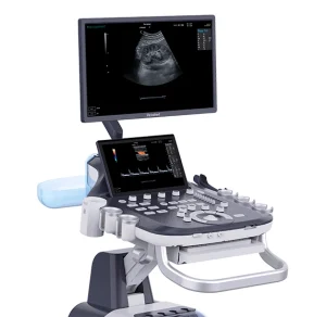 |  |  |  |  |  | ||||||||||
| SKU | SF1033560130124-1 | SF1033560012-8 | SF1033560075-2 | SF1033560075-4 | SF1033560012-20 | SF1033560012-11 | ||||||||||
| Rating | ||||||||||||||||
| Price |
| $13,900.00 | $6,542.00 | $1,991.00 | $4,620.00 |
| ||||||||||
| Stock | ||||||||||||||||
| Availability | ||||||||||||||||
| Add to cart | ||||||||||||||||
| Description | Shipped From Abroad
Delivery & Availability:
Typically 10-21 working days – excluding furniture and heavy/bulky equipment. Please contact us for further information.
| In Stock A feature-rich system inheriting the Wi-Sono high-end platform, the P15 uses an array of advanced tools to help enhance the image quality. It's a cost-effective, simplified console with an intuitive user interface and multiple intelligent functions. Delivery & Availability: Typically 2 working days – excluding furniture and heavy/bulky equipment. Please contact us for further information. | Shipped from Abroad It is a system with professional tool dedicated to exercise and resting ECG examination. Treadmill has 12 lead ECG modules. With ECG Analyzing Software. Delivery & Availability: Typically 21 working days – excluding furniture and heavy/bulky equipment. Please contact us for further information. | Shipped from Abroad The Holta Monitor allows quick analysis of ECG examination and detection, reviewing and editing capability in the qualitative assessment of VE, VT, Single SVE, PSVT, Pauses, Irregular Rhythm, VT, IVR, Brady - and Tachycardia, Couplets, ST-segment elevation and depression, Maximum, Minimum and averaged Heart Rates, artifacts Delivery & Availability: Typically 10 working days – excluding furniture and heavy/bulky equipment. Please contact us for further information. | Shipped from Abroad SonoScape has developed a new probe and function for the E1 Exp. With these additions the E1 Exp will bring users a more efficient examination experience with satisfying image quality and a smooth workflow. Delivery & Availability: Typically 5-7 working days – excluding furniture and heavy/bulky equipment. Please contact us for further information. | Shipped from Abroad Easily accomplish more with SonoScape’s new P50 ultrasound system. Incorporating single crystal clarity, automatic corrections and calculation, and user defined flexibility promises a confident diagnostic experience as well as opening new doors of opportunity for ultrasound use. Delivery & Availability: Typically 7-14 working days – excluding furniture and heavy/bulky equipment. Please contact us for further information. | ||||||||||
| Content | https://youtu.be/K2dzsICPG_s
Ultrasound General imaging Images
Convex Probe-Color Mode-Liver6
Convex Probe-Color Mode-Kidney 1
Convex Probe-Color Mode-Kidney 2 Ultrasound Carotid Images
Linear Probe-Color Mode-Carotid1
Linear Probe-Color Mode-Carotid2
Linear Probe-Color Mode-Carotid3 Ultrasound Cardiovascular Images
Phased Array Probe-Color Mode-Cardiac1
Phased Array Probe-Color Mode-Cardiac2
Phased Array Probe-Color Mode-Cardiac3 PU-MT241A Specification
| DETAILS
Super Wide-bandwidth Platform
Inheriting Wi-sono's ultra-wide system platform and with the advanced probe technology, high-resolution and deep penetration images are provided for precision medicine.
Spatial Compound Imaging
Spatial Compound Imaging utilizes several lines of sight for optimal contrast resolution, speckle reduction and border detection, with which P15 is ideal for superficial and abdominal imaging with better clarity and improved continuity of structures.
μ-Scan+
The new generation μ-Scan imaging technology gives you better image quality by reducing noise, improving signal strength and improving visualization.
Dynamic Color
Dynamic color improves upon already existing color Doppler technologies for a clearer capture of color flow and detailed visualization of even tiny veins with lower velocities.
Real-time Panoramic
With real-time panoramic, you can acquire an extended field of view for large organs or long vessels for easy measurement and diagnostic efficiency. Accomplished in real-time for the convenience of the sonographers, any mistake can also be easily back tracked and corrected without interrupting the scan.
3D/4D
Outstanding volume performance with speed and convenience makes P15 outshine others on volume imaging.
Tissue Doppler Imaging
Tissue Doppler Imaging allows clinical doctors to quantitatively evaluate local myocardial movements and functions, facilitating them with the ability to analyze and compare the motions of the different parts of the patient's heart.
Auto IMT
Quick measurement of intra-media vessel thickness ensures good reproducibility and high diagnostic efficiency.
Click Here To Download Catalogue | It is a system with professional tool dedicated to exercise and resting ECG examination. Treadmill has 12 lead ECG modules. With ECG Analyzing Software.
Technical Specification:
Click Here To Download Catalogue | The Holter Monitor allows quick analysis of ECG examination (arrhythmias and ST segment).
Technical specifications:
HolCARD 24W Software:
Click Here To Download Catalogue | DETAILS
Efficient Diagnosis
μ-Scan, Speckle Reduction & Edge Enhancement
Spatial Compound Imaging
PIH - Pure Inversion Harmonic
Wide Scan - Enlarged Image Area
Tissue-Specific Imaging
SR Flow
Ergonomic Designs
Up to 2 Transducer Ports
Light Weight and Compact
15.6 inch Anti-flickering HD LED Screen
Tilting Monitor Angle Adjustment
Backlit Keyboard and Intelligent Panel
Long-lasting Battery for 90 mins
Ease of Use
Quick Boot Up
Auto-Brightness Adjustment
Auto Image Optimization
Auto IMT
Auto Trace
Equipped Accessories
Wi-Fi and Bluetooth Available
DICOM
500GB Hard Disk
Height Adjustable Trolley
Durable, Carry-on Site Suitcase
Click Here To Download Catalogue | DETAILS
Powerful Compact Precision
Taking into consideration the evolving expectations and needs for ultrasound, the P50 is a slim and unobtrusive trolley system that is comfortable in tight, congested spaces with little room to work in. Providing everything you need for a comfortable examination in a small space for both you and your patient.
Single Crystal Transducer
Wideband single crystal probes greatly improve the signal ratio, acquire stunning images and provide superior sensitivity and resolution for both the near and far-fields.
μ-Scan+
The new generation μ-Scan imaging technologies give you better image quality by reducing noise, improving signal strength and improving visualization.
Dynamic Color
Dynamic colour improves upon already existing colour Doppler technologies for clear capture of colour flow and detail visualization of even tiny veins with lower velocities.
Solution for Radiology
P50, is a leading-edge ultrasound system that can meet the demands of any clinical setting. You can experience a superior performance in multi-dimensional imaging for a full range of clinical applications – abdominal, breast and cardiovascular.
C-xlasto Imaging
By understanding that tissue stiffness varies depending on the type of tissue, we can use C-xlasto Imaging to easily find abnormalities and tumours within soft tissue. The differences in tissue responses are detected and visualized in real-time by the elastography algorithms through different representations, which can be particularly helpful in analyzing breast, thyroid and musculoskeletal structures. Predominately used only in linear probes, SonoScape’s new transvaginal and bi-plane probe for gynaecology and urology are breaking the mould and expanding elastography applications.
Real-time Color Panoramic
With the combination of colour flow and real-time panoramic, visualizing the blood flow of an entire vein or artery is now an easy task. Accomplished in real-time for the convenience of the sonographers, any mistakes can also be easily backtracked and corrected without interrupting the scan.
Contrast Imaging
Contrast Imaging on P50 makes full use of the infra harmonic signal and second harmonic signal to improve the image resolution and deep penetration. What’s more, the Dynamic Acoustic Control technology effectively controls the acoustic pressure for the contrast agent, decreasing the required agent dose and assures uniform image quality, guaranteeing longer contrast agent duration and better lesion perfusion of delayed phase observation.
Solution for OB/GYN
P50 has superior image quality, automated measurement tools, and a variety of volume technologies to provide ideal solutions for clinical examinations such as pregnancy examinations, and gynecologic disease diagnosis. With a new 4D transvaginal probe, P50 helps you to see and detect fetal abnormalities and significantly improves your diagnostic confidence during your examinations.
S-Live Silhouette
A unique transparent 3D anatomical image of the fetus for improved initial anatomical review. By using this new application, the system can create completely different fetal images from conventional ultrasound images, which can depict the fetal's intracorporeal anatomical structure.
Pelvic Floor 4D
Working in conjunction with SonoScape’s latest transvaginal probes, trans-perineal 4D pelvic floor ultrasound provides a useful clinical assessment of the impact of vaginal delivery on the female anterior compartment. Allowing doctors to judge whether the pelvic organs prolapsed or not, the extent of prolapse, and determining whether the pelvic muscles tore correctly.
S-Guide
S-Guide gives the user an extensive list of example obstetric ultrasound images as reference guides and a convenient checklist system to keep track of their progress during their obstetrics examination.
Auto Face
Automatically removes masking layers in front of the fetus’s face for a clearer vision of the fetus’s face.
AVC Follicle
AVC Follicle automatically identifies how many follicles are present and calculates their individual volumes.
Solution for Cardiology
P50 provides clear 2D clinical images and Doppler sensitivity to assess critical cardiac performance. Compatible with SonoScape’s single crystal probes, the P50 can provide images with better resolution and penetration in Cardiac diagnosis.
Tissue Doppler Imaging
Tissue Doppler Imaging allows clinical doctors to quantitatively evaluate local myocardial movements and functions, facilitating them with the ability to analyze and compare the motions of the different parts of the patient’s heart.
Stress Echo
Stress echocardiography is the combination of 2D echocardiography with physical, pharmacological or electrical stress of the patient. It also then provides users with report management tools such as configurable template editor, multiple loops to select one for storage, wall motion scoring, stress echo report, etc
Auto IMT
Auto IMT is used when determining the level of vascular sclerosis present in the patient by automatically tracing and calculating the thickness of the carotid vessels. What distinguishes the P50 is that it provides an instant and accurate Mean and Max index at the touch of a single button.
Auto EF
Automated 2D Cardiac Quantification is a fully intelligent trace function for endocardium with 19 easily-adjustable points providing rapid access to proven 2D EF and volumes.
Click Here To Download Catalogue | ||||||||||
| Weight | N/A | N/A | N/A | N/A | N/A | N/A | ||||||||||
| Dimensions | N/A | N/A | N/A | N/A | N/A | N/A | ||||||||||
| Additional information |


Reviews
There are no reviews yet.