Q7 Wireless Ultrasound System
$0.00
Shipped From Abroad
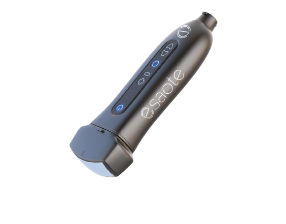
The ultra-compact wireless ultrasound system
The Q7 wireless ultrasound system takes convenience and portability to the next level. Its ultra-small size and light weight allow you to carry it right in your pocket, making it ideal for on-the-field use. This affordable and user-friendly device is the perfect tool to enhance patient outcomes and support your diagnosis.
Typically 10-21 working days – excluding furniture and heavy/bulky equipment. Please contact us for further information.
Description
The Esaote Q7 Wireless Ultrasound System is a wireless, pocket-sized ultrasound device engineered for versatility and portability. Its ultra-light weight and small footprint make it easily deployable in ambulances, field work or mobile clinics. It features six streamlined buttons for one-hand operation, supports multiple operating systems (Windows, Android, iOS) via a wireless connection, and boots in under 5 seconds. With support for B-mode, M-mode, PW Doppler, Colour Flow, and Power Doppler, it is tailored for whole-body scanning using interchangeable probe heads, delivering high flexibility
One device, all applications
Q7 integrates a suite of specialized applications tailored for whole-body scanning, empowering clinicians to tackle diverse clinical scenarios with a single device. Its wireless, ultra-portable design delivers imaging precision wherever it’s needed — clinic, bedside, or remote settings, with detailed imaging driven by TEI frequency and postprocessing filtering.
Lightweight precision with extended battery life
Q7 reimagines portability without sacrificing power. Weighing just 160 grams and small enough to slip into a pocket, it’s light enough to forget you’re carrying it—yet engineered to deliver uninterrupted high-performance scanning for over 4 hours. This featherweight powerhouse seamlessly transitions from clinic to field, ensuring critical tasks stay on track while setting a new standard in productivity. Think big capability, not bulk.
- Weight: 160g, with a linear probe head
- Size: 150x40x25 mm with linear probe head
- Battery time: 4 hours of scanning
Usability first
Enhance control and optimize images effortlessly with six buttons seamlessly integrated into Q7’s ergonomic design. The hand guiding the probe can simultaneously adjust settings, fine-tune imaging parameters, or capture critical data, all without breaking scanning rhythm. This intuitive one-handed operation eliminates disruptive pauses, letting clinicians prioritize precision over device management, and it transforms complex workflows into fluid, uninterrupted examinations, empowering you to deliver faster care.
Bridge devices, systems, and standards
- For those clinical settings requiring cable connection, Q7 offers fast and reliable USB data transfer (Windows® and Android® only).
- Q7 app is available for portable devices running on Windows®, Android®, and iOS®.
- The device offers flexible wireless dataflow with DICOM PACS and Worklist server query, including user authentication and auto-anonymized images for safe handling of clinical information.
- Wireless connectivity removes the unnecessary complications of cables, while allowing for fast and secure data transfer.
Swap probes in seconds, never miss a scan
Q7 eliminates downtime with its rapid-swap probe system, designed for seamless transitions between body parts. Effortlessly switch probe heads in seconds—no tools, no reboots, just click-and-go precision. The device’s near-instant reaction time ensures settings auto-adopt to the new probe, keeping workflows uninterrupted and clinicians laser-focused on the patient. Whether moving from cardiac to abdominal scans or pediatric to musculoskeletal exams, Q7 turns cumbersome setup into a relic of the past, redefining efficiency.
Quick Comparison
| Q7 Wireless Ultrasound System remove | Sonoscape P10 Ultrasound Machine remove | Sonoscape S8 Exp Portable Ultrasound remove | Sonoscape P50 Ultrasound Machine remove | ASPEL AsCARD Green ECG Machine remove | DrGem GXR-SD 400mA Floor Mounted Digital X-ray remove | |
|---|---|---|---|---|---|---|
| Name | Q7 Wireless Ultrasound System remove | Sonoscape P10 Ultrasound Machine remove | Sonoscape S8 Exp Portable Ultrasound remove | Sonoscape P50 Ultrasound Machine remove | ASPEL AsCARD Green ECG Machine remove | DrGem GXR-SD 400mA Floor Mounted Digital X-ray remove |
| Image |  |  |  |  |  |  |
| SKU | SF1033560012-7 | SF1033560012-15 | SF1033560012-11 | SF1033560075-9 | SF1033560074-5 | |
| Rating | ||||||
| Price |
| $9,350.00 | $9,350.00 |
|
|
|
| Stock | ||||||
| Availability | ||||||
| Add to cart | ||||||
| Description | Shipped From Abroad
The ultra-compact wireless ultrasound systemThe Q7 wireless ultrasound system takes convenience and portability to the next level. Its ultra-small size and light weight allow you to carry it right in your pocket, making it ideal for on-the-field use. This affordable and user-friendly device is the perfect tool to enhance patient outcomes and support your diagnosis. Delivery & Availability:
Typically 10-21 working days – excluding furniture and heavy/bulky equipment. Please contact us for further information.
| Shipped from Abroad The P10 color Doppler ultrasound system is a new generation product from SonoScape. It is designed to give high quality images, rich probe configurations, various clinical tools and automatic analysis software to provide you with comprehensive solutions for your growing demand for clinical applications. Delivery & Availability: Typically 5-7 working days – excluding furniture and heavy/bulky equipment. Please contact us for further information. | Shipped from Abroad With ultra-modern innovative design and the clinically-proven technologies, S8 Exp is portable ultrasound scanner well equipped as a low-physical-effort and enhanced-image-quality ultrasound scanner, which not only provides optimized solutions for versatile applications, but does help to improve the user-experience for both routine and non-traditional challenges. Delivery & Availability: Typically 5-7 working days – excluding furniture and heavy/bulky equipment. Please contact us for further information. | Shipped from Abroad Easily accomplish more with SonoScape’s new P50 ultrasound system. Incorporating single crystal clarity, automatic corrections and calculation, and user defined flexibility promises a confident diagnostic experience as well as opening new doors of opportunity for ultrasound use. Delivery & Availability: Typically 7-14 working days – excluding furniture and heavy/bulky equipment. Please contact us for further information. | Shipped from Abroad AsCARD Green v.06.101 is a 1-, 3-, 6- and 12-channel ECG unit which enables to make electrocardiogram in full 12 leads. Intended for ECG examinations of adult and paediatric patients aimed at identification of cardiological abnormalities, myocardial ischaemia or infarction. The device is intended for use in healthcare facilities by duly trained personnel. ECG examination may be recorded in manual or automatic mode with the ability to perform the analysis and interpretation. Delivery & Availability: Typically 10 working days – excluding furniture and heavy/bulky equipment. Please contact us for further information. | In Stock The GXR-SD Digital X-ray is a diagnostic digital radiography system that provides reliable high quality digital radiographic images with a reduced dose. The GXR-SD DR systems offer comprehensive digital solutions to all radiography needs, featuring ACQUIDR digital imaging system with stationary or portable digital flat-panel detectors as well as reliable high-frequency x-ray generators that are known worldwide for their excellent performance, lifetime and stability. Patient tables and wall stands are also offered. Delivery & Availability: Typically 21 working days – excluding furniture and heavy/bulky equipment. Please contact us for further information. |
| Content | The Esaote Q7 Wireless Ultrasound System is a wireless, pocket-sized ultrasound device engineered for versatility and portability. Its ultra-light weight and small footprint make it easily deployable in ambulances, field work or mobile clinics. It features six streamlined buttons for one-hand operation, supports multiple operating systems (Windows, Android, iOS) via a wireless connection, and boots in under 5 seconds. With support for B-mode, M-mode, PW Doppler, Colour Flow, and Power Doppler, it is tailored for whole-body scanning using interchangeable probe heads, delivering high flexibility
PORTABLE AND VERSATILE
One device, all applicationsQ7 integrates a suite of specialized applications tailored for whole-body scanning, empowering clinicians to tackle diverse clinical scenarios with a single device. Its wireless, ultra-portable design delivers imaging precision wherever it’s needed — clinic, bedside, or remote settings, with detailed imaging driven by TEI frequency and postprocessing filtering. ALWAYS WITH YOU
Lightweight precision with extended battery life
Q7 reimagines portability without sacrificing power. Weighing just 160 grams and small enough to slip into a pocket, it’s light enough to forget you’re carrying it—yet engineered to deliver uninterrupted high-performance scanning for over 4 hours. This featherweight powerhouse seamlessly transitions from clinic to field, ensuring critical tasks stay on track while setting a new standard in productivity. Think big capability, not bulk.
COMFORTABLE CONTROL
Usability firstEnhance control and optimize images effortlessly with six buttons seamlessly integrated into Q7's ergonomic design. The hand guiding the probe can simultaneously adjust settings, fine-tune imaging parameters, or capture critical data, all without breaking scanning rhythm. This intuitive one-handed operation eliminates disruptive pauses, letting clinicians prioritize precision over device management, and it transforms complex workflows into fluid, uninterrupted examinations, empowering you to deliver faster care. CONNECTIVITY WITHOUT LIMITS
Bridge devices, systems, and standards
NEAR-INSTANT REACTION TIME
Swap probes in seconds, never miss a scanQ7 eliminates downtime with its rapid-swap probe system, designed for seamless transitions between body parts. Effortlessly switch probe heads in seconds—no tools, no reboots, just click-and-go precision. The device’s near-instant reaction time ensures settings auto-adopt to the new probe, keeping workflows uninterrupted and clinicians laser-focused on the patient. Whether moving from cardiac to abdominal scans or pediatric to musculoskeletal exams, Q7 turns cumbersome setup into a relic of the past, redefining efficiency. | DETAILS
B + Compound
B + Compound utilizes several lines of sight for optimal contrast resolution, speckle reduction and border detection, with which P10 is ideal for superficial and abdominal imaging with better clarity and improved continuity of structures.
μ-Scan
The new generation μ-Scan imaging technology gives you better image quality by reducing noise, improving signal strength and improving visualization.
P10 offers a comprehensive selection of electronic probes to maximize its capabilities to meet a wide range of applications including abdomen, pediatric, OB/GYN, cardiovascular, musculoskeletal, etc. The advanced probe technologies also effectively enhance the image quality and confidence in reaching clinical diagnoses, even in difficult patients.
Convex Probe 3C-A
Ideal for an abundant of application such as abdomen, gynecology, obstetrics, urology and even abdomen biopsy.
Linear Probe L741
This linear probe is designed to satisfy vascular, breast, thyroid, and other small parts diagnosis, and its adjustable parameters could also present users a clear view of MSK and deep vessels.
Phase Array Probe 3P-A
For the purpose of adult and pediatric cardiology and emergency, the phase array probe provides elaborate presets for different exam modes, even for difficult patients.
Intracavitary Probe 6V1
Intracavitary probe could face application of gynecology, urology, prostate, and its temperature detection technology not only protects the patient but also extends the service life.
Click Here To Download Catalogue | Sonoscape S8 Exp Portable Ultrasound scannerDETAILS Agile and Versatile With ultra-modern innovative design and the clinically-proven technologies, S8 Exp Portable Ultrasound scanner is well equipped as a low-physical-effort and enhanced-image-quality ultrasound scanner, which not only provides optimized solutions for versatile applications but does help to improve the user experience for both routine and non-traditional challenges. Working with S8 Exp, it will trigger your unlimited reverie and endow you with endless charm. Carrying forward the classical design of SonoScape's portable ultrasound products, S8 Exp successfully combines the best ergonomics, attractive design and ease of use. This charismatic identity is also enhanced by a sophisticated color palette—with sedate grey as its interior paint color and pearl white as exterior cover, S8 Exp reveals a style of aristocrat and strong character among SonoScape's ultrasound systems. Workflow The S8 Exp is a portable ultrasound scanner that adapts to your workflow, whether you are in the consulting room, at the bedside, or at a remote location. With easy-to-use new platform designed for sonographers' needs and full connection interfaces for easy connectivity and data sharing, S8 Exp leads to improved user comfort and clinical outcome as well as patient throughput and working efficiency. Powerful Platform Embedded with SonoScape's core imaging technologies such as μ-scan, PHI and Spatial Compound, S8 Exp boasts exceptional 2D image, sensitive spectral, Color and Power Doppler, displaying well-defined anatomy and pathology and facilitating a highly optimized diagnostic user environment for conclusive diagnoses. Besides, S8 Exp offers a comprehensive selection of electronic probes to maximally extend its capabilities to meet a wide range of applications including the abdomen, pediatric, OB/GYN, cardiovascular, musculoskeletal, etc. The advanced probe technologies also effectively enhance the image quality and confidence in reaching clinical diagnoses even in difficult patients.Click Here To Download Catalogue | DETAILS
Powerful Compact Precision
Taking into consideration the evolving expectations and needs for ultrasound, the P50 is a slim and unobtrusive trolley system that is comfortable in tight, congested spaces with little room to work in. Providing everything you need for a comfortable examination in a small space for both you and your patient.
Single Crystal Transducer
Wideband single crystal probes greatly improve the signal ratio, acquire stunning images and provide superior sensitivity and resolution for both the near and far-fields.
μ-Scan+
The new generation μ-Scan imaging technologies give you better image quality by reducing noise, improving signal strength and improving visualization.
Dynamic Color
Dynamic colour improves upon already existing colour Doppler technologies for clear capture of colour flow and detail visualization of even tiny veins with lower velocities.
Solution for Radiology
P50, is a leading-edge ultrasound system that can meet the demands of any clinical setting. You can experience a superior performance in multi-dimensional imaging for a full range of clinical applications – abdominal, breast and cardiovascular.
C-xlasto Imaging
By understanding that tissue stiffness varies depending on the type of tissue, we can use C-xlasto Imaging to easily find abnormalities and tumours within soft tissue. The differences in tissue responses are detected and visualized in real-time by the elastography algorithms through different representations, which can be particularly helpful in analyzing breast, thyroid and musculoskeletal structures. Predominately used only in linear probes, SonoScape’s new transvaginal and bi-plane probe for gynaecology and urology are breaking the mould and expanding elastography applications.
Real-time Color Panoramic
With the combination of colour flow and real-time panoramic, visualizing the blood flow of an entire vein or artery is now an easy task. Accomplished in real-time for the convenience of the sonographers, any mistakes can also be easily backtracked and corrected without interrupting the scan.
Contrast Imaging
Contrast Imaging on P50 makes full use of the infra harmonic signal and second harmonic signal to improve the image resolution and deep penetration. What’s more, the Dynamic Acoustic Control technology effectively controls the acoustic pressure for the contrast agent, decreasing the required agent dose and assures uniform image quality, guaranteeing longer contrast agent duration and better lesion perfusion of delayed phase observation.
Solution for OB/GYN
P50 has superior image quality, automated measurement tools, and a variety of volume technologies to provide ideal solutions for clinical examinations such as pregnancy examinations, and gynecologic disease diagnosis. With a new 4D transvaginal probe, P50 helps you to see and detect fetal abnormalities and significantly improves your diagnostic confidence during your examinations.
S-Live Silhouette
A unique transparent 3D anatomical image of the fetus for improved initial anatomical review. By using this new application, the system can create completely different fetal images from conventional ultrasound images, which can depict the fetal's intracorporeal anatomical structure.
Pelvic Floor 4D
Working in conjunction with SonoScape’s latest transvaginal probes, trans-perineal 4D pelvic floor ultrasound provides a useful clinical assessment of the impact of vaginal delivery on the female anterior compartment. Allowing doctors to judge whether the pelvic organs prolapsed or not, the extent of prolapse, and determining whether the pelvic muscles tore correctly.
S-Guide
S-Guide gives the user an extensive list of example obstetric ultrasound images as reference guides and a convenient checklist system to keep track of their progress during their obstetrics examination.
Auto Face
Automatically removes masking layers in front of the fetus’s face for a clearer vision of the fetus’s face.
AVC Follicle
AVC Follicle automatically identifies how many follicles are present and calculates their individual volumes.
Solution for Cardiology
P50 provides clear 2D clinical images and Doppler sensitivity to assess critical cardiac performance. Compatible with SonoScape’s single crystal probes, the P50 can provide images with better resolution and penetration in Cardiac diagnosis.
Tissue Doppler Imaging
Tissue Doppler Imaging allows clinical doctors to quantitatively evaluate local myocardial movements and functions, facilitating them with the ability to analyze and compare the motions of the different parts of the patient’s heart.
Stress Echo
Stress echocardiography is the combination of 2D echocardiography with physical, pharmacological or electrical stress of the patient. It also then provides users with report management tools such as configurable template editor, multiple loops to select one for storage, wall motion scoring, stress echo report, etc
Auto IMT
Auto IMT is used when determining the level of vascular sclerosis present in the patient by automatically tracing and calculating the thickness of the carotid vessels. What distinguishes the P50 is that it provides an instant and accurate Mean and Max index at the touch of a single button.
Auto EF
Automated 2D Cardiac Quantification is a fully intelligent trace function for endocardium with 19 easily-adjustable points providing rapid access to proven 2D EF and volumes.
Click Here To Download Catalogue | AsCARD Green v.06.101 is a 1-, 3-, 6- and 12-channel ECG unit which enables to make electrocardiogram in full 12 leads. Intended for ECG examinations of adult and paediatric patients aimed at identification of cardiological abnormalities, myocardial ischaemia or infarction. The device is intended for use in healthcare facilities by duly trained personnel. ECG examination may be recorded in manual or automatic mode with the ability to perform the analysis and interpretation.
Electrocardiograph is based on advanced microprocessor technology .It is equipped with a thermal printer with high-resolution head and 4,3" LCD display. A touch panel and high-tech membrane keyboard makes this device intuitive in usage and its menu navigation exceptionally easy. This light-weight, small-footprint and battery powered cause that device can be easily transported to any location. With plastic casing and foil covered keyboard, the device is neat and easy to clean.
Technical Specifications:
Click Here To Catalogue Download | DrGem GXR-SD 400mA Floor Mounted Digital X-ray system matches with a radiographic room which perfectly fits your workow and can be easily upgraded to DR system with the help of DR interface and PC interface in GXR generator as well as Bucky suitable to Flat Panel Detector. GXR X-ray system is equipped with a high frequency X-ray generator which consistently produces high quality radiograph in favor of high quality X-ray output with a very small kV ripple and accurate mA and mAs. GXR X-ray system is designed to provide convenience to operator and comfort to patient
Features of DrGem GXR-SD 400mA Floor Mounted Digital X-ray:
Click Here To Download Catalogue |
| Weight | N/A | N/A | N/A | N/A | N/A | N/A |
| Dimensions | N/A | N/A | N/A | N/A | N/A | N/A |
| Additional information |


Reviews
There are no reviews yet.