New
ri-scope® retinoscope
$0.00
Shipped From Abroad
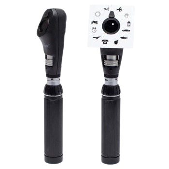
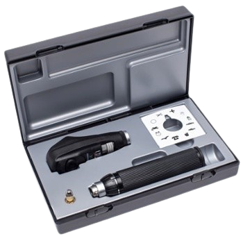
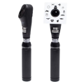
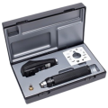
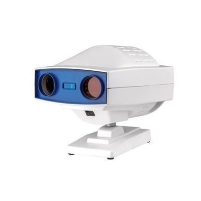
The ri-scope® retinoscope, also referred to as the skiascope, measures the refractive power of the eye. Refractive flaws, such as near- and far-sightedness, as well as astigmatism, can be detected and diagnosed. Riester offers the split-light retinoscope (glow is a stitch) model.
Delivery & Availability:
Typically 10-21 working days – excluding furniture and heavy/bulky equipment. Please contact us for further information.
Typically 10-21 working days – excluding furniture and heavy/bulky equipment. Please contact us for further information.
Description
This retinoscope simplifies the diagnosis of astigmatic refractive flaws using a slit-light beam. The line image can be focused and precisely turned 360° using a knurled thumb screw, with the angle displayed on an integrated scale. It includes two fixation cards for dynamic retinoscopy, features an integrated eyeglass protection, and has a dust-proof casing made of impact-resistant ABS plastic.
Features and benefits
- Available with XL 3.5 V xenon lamp or HL 2.5 V halogen lamp
- The slit-light retinoscope, with a light beam in the form of a line, simplifies the detection and diagnosis of astigmatic refractive flaws. The reflex in the form of a line is moved vertically to the axis across the pupils of the patient with a slight oscillating movement. A shadow moves in the same or opposite direction.
- Co-movement (plus lines): The patient is farsighted.
- Counter-movement (minus lines): The patient is near-sighted.
- Easy to operate with a knurled thumb screw. The line and spot image can be focused with the controls and turned 360°, with the angle read-out displayed on the integrated scale.
- A holder for hanging and placement of the fixation card into position for dynamic retinoscopy.
- Delivered with two fixation cards. The patient’s eye can adjust optimally to the distance to the retinoscope.
- Integrated eyeglass protection
- Bayonet fitting for quick and secure attachment to the handle
- Dust-proof, very sturdy and light casing made of impact-resistant ABS plastic
Specifications
| ri-scope® retinoscope | |
|---|---|
| Dimension |  |
| The lamp for ri-scope® retinoscope | Slit – HL 2.5 V, 440 mA, mean life span 15 h Slit – XL 3.5 V, 690 mA, mean life span 50 h |
| Battery | 2 Alkaline AA / C 1.5 V batteries. 1 Li-Ion battery for C-handle |
| Ambient temperature | 0 °C to 40 °C |
| Relative humidity | 30% to 70% non-condensing |
| Storage/ Transport ambient temperature | -10 °C to 55 °C |
| Storage/ Transport relative humidity | 30% to 85% non-condensing |
| Air pressure/ Transport relative humidity | 800 hPa – 1100 hPa |
Quick Comparison
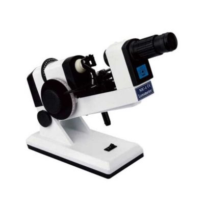
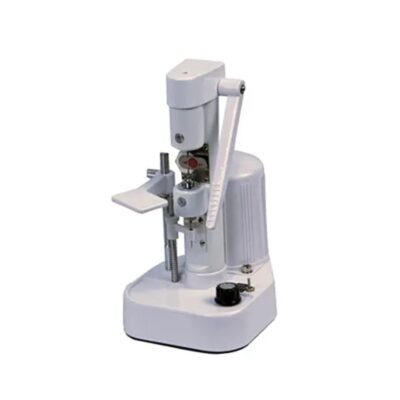
| ri-scope® retinoscope remove | Portable Fundus Camera remove | Automatic Computer Goldmann (Visual Field Analyzer) remove | Binocular Indirect Ophthalmoscope remove | ENT/Neurosurgery Operating Microscope remove | Ear Irrigation and acumen removal remove | |||||||||||||||||||||||||||||||||||||||||||||||||||||||||||||||||||||||||||||||||||||||||||||||||||||||
|---|---|---|---|---|---|---|---|---|---|---|---|---|---|---|---|---|---|---|---|---|---|---|---|---|---|---|---|---|---|---|---|---|---|---|---|---|---|---|---|---|---|---|---|---|---|---|---|---|---|---|---|---|---|---|---|---|---|---|---|---|---|---|---|---|---|---|---|---|---|---|---|---|---|---|---|---|---|---|---|---|---|---|---|---|---|---|---|---|---|---|---|---|---|---|---|---|---|---|---|---|---|---|---|---|---|---|---|---|
| Name | ri-scope® retinoscope remove | Portable Fundus Camera remove | Automatic Computer Goldmann (Visual Field Analyzer) remove | Binocular Indirect Ophthalmoscope remove | ENT/Neurosurgery Operating Microscope remove | Ear Irrigation and acumen removal remove | ||||||||||||||||||||||||||||||||||||||||||||||||||||||||||||||||||||||||||||||||||||||||||||||||||||||
| Image | 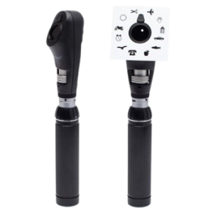 | 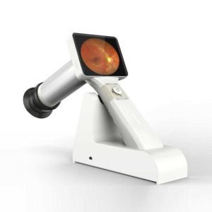 |  | 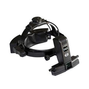 | 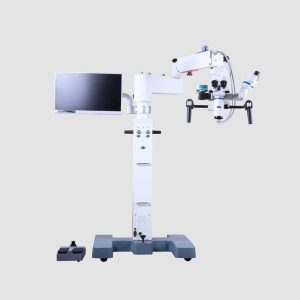 | 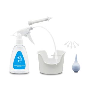 | ||||||||||||||||||||||||||||||||||||||||||||||||||||||||||||||||||||||||||||||||||||||||||||||||||||||
| SKU | SF1033560107-23 | SF103356013013 | SF1033560107-4 | SF1033560109-1 | SF103356013012 | |||||||||||||||||||||||||||||||||||||||||||||||||||||||||||||||||||||||||||||||||||||||||||||||||||||||
| Rating | ||||||||||||||||||||||||||||||||||||||||||||||||||||||||||||||||||||||||||||||||||||||||||||||||||||||||||||
| Price |
| $2,310.00 | $3,850.00 | $880.00 |
|
| ||||||||||||||||||||||||||||||||||||||||||||||||||||||||||||||||||||||||||||||||||||||||||||||||||||||
| Stock | ||||||||||||||||||||||||||||||||||||||||||||||||||||||||||||||||||||||||||||||||||||||||||||||||||||||||||||
| Availability | ||||||||||||||||||||||||||||||||||||||||||||||||||||||||||||||||||||||||||||||||||||||||||||||||||||||||||||
| Add to cart | ||||||||||||||||||||||||||||||||||||||||||||||||||||||||||||||||||||||||||||||||||||||||||||||||||||||||||||
| Description | Shipped From Abroad
The ri-scope® retinoscope, also referred to as the skiascope, measures the refractive power of the eye. Refractive flaws, such as near- and far-sightedness, as well as astigmatism, can be detected and diagnosed. Riester offers the split-light retinoscope (glow is a stitch) model.
Delivery & Availability:
Typically 10-21 working days – excluding furniture and heavy/bulky equipment. Please contact us for further information.
| 83Shipped from abroad
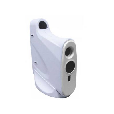
This is a portable medical camera for fundus imaging, diagnosis, and especially for fundus disease screening. It's compact, easy to obtain high definition fundus image. It can be conveniently applied to rapid screening, out diagnosis, bedside diagnosis and remote medical treatment, etc.
| In Stock
Features:
The Bio-1000 automated perimeter absorbs the advantages of international advanced perimetry devices. It comprises the highly integrated computer, optics, machinery and electronics systems.
Delivery & Availability:
Typically 7-14 working days – excluding furniture and heavy/bulky equipment. Please contact us for further information.
| Shipped from abroad
Super lightweight design, reduce fatigue, operation is very convenient.
| Shipped from abroad
Corder Microscope has Fluid, Responsive and Accurate.Fluid. Responsive. Accurate. These were a few of the principles guiding every phase in the design of the Corder Microscope. With the choicest mechanical machined components, the Corder Microscope has the grace and agility to adjust to every desired position on command. Well designed Apochromatic optics treated with Corder's Mcoatings produce true-to life sharp images with high depth, definition and contrast. | In Stock
Features:
●Professional
Same ear wax removal tool as those used by doctors, you can easily eliminate ear wax buildup at home, really save your money and time on medical visiting. Safe and Environmentally Friendly.
●Quick & Easy
This ear wax removal kit is a quick, effective treatment for excess ear wax buildup. Fill the bottle with solution, Twist on the disposable tip, Use the trigger handle to spray solution into the ear canal. So Easy.
Delivery & Availability:
Typically 7-14 working days – excluding furniture and heavy/bulky equipment. Please contact us for further information.
| ||||||||||||||||||||||||||||||||||||||||||||||||||||||||||||||||||||||||||||||||||||||||||||||||||||||
| Content | This retinoscope simplifies the diagnosis of astigmatic refractive flaws using a slit-light beam. The line image can be focused and precisely turned 360° using a knurled thumb screw, with the angle displayed on an integrated scale. It includes two fixation cards for dynamic retinoscopy, features an integrated eyeglass protection, and has a dust-proof casing made of impact-resistant ABS plastic.
Features and benefits
Specifications
| Portable Fundus Camera is a portable medical camera for fundus imaging, diagnosis, and especially for fundus disease screening. It's compact, easy to obtain high definition fundus image. It can be conveniently applied to rapid screening, out diagnosis, bedside diagnosis and remote medical treatment, etc.
Features of Portable Fundus Camera:
| The Bio-1000 automated perimeter absorbs the advantages of international advanced perimetry devices. It comprises the highly integrated computer, optics, machinery and electronics systems. Incorporated with the advanced configuration, comprehensive software inspection categories, and strictly in accordance with international Goldman standard, it provide scientific means for glaucoma, fundus disease, visual pathway injury and neurological diseases.
Feature:
* Comprehensive real-time monitoring,Heiji-krakau physiological blind spot monitoring,gaze tracking/head position tracking,automatic measurement of pupil diameter, reduce the impact of pupil effect on visual field detection.
* Personalized design,accurate clinical analysis,accurate and repid examination strategy.
* Under international Goldman standard,providing a variety of classic test procedures and report analysis.
Technical Specification:
Click Here To Download Catalogue | Ophthalmoscope Features:
| Features:Corder Microscope has Fluid, Responsive and Accurate.Fluid. Responsive. Accurate. These were a few of the principles guiding every phase in the design of the Corder Microscope. With the choicest mechanical machined components, the Corder Microscope has the grace and agility to adjust to every desired position on command. Well designed Apochromatic optics treated with Corder's Mcoatings produce true-to life sharp images with high depth, definition and contrast. More comfortable operation Tiltable binocular tubes available, which can incline more than 60° depending on the posture and physique of the operating surgeon. Movable range: 30° (straight) to 90° (inclined) Corder microscope configured with XYZ motorized movement operated through a comfortable foot /Handle control, a veryeffective co-axial illumnation and 50W halogen light source makes it ideal for Neuro surgeries.Doctor-patient communication is easierTo address digital documentation needs, a host of digital SLR, video camera, and CCD adapters are made available with the ProLine in addition to Corder's proprietary iVu multi-functional imaging solution. 1080P full hd image quality, efficient image management during the operation. Integrate your digital workflow to facilitate case management and facilitate more intuitive patient communication. Technical Permeants: Magnification: motorized zoom system, 1:6 zoom ratio, magnification 3x~16x Focusing range: 50mm Binocular tube: 30°~90° tiltable tube ,(0° ~200° optional) Eyepiece: 12.5x / 10x Objective lens: F 300mm(175mm, 250mm, 350mm optional) pupil distance: 55mm~75mm diopter adjustment: +6D ~ -6D Field of view: Φ74~Φ12mm X-Y translator: Motorized by foot switch or handle controller, ±30mm Assistant tube: 360° Rotating assistant tube Reset functions: YES Illumination System: Coaxial illumination Light source: Halogen lamp Light intensity adjustment: Continuous brightness adjustment 0-100000lux Fiber optic illumination: Dual fiber Field of illumination: Φ50mm Filter: Red free filter, small spot Accessories CCD Camera system: Beam splitter, CCD adapter, CCD, Display XENON LAMP: 150000lux Integrated Video Adapter: SONY / CANON CameraClick Here To Download Catalogue | Features: ●Professional Same ear wax removal tool as those used by doctors, you can easily eliminate ear wax buildup at home, really save your money and time on medical visiting. Safe and Environmentally Friendly. ●Quick & Easy This ear wax removal kit is a quick, effective treatment for excess ear wax buildup. Fill the bottle with solution, Twist on the disposable tip, Use the trigger handle to spray solution into the ear canal. So Easy. ●Standard Capacity of the ear cleaner solution bottle is 10.6Oz, it has the most suitable size to hold in hand. Working at condition 32-122℉(0-50℃). Recommend to fill 1/5 of the bottle with OTC hydrogen peroxide, and 4/5 with very warm water. ●Complete Ear Washer System Our earwax removal kit comes with 1× Ear Washer Bottle, 1× Wash Basin, 1× Rubber Bulb, 1× Short Injection Head, 1× Long Hose Injection Head, 5× Disposable Tip, 1× User Manual. | ||||||||||||||||||||||||||||||||||||||||||||||||||||||||||||||||||||||||||||||||||||||||||||||||||||||
| Weight | N/A | N/A | N/A | N/A | N/A | N/A | ||||||||||||||||||||||||||||||||||||||||||||||||||||||||||||||||||||||||||||||||||||||||||||||||||||||
| Dimensions | N/A | N/A | N/A | N/A | N/A | N/A | ||||||||||||||||||||||||||||||||||||||||||||||||||||||||||||||||||||||||||||||||||||||||||||||||||||||
| Additional information |


Reviews
There are no reviews yet.