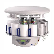
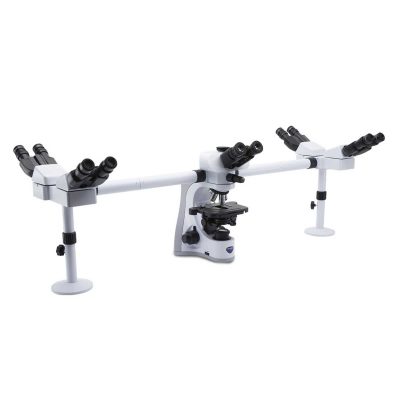
Spin Tissue Processor STP 120
$6,050.00
Shipped From Abroad
Myr Spin Tissue Processor STP 120 has been developed to meet the requirements of every single laboratory. The technology and the unique processing method of the STP 120 qualifies it as the most successful Spin Tissue Processors ever. With more than 3.500 units installed around the world confirms its leading position.
Typically 10-21 working days – excluding furniture and heavy/bulky equipment. Please contact us for further information.
Description
Features
- A worldwide unique technique
Tissue processing is a technique than uses alcohols to remove water from tissues and replace it with a medium that allows sectioning of tissue. Several methods are used to achieve this. Myr Spin Tissue Processor STP 120 fulfills it in a patented and unique technique that combines several movements for the tissue to achieve perfect infiltration results. This is possible thanks to the world’s best spin processing method.
- ROTATIONAL AGITATION
The basket with the cassettes is immersed into the reagent vessel. In this position, the basket turns at 60 rpm and changes the rotational direction every 60 seconds. The rotational agitation achieves a perfect infiltration of tissue, a homogeneous mixture of the reagents and a reduction of processing time. To get better results, the user can start optionally a shaking process.
- SHAKING
This movement can be optionally activated on the control panel and allows the basket to perform an up-down movement inside the vessel that combined with the rotational agitation fulfills a helicoidal movement that increases infiltration quality at a high degree of precision. At the end of this process, baskets start centrifuging.
- CENTRIFUGING
This function is activated as soon as the infiltration time comes to an end. The basket rises above the reagent’s level but rests inside the vessel. For a period of 60 seconds, it starts whirling at 210 rpm and changes the rotational direction every 15 seconds. This process allows the tissue to be optimally drained and avoids carry-over of reagents from one vessel to another.
Tissue processing achieves by this method almost similar results to those obtained with vacuum systems!
- Ergonomic control panel
The buttons of the control panel are arranged ergonomically for easy handling. The LCD display shows all the parameters throughout the process, such as program number, vessel, remaining time, start delay, total duration of the program, rotational agitation and shaking, basket’s centrifuging, temperature of paraffin baths, date and time.
- Easy handling
The instrument has the capacity to store up to 10 different programs that can be freely set up by the user. Each one of the programs can be started in immediate or delayed mode, without a time limit. The instrument can easily be rotated via the four rollers mounted on the base. This allows the user to have fast and safe access to each one of the vessels.
- Maximum safety standards for the user and the specimen
The individual cover for each one of the vessels reduces the emission of vapors to a minimum. The STP 120-2 and STP 120-3 versions incorporate a fume extraction system that – through a fan and an active charcoal filter – cleanse the vapors before being discharged into the air. Optional adaptor for the laboratory general extraction system.
In case of a power shutdown, the specimens are automatically lowered inside the vessel by means of a battery to protect them against desiccation and solid paraffins. Once power is restored, the instrument resumes the program at the same point in which it was interrupted. lf it is a long power failure and the paraffin baths become solidified, the safety program will be activated. The instrument will then wait for the baths to be fully liquified before going ahead with the change to the paraffin baths.
Emergency motions can be implemented through the battery, such as moving the basket up and down or station change (as long as the basket is not inside a solidified paraffin bath). The instrument is also equipped with an emergency stop button. It is possible to interrupt a program for reloading or advanced unloading of samples.
- Alarms during the process
If the specimens remain inside a station for a longer time than the programmed time, e.g. due to a power failure, the display will show a message indicating the station number and the overtime spent in that station compared with the initially programmed time. The acoustic and visual alarms can easily be identified by the user. The keyboard can be locked by the user to avoid an inadvertent change of the process parameters during operation.
Technical Data Spin Tissue Processor STP 120
| Power requirements | ||
|---|---|---|
| Nominal voltage | 100 – 120 V | 220-240 V AC (±10%) |
| Network’s frequency | 50/60 Hz | |
| Consumption | 400 VA | |
| Fuses | 115 V (2xT4A) | 230 V (2xT2A) |
| Accumulator Nickel-Cadmium | 12 V 600 mA | |
| Capacity | ||
| Reagent stations | ||
| Number of vessels | 10 (9 if 3 paraffin baths are used) | |
| Volume per vessel | 1,8 l | |
| Paraffin stations | ||
| Number | 2 (optionally 3) | |
| Volume | 1,8 l | |
| Nominal voltage | 24 V AC | |
| Nominal power per station | 100 VA | |
| Temperature setting range (temperature increase by 1 degree celsius) | 45 – 70 °C | |
| Overtemperature release | 75 °C (± 4 °C) | |
| Cassette baskets | ||
| Number of baskets | 1 (optionally 2) | |
| Basket capacity | 120 cassettes (optionally 240) | |
| Programming | ||
| Number of programs | 10 (selectable) | |
| Infiltration time per station | from 1 m to 99 h 59 m | |
| Rotational agitation | selectable | |
| Shaking | selectable | |
| Centrifuging time | selectable | |
| Program start delay | selectable without time limit | |
| Dimensions | ||
| Diameter | 850 mm | |
| Height | 500 – 700 mm | |
| Diameter of the roller’s circle | 670 mm | |
| Weight | ||
| Including packaging | 130 kg | |
| Net (fully equipped) | 70 kg |
Optional accessories
| Spiral support for a second cassette basket (double processing capacity) |
| Additional basket |
| Additional paraffin bath (necessary when operating the processor with two baskets) |
| Fume extraction system with active charcoal filter |
Click here to download Catalogue
Quick Comparison
| Spin Tissue Processor STP 120 remove | Optika B-159R-PL Binocular Microscope remove | Hawksley 1400 Haematocrit Centrifuge remove | OPTIKA B-383POL Trinocular Polarizing Microscope remove | Chemical Weighing Balance remove | Optika B-510ASB Trinocular Phase Contrast Microscope remove | ||||||||||||||||||||||||||||||||||||||||||||||||||||||||||||||||||||||||||||||||||||||||||||||||||||||||||||||||||||||||||||||||||||||||||||||||||||||||||||||||||||||||||||||||||||||||||||||||||||||||||||||||||||||||||||||||||||||||||||||||||||||||||||||||||||||||||||||||||||||||||||||||||||||||||||||||||||||||||||||||||||||||||||||||||||||||||||||||||||||||||||||||||||||||||||||||||||||||||||||||||||||||||||||||||||||||||||||||||||||||||||||||||||||||||||||||||||||||||||||||||||||||||||||||||||||||||||||||||||||||||||||||||||||||||||||||||||||||||||||||||||||||||||||||||||||||||||||||||||||||
|---|---|---|---|---|---|---|---|---|---|---|---|---|---|---|---|---|---|---|---|---|---|---|---|---|---|---|---|---|---|---|---|---|---|---|---|---|---|---|---|---|---|---|---|---|---|---|---|---|---|---|---|---|---|---|---|---|---|---|---|---|---|---|---|---|---|---|---|---|---|---|---|---|---|---|---|---|---|---|---|---|---|---|---|---|---|---|---|---|---|---|---|---|---|---|---|---|---|---|---|---|---|---|---|---|---|---|---|---|---|---|---|---|---|---|---|---|---|---|---|---|---|---|---|---|---|---|---|---|---|---|---|---|---|---|---|---|---|---|---|---|---|---|---|---|---|---|---|---|---|---|---|---|---|---|---|---|---|---|---|---|---|---|---|---|---|---|---|---|---|---|---|---|---|---|---|---|---|---|---|---|---|---|---|---|---|---|---|---|---|---|---|---|---|---|---|---|---|---|---|---|---|---|---|---|---|---|---|---|---|---|---|---|---|---|---|---|---|---|---|---|---|---|---|---|---|---|---|---|---|---|---|---|---|---|---|---|---|---|---|---|---|---|---|---|---|---|---|---|---|---|---|---|---|---|---|---|---|---|---|---|---|---|---|---|---|---|---|---|---|---|---|---|---|---|---|---|---|---|---|---|---|---|---|---|---|---|---|---|---|---|---|---|---|---|---|---|---|---|---|---|---|---|---|---|---|---|---|---|---|---|---|---|---|---|---|---|---|---|---|---|---|---|---|---|---|---|---|---|---|---|---|---|---|---|---|---|---|---|---|---|---|---|---|---|---|---|---|---|---|---|---|---|---|---|---|---|---|---|---|---|---|---|---|---|---|---|---|---|---|---|---|---|---|---|---|---|---|---|---|---|---|---|---|---|---|---|---|---|---|---|---|---|---|---|---|---|---|---|---|---|---|---|---|---|---|---|---|---|---|---|---|---|---|---|---|---|---|---|---|---|---|---|---|---|---|---|---|---|---|---|---|---|---|---|---|---|---|---|---|---|---|---|---|---|---|---|---|---|---|---|---|---|---|---|---|---|---|---|---|---|---|---|---|---|---|---|---|---|---|---|---|---|---|---|---|---|---|---|---|---|---|---|---|---|---|---|---|---|---|---|---|---|---|---|---|---|---|---|---|---|---|---|---|---|---|---|---|---|---|---|---|---|---|---|---|---|---|---|---|---|---|---|---|---|---|---|---|---|---|---|---|---|---|---|---|---|---|---|---|---|---|---|---|---|---|---|---|---|---|---|---|---|---|---|---|---|---|---|---|---|---|---|---|---|---|---|---|---|---|---|---|---|---|---|---|---|---|---|---|---|---|---|---|---|---|---|---|---|---|---|---|---|---|---|---|---|---|---|---|---|---|---|---|---|---|
| Name | Spin Tissue Processor STP 120 remove | Optika B-159R-PL Binocular Microscope remove | Hawksley 1400 Haematocrit Centrifuge remove | OPTIKA B-383POL Trinocular Polarizing Microscope remove | Chemical Weighing Balance remove | Optika B-510ASB Trinocular Phase Contrast Microscope remove | |||||||||||||||||||||||||||||||||||||||||||||||||||||||||||||||||||||||||||||||||||||||||||||||||||||||||||||||||||||||||||||||||||||||||||||||||||||||||||||||||||||||||||||||||||||||||||||||||||||||||||||||||||||||||||||||||||||||||||||||||||||||||||||||||||||||||||||||||||||||||||||||||||||||||||||||||||||||||||||||||||||||||||||||||||||||||||||||||||||||||||||||||||||||||||||||||||||||||||||||||||||||||||||||||||||||||||||||||||||||||||||||||||||||||||||||||||||||||||||||||||||||||||||||||||||||||||||||||||||||||||||||||||||||||||||||||||||||||||||||||||||||||||||||||||||||||||||||||||||||
| Image | |  | 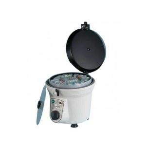 |  | 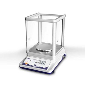 | 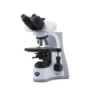 | |||||||||||||||||||||||||||||||||||||||||||||||||||||||||||||||||||||||||||||||||||||||||||||||||||||||||||||||||||||||||||||||||||||||||||||||||||||||||||||||||||||||||||||||||||||||||||||||||||||||||||||||||||||||||||||||||||||||||||||||||||||||||||||||||||||||||||||||||||||||||||||||||||||||||||||||||||||||||||||||||||||||||||||||||||||||||||||||||||||||||||||||||||||||||||||||||||||||||||||||||||||||||||||||||||||||||||||||||||||||||||||||||||||||||||||||||||||||||||||||||||||||||||||||||||||||||||||||||||||||||||||||||||||||||||||||||||||||||||||||||||||||||||||||||||||||||||||||||||||||
| SKU | SF1033560130128-3 | SF1033560098-4 | SF1033560084-118 | SF1033560098-16 | SF1033560084-190 | SF1033560098-9 | |||||||||||||||||||||||||||||||||||||||||||||||||||||||||||||||||||||||||||||||||||||||||||||||||||||||||||||||||||||||||||||||||||||||||||||||||||||||||||||||||||||||||||||||||||||||||||||||||||||||||||||||||||||||||||||||||||||||||||||||||||||||||||||||||||||||||||||||||||||||||||||||||||||||||||||||||||||||||||||||||||||||||||||||||||||||||||||||||||||||||||||||||||||||||||||||||||||||||||||||||||||||||||||||||||||||||||||||||||||||||||||||||||||||||||||||||||||||||||||||||||||||||||||||||||||||||||||||||||||||||||||||||||||||||||||||||||||||||||||||||||||||||||||||||||||||||||||||||||||||
| Rating | |||||||||||||||||||||||||||||||||||||||||||||||||||||||||||||||||||||||||||||||||||||||||||||||||||||||||||||||||||||||||||||||||||||||||||||||||||||||||||||||||||||||||||||||||||||||||||||||||||||||||||||||||||||||||||||||||||||||||||||||||||||||||||||||||||||||||||||||||||||||||||||||||||||||||||||||||||||||||||||||||||||||||||||||||||||||||||||||||||||||||||||||||||||||||||||||||||||||||||||||||||||||||||||||||||||||||||||||||||||||||||||||||||||||||||||||||||||||||||||||||||||||||||||||||||||||||||||||||||||||||||||||||||||||||||||||||||||||||||||||||||||||||||||||||||||||||||||||||||||||||||||
| Price | $6,050.00 |
| $690.00 |
| $658.00 |
| |||||||||||||||||||||||||||||||||||||||||||||||||||||||||||||||||||||||||||||||||||||||||||||||||||||||||||||||||||||||||||||||||||||||||||||||||||||||||||||||||||||||||||||||||||||||||||||||||||||||||||||||||||||||||||||||||||||||||||||||||||||||||||||||||||||||||||||||||||||||||||||||||||||||||||||||||||||||||||||||||||||||||||||||||||||||||||||||||||||||||||||||||||||||||||||||||||||||||||||||||||||||||||||||||||||||||||||||||||||||||||||||||||||||||||||||||||||||||||||||||||||||||||||||||||||||||||||||||||||||||||||||||||||||||||||||||||||||||||||||||||||||||||||||||||||||||||||||||||||||
| Stock | |||||||||||||||||||||||||||||||||||||||||||||||||||||||||||||||||||||||||||||||||||||||||||||||||||||||||||||||||||||||||||||||||||||||||||||||||||||||||||||||||||||||||||||||||||||||||||||||||||||||||||||||||||||||||||||||||||||||||||||||||||||||||||||||||||||||||||||||||||||||||||||||||||||||||||||||||||||||||||||||||||||||||||||||||||||||||||||||||||||||||||||||||||||||||||||||||||||||||||||||||||||||||||||||||||||||||||||||||||||||||||||||||||||||||||||||||||||||||||||||||||||||||||||||||||||||||||||||||||||||||||||||||||||||||||||||||||||||||||||||||||||||||||||||||||||||||||||||||||||||||||||
| Availability | |||||||||||||||||||||||||||||||||||||||||||||||||||||||||||||||||||||||||||||||||||||||||||||||||||||||||||||||||||||||||||||||||||||||||||||||||||||||||||||||||||||||||||||||||||||||||||||||||||||||||||||||||||||||||||||||||||||||||||||||||||||||||||||||||||||||||||||||||||||||||||||||||||||||||||||||||||||||||||||||||||||||||||||||||||||||||||||||||||||||||||||||||||||||||||||||||||||||||||||||||||||||||||||||||||||||||||||||||||||||||||||||||||||||||||||||||||||||||||||||||||||||||||||||||||||||||||||||||||||||||||||||||||||||||||||||||||||||||||||||||||||||||||||||||||||||||||||||||||||||||||||
| Add to cart | |||||||||||||||||||||||||||||||||||||||||||||||||||||||||||||||||||||||||||||||||||||||||||||||||||||||||||||||||||||||||||||||||||||||||||||||||||||||||||||||||||||||||||||||||||||||||||||||||||||||||||||||||||||||||||||||||||||||||||||||||||||||||||||||||||||||||||||||||||||||||||||||||||||||||||||||||||||||||||||||||||||||||||||||||||||||||||||||||||||||||||||||||||||||||||||||||||||||||||||||||||||||||||||||||||||||||||||||||||||||||||||||||||||||||||||||||||||||||||||||||||||||||||||||||||||||||||||||||||||||||||||||||||||||||||||||||||||||||||||||||||||||||||||||||||||||||||||||||||||||||||||
| Description | Shipped From Abroad
Myr Spin Tissue Processor STP 120 has been developed to meet the requirements of every single laboratory. The technology and the unique processing method of the STP 120 qualifies it as the most successful Spin Tissue Processors ever. With more than 3.500 units installed around the world confirms its leading position.
Delivery & Availability:
Typically 10-21 working days – excluding furniture and heavy/bulky equipment. Please contact us for further information.
| Shipped from Abroad
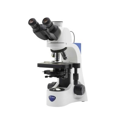
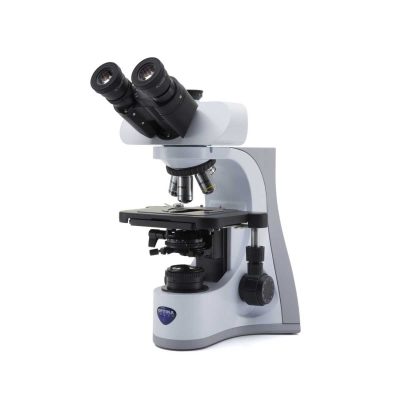
Cordless binocular microscope ideal for students and primary schools, with four PLAN achromatic lenses (1000x), FN 18 high eyepointeyepieces, finite optical system, coaxial focusing, mechanical stage, Abbe condenser and powerful, uniform, white color temperature 1 W X-LED1 illumination.
| In stock
| Shipped from Abroad
OPTIKA polarizing microscopes offer a complete system for your laboratory analysis, including polarizer and analyzer filters, Bertrand lens for conoscopic observation, compensator plates, and high-precision rotatable stages. Also available the X-LED illumination system, a high-intensity light source that delivers bright and clear images.
| In stock
This is a 210g/0.001g Analytical electronic chemical balance for industrial and scientific field.
| Shipped from Abroad
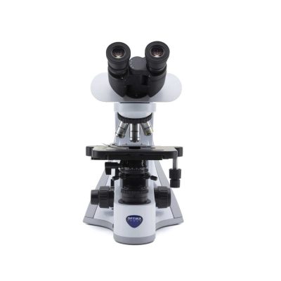
Advanced routine laboratory microscope for brightfield and phase contrast observations with IOS W-PLAN objectives and rackless stage. Ideal for Asbestos analysis in accordance to international rules with 12.5x eyepieces and Walton & Becket graticule to perform perfect asbestos fibers analysis at a glance. The high-efficiency X-LED3 makes it reliable for all transmitted light observations.
| |||||||||||||||||||||||||||||||||||||||||||||||||||||||||||||||||||||||||||||||||||||||||||||||||||||||||||||||||||||||||||||||||||||||||||||||||||||||||||||||||||||||||||||||||||||||||||||||||||||||||||||||||||||||||||||||||||||||||||||||||||||||||||||||||||||||||||||||||||||||||||||||||||||||||||||||||||||||||||||||||||||||||||||||||||||||||||||||||||||||||||||||||||||||||||||||||||||||||||||||||||||||||||||||||||||||||||||||||||||||||||||||||||||||||||||||||||||||||||||||||||||||||||||||||||||||||||||||||||||||||||||||||||||||||||||||||||||||||||||||||||||||||||||||||||||||||||||||||||||||
| Content | Features
Tissue processing achieves by this method almost similar results to those obtained with vacuum systems!
Emergency motions can be implemented through the battery, such as moving the basket up and down or station change (as long as the basket is not inside a solidified paraffin bath). The instrument is also equipped with an emergency stop button. It is possible to interrupt a program for reloading or advanced unloading of samples.
Technical Data Spin Tissue Processor STP 120
Click here to download Catalogue | Cordless binocular microscope ideal for students and primary schools, with four PLAN achromatic lenses (1000x), FN 18 high eyepointeyepieces, finite optical system, coaxial focusing, mechanical stage, Abbe condenser and powerful, uniform, white color temperature 1 W X-LED1 illumination. Slim and easy to carry, it is equipped with all the main controls to start learning how to use an advanced microscope and with long lasting LED illumination to provide over 20 years of use. The exclusive Li-Ion battery ensures unparalleled duration and fast recharge.
Features:
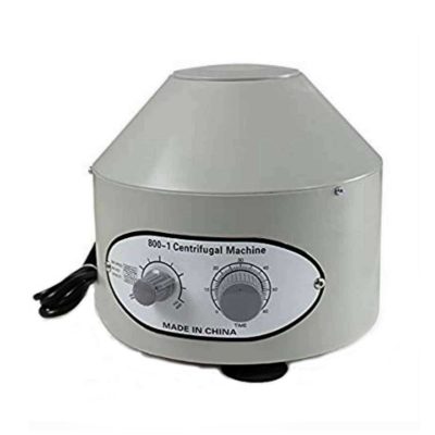
Click Here To Download Catalogue | The Hawksley Haematospin 1400 Centrifuges are built in the UK and are made from durable and high-quality components. These centrifuges are ideal for veterinary laboratories and practices specifically designed to produce reliable and accurate results on blood and urine. One of the main features of this centrifuge is that it takes both 2ml microtubes and 75mm microhaematocrits. The classic design of these centrifuges is used worldwide with guaranteed support for all servicing and parts.
Technical specifications:
Click Here To Download Catalogue | Observation mode: Brightfield, transmitted polarized light and conoscopy.
Bertrand lens and polarizing attachment: Swing-out type with centering mechanism for observation in conoscopy/orthoscopy. Rotatable analyzer from 0° to 90° with graduated scale. Tint plates: 1° order red (λ); λ/4; Quartz wedge.
Head: Trinocular (fixed 50/50), 30° inclined, 360° rotating.
Interpupillary distance: Adjustable between 48 and 75 mm.
Dioptric adjustment: On the left eyepiece tube.
Eyepieces: WF10x/20 mm, high eye-point and secured by screw. One with crosshair.
Nosepiece: Quadruple revolving nosepiece, rotation on ball bearings. Centering system for each objective.
Objectives (strain-free):
IOS N-PLAN POL 4x/0.10
IOS N-PLAN POL 10x/0.25
IOS N-PLAN POL 40x/0.65
IOS N-PLAN POL 60x/0.80
All with anti-fungus treatment.
Specimen stage: Rotatable stage with locking mechanism. Vernier scale with accuracy 0.1 mm. Diameter 160 mm. Specimen slide clamps.
Focusing: Coaxial coarse (adjustable tension) and fine focusing mechanism with limit stop to prevent the contact between objective and specimen.
Condenser: Abbe N.A. 1.25, with objective-coded iris diaphragm, focusable and centerable. With rotating polarizing filter.
Transmitted illumination (Fixed Koehler type): X-LED3 with white 3.6 W LED (6.300 K) with brightness control. Multi-plug 100-240Vac/6Vdc external power supply.
Click Here To Download Catalogue | This is a 210g/0.001g Analytical electronic chemical balance for industrial and scientific field.
| Advanced routine laboratory microscope for brightfield and phase contrast observations with IOS W-PLAN objectives and rackless stage. Ideal for Asbestos analysis in accordance to international rules with 12.5x eyepieces and Walton & Becket graticule to perform perfect asbestos fibers analysis at a glance. The high-efficiency X-LED3 makes it reliable for all transmitted light observations.
Features:
| |||||||||||||||||||||||||||||||||||||||||||||||||||||||||||||||||||||||||||||||||||||||||||||||||||||||||||||||||||||||||||||||||||||||||||||||||||||||||||||||||||||||||||||||||||||||||||||||||||||||||||||||||||||||||||||||||||||||||||||||||||||||||||||||||||||||||||||||||||||||||||||||||||||||||||||||||||||||||||||||||||||||||||||||||||||||||||||||||||||||||||||||||||||||||||||||||||||||||||||||||||||||||||||||||||||||||||||||||||||||||||||||||||||||||||||||||||||||||||||||||||||||||||||||||||||||||||||||||||||||||||||||||||||||||||||||||||||||||||||||||||||||||||||||||||||||||||||||||||||||
| Weight | N/A | N/A | N/A | N/A | N/A | N/A | |||||||||||||||||||||||||||||||||||||||||||||||||||||||||||||||||||||||||||||||||||||||||||||||||||||||||||||||||||||||||||||||||||||||||||||||||||||||||||||||||||||||||||||||||||||||||||||||||||||||||||||||||||||||||||||||||||||||||||||||||||||||||||||||||||||||||||||||||||||||||||||||||||||||||||||||||||||||||||||||||||||||||||||||||||||||||||||||||||||||||||||||||||||||||||||||||||||||||||||||||||||||||||||||||||||||||||||||||||||||||||||||||||||||||||||||||||||||||||||||||||||||||||||||||||||||||||||||||||||||||||||||||||||||||||||||||||||||||||||||||||||||||||||||||||||||||||||||||||||||
| Dimensions | N/A | N/A | N/A | N/A | N/A | N/A | |||||||||||||||||||||||||||||||||||||||||||||||||||||||||||||||||||||||||||||||||||||||||||||||||||||||||||||||||||||||||||||||||||||||||||||||||||||||||||||||||||||||||||||||||||||||||||||||||||||||||||||||||||||||||||||||||||||||||||||||||||||||||||||||||||||||||||||||||||||||||||||||||||||||||||||||||||||||||||||||||||||||||||||||||||||||||||||||||||||||||||||||||||||||||||||||||||||||||||||||||||||||||||||||||||||||||||||||||||||||||||||||||||||||||||||||||||||||||||||||||||||||||||||||||||||||||||||||||||||||||||||||||||||||||||||||||||||||||||||||||||||||||||||||||||||||||||||||||||||||
| Additional information | |||||||||||||||||||||||||||||||||||||||||||||||||||||||||||||||||||||||||||||||||||||||||||||||||||||||||||||||||||||||||||||||||||||||||||||||||||||||||||||||||||||||||||||||||||||||||||||||||||||||||||||||||||||||||||||||||||||||||||||||||||||||||||||||||||||||||||||||||||||||||||||||||||||||||||||||||||||||||||||||||||||||||||||||||||||||||||||||||||||||||||||||||||||||||||||||||||||||||||||||||||||||||||||||||||||||||||||||||||||||||||||||||||||||||||||||||||||||||||||||||||||||||||||||||||||||||||||||||||||||||||||||||||||||||||||||||||||||||||||||||||||||||||||||||||||||||||||||||||||||||||||


Reviews
There are no reviews yet.