Ultrasonic Vascular Doppler Detector Bi
$7,480.00
Shipped From Abroad
Feature:
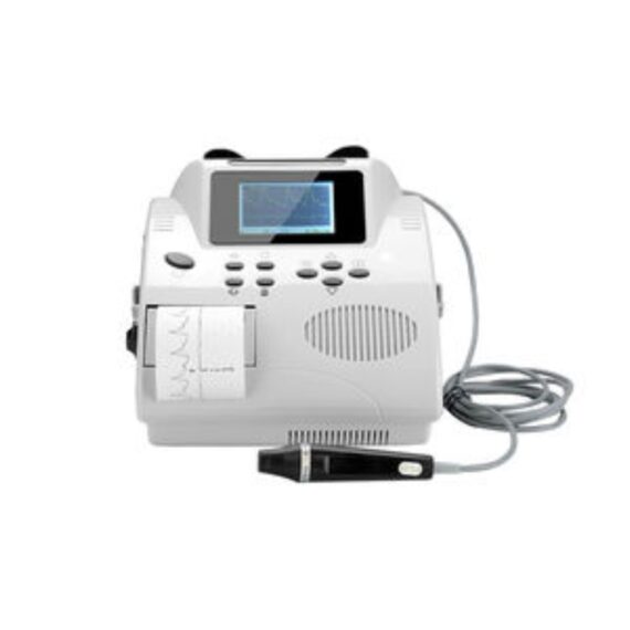
620VP TFT LCD displays the wave of the instant blood flow average velocity and strength. It prints the monitoring blood flow curve simultaneously;
Detect the blood stream status of arterial/venous by 8MHz probe;
Detect the blood flow average velocity, detect the result of fingers/toes and part of body’s vein anatomies operation;
BV-620VP(TFT) portable bidirectional vascular Doppler with large color LCD screen;
Built-in ARM microprocessor & real-time displays blood flow velocity waveform;
Delivery & Availability:
Typically 10-21 working days – excluding furniture and heavy/bulky equipment. Please contact us for further information.
Description
Feature:
620VP TFT LCD displays the wave of the instant blood flow average velocity and strength. It prints the monitoring blood flow curve simultaneously;
Detect the blood stream status of arterial/venous by 8MHz probe;
Detect the blood flow average velocity, detect the result of fingers/toes and part of body’s vein anatomies operation;
BV-620VP(TFT) portable bidirectional vascular Doppler with large color LCD screen;
Built-in ARM microprocessor & real-time displays blood flow velocity waveform;
Can store 50 detected waveforms;
High-speed USB/RS -232 port, can be connected directly to computer to analyze, store, and print the waveform data;
Detect peak and average blood speed
Detect blood flow of peripheral vessel
Detect subsection systolic pressure
Detect phlebostenosis, vein occlusions
Detect Systolic pressure of toes and fingers
Detect blood speed during recovery
Detect the pulse rate and show PR simultaneously
Specification:
Frequency range:Main unit:200±80~5000±1000HZ
Probe:350±80~2500±500HZ
External output:loudspeaker, single channel with earphone jack
Emissive waveform: sine wave
Ultrasonic frequency:5.0MHz±10% 、8.0MHz ±10%
Ultrasonic frequency average intensity: < 50mW/cm2
Velocity measure range:0~100 cm /s.
Test error:≤20%(relative error)
Overall sensitivity:> 100dB
Printing speed:40cm/min,60cm/min ,display synchronize with print
Frequency mode display range:0.2 KHz~7.0 KHz
LCD Display:128 X 64 dots, LCD cursor indicates blood stream speed, double display mode for the spectrum and velocity.
Power indication: blue LED indication
Charge indication: the charge indicator is yellow when charging and become green after full charge
Alarm indication: the indicator light is red when the lack of battery
Lack of paper indication: When the recorder is lack of paper, the indication light is red
Outline dimension:210*220*105 (mm)
Net weight:1.9 Kg
Environment condition:
Temperature:+5℃ ~ +40℃ ,
Humidity:<80%,
Atmospheric pressure:86kPa ~ 106kPa.
Transport and storage environment:
Temperature:-10℃ ~ +55℃ ,
Humidity:<80%,
Atmospheric pressure:86kPa ~ 106kPa
Quick Comparison
| Ultrasonic Vascular Doppler Detector Bi remove | Mayo Table remove | Pedal Bin remove | Stainless Steel Hospital Two Step Stool remove | Urine Analyzer remove | OPTIKA B-383DK Trinocular Darkfeld Microscope remove | |||||||||||||||||||||
|---|---|---|---|---|---|---|---|---|---|---|---|---|---|---|---|---|---|---|---|---|---|---|---|---|---|---|
| Name | Ultrasonic Vascular Doppler Detector Bi remove | Mayo Table remove | Pedal Bin remove | Stainless Steel Hospital Two Step Stool remove | Urine Analyzer remove | OPTIKA B-383DK Trinocular Darkfeld Microscope remove | ||||||||||||||||||||
| Image |  | 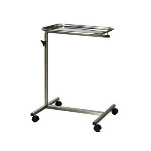 |  |  | 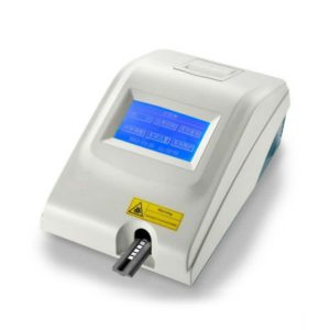 | 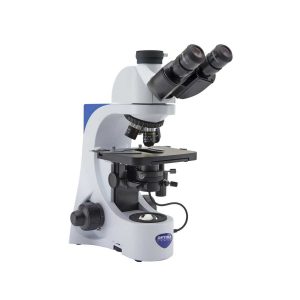 | ||||||||||||||||||||
| SKU | SF103356013038 | SF1033560084-239 | SF1033560084-201 | SF1033560084-204 | SF1033560084-224 | SF1033560098-14 | ||||||||||||||||||||
| Rating | ||||||||||||||||||||||||||
| Price | $7,480.00 | $48.00 | $17.00 | $27.00 | $540.00 |
| ||||||||||||||||||||
| Stock | ||||||||||||||||||||||||||
| Availability | ||||||||||||||||||||||||||
| Add to cart | ||||||||||||||||||||||||||
| Description | Shipped From Abroad
Feature:
620VP TFT LCD displays the wave of the instant blood flow average velocity and strength. It prints the monitoring blood flow curve simultaneously;
Detect the blood stream status of arterial/venous by 8MHz probe;
Detect the blood flow average velocity, detect the result of fingers/toes and part of body's vein anatomies operation;
BV-620VP(TFT) portable bidirectional vascular Doppler with large color LCD screen;
Built-in ARM microprocessor & real-time displays blood flow velocity waveform;
Delivery & Availability:
Typically 10-21 working days – excluding furniture and heavy/bulky equipment. Please contact us for further information.
| In stock
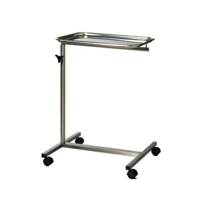
Material Stainless: Steel
Height Adjustable: Yes
Number of Wheel: 4
Height: 3-4 Feet
| In stock
| In stock
Features:
| In stock
Used for Clinical Diagnostics.
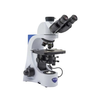
| Shipped from Abroad B-383DK - Darkfield Microscope Laboratory upright microscope for brightfield and darkfield observations with N-PLAN objectives (and W-PLAN 100x with iris) for biology and especially darkfield fresh blood analysis and the exclusive X-LED3 illumination system. The special condenser with integrated, exclusive X-LED3 illuminator replaces any other external and expensive lighting source required for these applications and is ideal for great-looking, rich and high-quality specimen view. Delivery & Availability: Typically 21 working days – excluding furniture and heavy/bulky equipment. Please contact us for further information. | ||||||||||||||||||||
| Content | Feature: 620VP TFT LCD displays the wave of the instant blood flow average velocity and strength. It prints the monitoring blood flow curve simultaneously; Detect the blood stream status of arterial/venous by 8MHz probe; Detect the blood flow average velocity, detect the result of fingers/toes and part of body's vein anatomies operation; BV-620VP(TFT) portable bidirectional vascular Doppler with large color LCD screen; Built-in ARM microprocessor & real-time displays blood flow velocity waveform; Can store 50 detected waveforms; High-speed USB/RS -232 port, can be connected directly to computer to analyze, store, and print the waveform data; Detect peak and average blood speed Detect blood flow of peripheral vessel Detect subsection systolic pressure Detect phlebostenosis, vein occlusions Detect Systolic pressure of toes and fingers Detect blood speed during recovery Detect the pulse rate and show PR simultaneously Specification: Frequency range:Main unit:200±80~5000±1000HZ Probe:350±80~2500±500HZ External output:loudspeaker, single channel with earphone jack Emissive waveform: sine wave Ultrasonic frequency:5.0MHz±10% 、8.0MHz ±10% Ultrasonic frequency average intensity: < 50mW/cm2 Velocity measure range:0~100 cm /s. Test error:≤20%(relative error) Overall sensitivity:> 100dB Printing speed:40cm/min,60cm/min ,display synchronize with print Frequency mode display range:0.2 KHz~7.0 KHz LCD Display:128 X 64 dots, LCD cursor indicates blood stream speed, double display mode for the spectrum and velocity. Power indication: blue LED indication Charge indication: the charge indicator is yellow when charging and become green after full charge Alarm indication: the indicator light is red when the lack of battery Lack of paper indication: When the recorder is lack of paper, the indication light is red Outline dimension:210*220*105 (mm) Net weight:1.9 Kg Environment condition: Temperature:+5℃ ~ +40℃ , Humidity:<80%, Atmospheric pressure:86kPa ~ 106kPa. Transport and storage environment: Temperature:-10℃ ~ +55℃ , Humidity:<80%, Atmospheric pressure:86kPa ~ 106kPa |
|
| Features:
| Used for Clinical Diagnostics.
| Observation mode: Brightfield, oil immersion darkfield.
Head: Trinocular (fixed 50/50), 30° inclined, 360° rotating.
Interpupillary distance: Adjustable between 48 and 75 mm.
Dioptric adjustment: On the left eyepiece tube.
Eyepieces: WF10x/20 mm, high eye-point and secured by screw.
Nosepiece: Quintuple revolving nosepiece, rotation on ball bearings.
Objectives:
N-PLAN 4x/0.10
N-PLAN 10x/0.25
N-PLAN 40x/0.65
W-PLAN 100x/1.25 (oil) with iris
All with anti-fungus treatment.
Specimen stage: Double layer rackless mechanical stage, 150×139 mm, 75×33 mm X-Y range.
Focusing: Coaxial coarse (adjustable tension) and fine focusing mechanism with limit stop to prevent the contact between objective and specimen.
Brightfield condenser: Abbe N.A. 1.25, with objective-coded iris diaphragm, focusable and centerable.
Darkfieldfield condenser: Darkfield N.A. 1.36 (oil immersion) with built-in X-LED3.
Transmitted illumination (Fixed Koehler type): X-LED3 with white 3.6 W LED (6,300K) with brightness control. Multi-plug 100-240Vac/6Vdc external power supply.
Click Here To Download Catalogue | ||||||||||||||||||||
| Weight | N/A | N/A | N/A | N/A | N/A | N/A | ||||||||||||||||||||
| Dimensions | N/A | N/A | N/A | N/A | N/A | N/A | ||||||||||||||||||||
| Additional information |

Reviews
There are no reviews yet.