

Anke Anatom 64 Clarity Multi-Slice Spiral CT Scan
$0.00
Shipped from Abroad
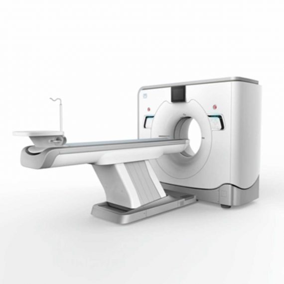
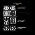
The ANATOM 64 CT scanner is the latest innovation for cardiac imaging based on Precision Platform system. The excellent design of Ahart technology which innovatively combined single spiral scan + gated imaging + mA modulation for easy heart imaging at extremely low radiation dose. We provide you ANATOM 64 Clarity/Precision of two models which are low/high configurations for preferences. It also offers you conventional clinical applications of low dose, better image quality and faster exams.
Delivery & Availability:
Typically 90 working days – excluding furniture and heavy/bulky equipment. Please contact us for further information.
Description
The ANATOM 64 CT scanner is the latest innovation for cardiac imaging based on Precision Platform system. The excellent design of Ahart technology which innovatively combined single spiral scan + gated imaging + mA modulation for easy heart imaging at extremely low radiation dose. We provide you ANATOM 64 Clarity/Precision of two models which are low/high configurations for preferences. It also offers you conventional clinical applications of low dose, better image quality and faster exams.
Features:
- Modularized OptiWave HD detector features low-cost & easy maintenance, high spatial resolution and long lifetime
- Admir3D iterative technology delivers optimal dose efficiency and noise reduction without compromising image quality
- High configurations of main components ensure the best results and maximum patient throughput
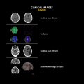
- Uniquely and creatively uses 140kV and 80kV dual energy scan mode for brain imaging on 16-slice CT to offers you extraordinary image quality both in low and high density resolutions
- AdoseTM mA modulation ensures you low dose imaging without compromising image quality particularly useful in pediatric applications
- Equipped with dedicated Abast and Amast for bone and metal artifacts
- The brilliant Ahart technology enables you to experience so easy and low-dose cardiac imaging applications
Technical Specifications:
| Model | ANATOM 64 Precision | ANATOM 64 Fit |
| Rack type | Low pressure slip ring | Low pressure slip ring |
| Scan aperture | 70cm | 70cm |
| Rack Physical inclination | ± 30 ° | N.A |
| Rack digital inclination | ± 50 ° | ± 50 ° |
| cooling method | Air-cooled | Air-cooled |
| Focus to the center distance | 56 cm | 53 cm |
| Maximum power (non-equivalent) | 80kW | 42kW |
| Votage (kV) | 80kV / 100kV / 120kV / 140kV | 70kV / 80kV / 100kV / 120kV /
140kV |
| Heat capacity | 8MHU | 5.0MHU |
| Heat dissipation rate | 931 kHU / min | 748kHU / min |
| cooling method | Oil cool | Oil cool |
| Large focus size | 1.1mm × 1.2mm | 1.2mm × 1.4mm |
| Small focus size | 0.6mm × 1.2mm | 0.7mm × 0.8mm |
| Tube current range | 10-670mA | 10-350mA |
| Detector type | Optiwave detectors | Optiwave detectors |
| Number of Z-axis | 32 | 32 |
| The width of the Z-axis | 20mm | 20mm |
| The number of elements per row | 912 | 848 |
| Total number of detectors | 29184 | 27136 |
| Acquisition mode | 64×0.625, 32×0.625,
16×0.625 |
64×0.625, 32×0.625,
16×0.625 |
| Scanning range | 1800mm | 1800mm |
| Horizontal positioning accuracy | ± 0.25mm | ± 0.25mm |
| weight capacity | 205kg | 205kg |
| Minimum height | 43cm | 43cm |
| Anti – collision protection device | Yes | Yes |
| Foot control switch | Yes | Yes |
| IV rack | Yes | Yes |
| CPU | 3.5GHz | 3.5GHz |
| RAM | 16 GB × 4 | 16 GB × 4 |
| Hard drive capacity | 1T × 2 | 1T × 2 |
| Display size | 24 inch LCD monitor | 24 inch LCD monitor |
| Display resolution | 1920 × 1200 | 1920 × 1200 |
| Computer operating system | Windows 7 | Windows 7 |
| Image reconstruction speed | 65 frames/ second | 65 frames/ second |
| Number of image store | 1000000 | 1000000 |
| Data external storage mode | CD / DVD / USB | CD / DVD / USB |
| Minimum Scan Time of 360 degree | 0.39sec | 0.75sec |
| Sub-millimeter acquisition layers | 64 | 64 |
| Double sub-millimeter acquisition
layers |
64 | 64 |
| Thinnest acquisition thickness | 0.625mm | 0.625mm |
| The thinnest reconstruction
thickness |
0.3125mm | 0.625mm |
| Conventional reconstruction
thickness (mm) |
0.3125 mm, 0.625 mm, 1.25 mm, 2.5
mm, 5.0 mm, 7.5 mm, 10 mm |
0.625 mm, 1.25 mm, 2.5 mm, 5.0
mm, 7.5 mm, 10 mm |
| The reconstruction matrix | 512 x 512, 1024 x 1024 | 512 x 512, 1024 x 1024 |
| Display matrix | 1024 × 1024 | 1024 × 1024 |
| Max FOV | 52cm | 50cm |
| The maximum display field of view | 70cm | 50cm |
| Maximum scan length | 1800mm | 1800mm |
| Maximum continuous helix scan
time |
120s | 120s |
| Pitch range | 0.5-1.5 | 0.5, 1.0, 1.5 |
| High contrast resolution | 21 Lp / cm @ 0% MTF | 21 Lp / cm @ 0% MTF |
| Low contrast resolution | 2mm @ 0.3% | 2mm @ 0.3% |
| Image noise | ≤ 0.25 | ≤ 0.29 |
| MPR | Yes | Yes |
| CPR | Yes | Yes |
| SSD | Yes | Yes |
| VR | Yes | Yes |
| MIP | Yes | Yes |
| MinIP | Yes | Yes |
| Virtual endoscopy | Yes | Yes |
| CT angiography | Yes | Yes |
| Tissue segmentation | Yes | Yes |
| One-key bone removal | Yes | Yes |
| Automatically patient table removal | Yes | Yes |
| Contrast Agent Automatic Tracking
Technology- bolus tracking |
Yes | Yes |
| Automatic linkage trigger
technology |
Yes | Yes |
| Cine mode display | Yes | Yes |
| Bone artifact suppression technique | AbastTM | AbastTM |
| Metal artifact suppression technique | AbastTM | AbastTM |
| Iterative reconstruction technique | Admir3D global iteration | Admir3D full-domain iteration |
| Low – dose children ‘s scanning
technology |
Yes | Yes |
| Low – dose lung scan | Yes | Yes |
| Gray matter enhancement
technology |
AccuHead | AccuHead |
| High resolution imaging of the lung | AccuLung | AccuLung |
| Inner ear high resolution imaging | AccuOtica | AccuOtica |
| Body high resolution imaging | AccuBody | AccuBody |
| Bone high resolution imaging | AccuBone | AccuBone |
| Head dual-energy imaging | Ahead | Ahead |
| CT perfusion imaging | Optional | Optional |
| Quantitative analysis of blood
vessels |
Optional | Optional |
| Heart coronary artery imaging | Aheart | N.A |
| ECG gated | Yes | N.A |
| Low dose cardiac scan | Yes | N.A |
| Green energy saving technology | AccuSaving | AccuSaving |
| Dual-energy scan technology | Optional | Optional |
Click Here To Download Catalogue
Additional information
| Model | Advanced, Advanced Plus, Basic, Smart |
|---|
Quick Comparison
| Settings | Anke Anatom 64 Clarity Multi-Slice Spiral CT Scan remove | Sonoscape P10 Ultrasound Machine remove | ASPEL AsCARD Grey ECG Machine remove | DrGem Diamond All-In-One Digital X-ray Machine remove | DrGem Ceiling Mounted Digital X-ray remove | Sonoscape P15 Ultrasound Machine With Four Probes remove | ||||||||||||||||||||||||||||||||||||||||||||||||||||||||||||||||||||||||||||||||||||||||||||||||||||||||||||||||||||||||||||||||||||||||||||||||||||||||||||||||||||||||||||||||||||||||||||||||||||||||||||||||||||||||||||||||||||||||||||||||||||||||||||||||||||||||||||||||||||||||||
|---|---|---|---|---|---|---|---|---|---|---|---|---|---|---|---|---|---|---|---|---|---|---|---|---|---|---|---|---|---|---|---|---|---|---|---|---|---|---|---|---|---|---|---|---|---|---|---|---|---|---|---|---|---|---|---|---|---|---|---|---|---|---|---|---|---|---|---|---|---|---|---|---|---|---|---|---|---|---|---|---|---|---|---|---|---|---|---|---|---|---|---|---|---|---|---|---|---|---|---|---|---|---|---|---|---|---|---|---|---|---|---|---|---|---|---|---|---|---|---|---|---|---|---|---|---|---|---|---|---|---|---|---|---|---|---|---|---|---|---|---|---|---|---|---|---|---|---|---|---|---|---|---|---|---|---|---|---|---|---|---|---|---|---|---|---|---|---|---|---|---|---|---|---|---|---|---|---|---|---|---|---|---|---|---|---|---|---|---|---|---|---|---|---|---|---|---|---|---|---|---|---|---|---|---|---|---|---|---|---|---|---|---|---|---|---|---|---|---|---|---|---|---|---|---|---|---|---|---|---|---|---|---|---|---|---|---|---|---|---|---|---|---|---|---|---|---|---|---|---|---|---|---|---|---|---|---|---|---|---|---|---|---|---|---|---|---|---|---|---|---|---|---|---|---|---|---|---|---|---|---|---|---|---|---|---|---|---|---|
| Name | Anke Anatom 64 Clarity Multi-Slice Spiral CT Scan remove | Sonoscape P10 Ultrasound Machine remove | ASPEL AsCARD Grey ECG Machine remove | DrGem Diamond All-In-One Digital X-ray Machine remove | DrGem Ceiling Mounted Digital X-ray remove | Sonoscape P15 Ultrasound Machine With Four Probes remove | ||||||||||||||||||||||||||||||||||||||||||||||||||||||||||||||||||||||||||||||||||||||||||||||||||||||||||||||||||||||||||||||||||||||||||||||||||||||||||||||||||||||||||||||||||||||||||||||||||||||||||||||||||||||||||||||||||||||||||||||||||||||||||||||||||||||||||||||||||||||||||
| Image |  |  |  |  |  |  | ||||||||||||||||||||||||||||||||||||||||||||||||||||||||||||||||||||||||||||||||||||||||||||||||||||||||||||||||||||||||||||||||||||||||||||||||||||||||||||||||||||||||||||||||||||||||||||||||||||||||||||||||||||||||||||||||||||||||||||||||||||||||||||||||||||||||||||||||||||||||||
| SKU | SF1033560092-2 | SF1033560012-7 | SF1033560075-5 | SF1033560074-3 | SF1033560074-4 | SF1033560012-8 | ||||||||||||||||||||||||||||||||||||||||||||||||||||||||||||||||||||||||||||||||||||||||||||||||||||||||||||||||||||||||||||||||||||||||||||||||||||||||||||||||||||||||||||||||||||||||||||||||||||||||||||||||||||||||||||||||||||||||||||||||||||||||||||||||||||||||||||||||||||||||||
| Rating | ||||||||||||||||||||||||||||||||||||||||||||||||||||||||||||||||||||||||||||||||||||||||||||||||||||||||||||||||||||||||||||||||||||||||||||||||||||||||||||||||||||||||||||||||||||||||||||||||||||||||||||||||||||||||||||||||||||||||||||||||||||||||||||||||||||||||||||||||||||||||||||||||
| Price |
| $9,350.00 | $1,166.00 |
|
| $13,900.00 | ||||||||||||||||||||||||||||||||||||||||||||||||||||||||||||||||||||||||||||||||||||||||||||||||||||||||||||||||||||||||||||||||||||||||||||||||||||||||||||||||||||||||||||||||||||||||||||||||||||||||||||||||||||||||||||||||||||||||||||||||||||||||||||||||||||||||||||||||||||||||||
| Stock | ||||||||||||||||||||||||||||||||||||||||||||||||||||||||||||||||||||||||||||||||||||||||||||||||||||||||||||||||||||||||||||||||||||||||||||||||||||||||||||||||||||||||||||||||||||||||||||||||||||||||||||||||||||||||||||||||||||||||||||||||||||||||||||||||||||||||||||||||||||||||||||||||
| Availability | ||||||||||||||||||||||||||||||||||||||||||||||||||||||||||||||||||||||||||||||||||||||||||||||||||||||||||||||||||||||||||||||||||||||||||||||||||||||||||||||||||||||||||||||||||||||||||||||||||||||||||||||||||||||||||||||||||||||||||||||||||||||||||||||||||||||||||||||||||||||||||||||||
| Add to cart | ||||||||||||||||||||||||||||||||||||||||||||||||||||||||||||||||||||||||||||||||||||||||||||||||||||||||||||||||||||||||||||||||||||||||||||||||||||||||||||||||||||||||||||||||||||||||||||||||||||||||||||||||||||||||||||||||||||||||||||||||||||||||||||||||||||||||||||||||||||||||||||||||
| Description | Shipped from Abroad
The ANATOM 64 CT scanner is the latest innovation for cardiac imaging based on Precision Platform system. The excellent design of Ahart technology which innovatively combined single spiral scan + gated imaging + mA modulation for easy heart imaging at extremely low radiation dose. We provide you ANATOM 64 Clarity/Precision of two models which are low/high configurations for preferences. It also offers you conventional clinical applications of low dose, better image quality and faster exams.
Delivery & Availability: Typically 90 working days – excluding furniture and heavy/bulky equipment. Please contact us for further information. | Shipped from Abroad The P10 color Doppler ultrasound system is a new generation product from SonoScape. It is designed to give high quality images, rich probe configurations, various clinical tools and automatic analysis software to provide you with comprehensive solutions for your growing demand for clinical applications. Delivery & Availability: Typically 5-7 working days – excluding furniture and heavy/bulky equipment. Please contact us for further information. | Shipped from Abroad Electrocardiograph AsCARD Grey v.07.225 - is a 1, 3, 6, 12 channel ECG unit which enables to make electrocardiogram in full 12 leads. It is intended to conduct ECG examinations of adults and paediatric patients in all types of health care centres. ECG examination may be recorded in manual or automatic mode, with the possibility of analysis and interpretation. The device can be powered from 100 V ÷ 240 V mains supply or by an internal battery. Delivery & Availability: Typically 10 working days – excluding furniture and heavy/bulky equipment. Please contact us for further information. | Shipped from Abroad DrGem Diamond All-In-One Digital X-ray Machine is a fully automatic digital radiography system providing state-of-the-art image quality, image processing and user interface. With a wide selection of anatomical studies on the imaging software, DIAMOND automatically sets up the x-ray generator’s preprogrammed exposure technique settings, motorized radiographic stand positioning, x-ray collimation and post-image processing for the selected study. Specifically designed to increase workflow, this fully digital system offers convenient auto-positioning and advanced image processing to achieve big performance with little effort. Delivery & Availability: Typically 21 working days – excluding furniture and heavy/bulky equipment. Please contact us for further information. | In Stock The GXR-SD is a diagnostic digital radiography system that provides reliable high quality digital radiographic images with a reduced dose. The GXR-SD DR systems offer comprehensive digital solutions to all radiography needs, featuring ACQUIDR digital imaging system with stationary or portable digital flat-panel detectors as well as reliable high-frequency x-ray generators that are known worldwide for their excellent performance, lifetime and stability. Patient tables and wall stands are also offered. Delivery & Availability: Typically 21 working days – excluding furniture and heavy/bulky equipment. Please contact us for further information. | In Stock A feature-rich system inheriting the Wi-Sono high-end platform, the P15 uses an array of advanced tools to help enhance the image quality. It's a cost-effective, simplified console with an intuitive user interface and multiple intelligent functions. Delivery & Availability: Typically 2 working days – excluding furniture and heavy/bulky equipment. Please contact us for further information. | ||||||||||||||||||||||||||||||||||||||||||||||||||||||||||||||||||||||||||||||||||||||||||||||||||||||||||||||||||||||||||||||||||||||||||||||||||||||||||||||||||||||||||||||||||||||||||||||||||||||||||||||||||||||||||||||||||||||||||||||||||||||||||||||||||||||||||||||||||||||||||
| Content | The ANATOM 64 CT scanner is the latest innovation for cardiac imaging based on Precision Platform system. The excellent design of Ahart technology which innovatively combined single spiral scan + gated imaging + mA modulation for easy heart imaging at extremely low radiation dose. We provide you ANATOM 64 Clarity/Precision of two models which are low/high configurations for preferences. It also offers you conventional clinical applications of low dose, better image quality and faster exams.
Features:
Click Here To Download Catalogue | DETAILS
B + Compound
B + Compound utilizes several lines of sight for optimal contrast resolution, speckle reduction and border detection, with which P10 is ideal for superficial and abdominal imaging with better clarity and improved continuity of structures.
μ-Scan
The new generation μ-Scan imaging technology gives you better image quality by reducing noise, improving signal strength and improving visualization.
P10 offers a comprehensive selection of electronic probes to maximize its capabilities to meet a wide range of applications including abdomen, pediatric, OB/GYN, cardiovascular, musculoskeletal, etc. The advanced probe technologies also effectively enhance the image quality and confidence in reaching clinical diagnoses, even in difficult patients.
Convex Probe 3C-A
Ideal for an abundant of application such as abdomen, gynecology, obstetrics, urology and even abdomen biopsy.
Linear Probe L741
This linear probe is designed to satisfy vascular, breast, thyroid, and other small parts diagnosis, and its adjustable parameters could also present users a clear view of MSK and deep vessels.
Phase Array Probe 3P-A
For the purpose of adult and pediatric cardiology and emergency, the phase array probe provides elaborate presets for different exam modes, even for difficult patients.
Intracavitary Probe 6V1
Intracavitary probe could face application of gynecology, urology, prostate, and its temperature detection technology not only protects the patient but also extends the service life.
Click Here To Download Catalogue |
Electrocardiograph AsCARD Grey v.07.225 - is a 1, 3, 6, 12 channel ECG unit which enables to make electrocardiogram in full 12 leads. It is intended to conduct ECG examinations of adults and paediatric patients in all types of health care centres. ECG examination may be recorded in manual or automatic mode, with the possibility of analysis and interpretation. The device can be powered from 100 V ÷ 240 V mains supply or by an internal battery.
Technical Specification:1. Visualisation of 1, 3, 6 or 12 ECG waveforms, analysis results and interpretations, examinations stored in memory.
2. Recording of 12 standard leads.
3. Print out in 1, 3, 6 or 12 ECG waveforms mode. Printing of a selected group:
Click Here To Download Catalogue | DrGem Diamond All-In-One Digital X-ray Machine is a fully automatic digital radiography system providing state-of-the-art image quality, image processing and user interface. With a wide selection of anatomical studies on the imaging software, DIAMOND automatically sets up the x-ray generator’s pre-programmed exposure technique settings, motorized radiographic stand positioning, x-ray collimation and post-image processing for the selected study. Specifically designed to increase workflow, this fully digital system offers convenient auto-positioning and advanced image processing to achieve big performance with little effort.
Features of DrGem Diamond All-In-One Digital X-ray Machine:
Outstanding Image Quality -
Digital radiography via at panel detector improves your workflow, exam speed and comfort with efficiency. Digital at panel detector with Csl screen provides excellent spatial resolution, MTF, DQE and stability based on ne pixel pitch. A 3-field ion-chamber is provided for AEC function.
Automatic Collimation –
Automatic x-ray eld size control of the motorized collimator corresponds to dierent SIDs. Includes user adjustable lamp timer with on/oswitch.
Automatic Positioning –
Click Here To Download Catalogue | DrGem Ceiling Mounted Digital X-ray is a diagnostic digital radiography system that provides reliable high quality digital radiographic images with a reduced dose. The GXR-SD DR systems offer comprehensive digital solutions to all radiography needs, featuring ACQUIDR digital imaging system with stationary or portable digital flat-panel detectors as well as reliable high-frequency x-ray generators that are known worldwide for their excellent performance, lifetime and stability. Patient tables and wall stands are also offered.
Features:
Click Here To Download Catalogue | DETAILS
Super Wide-bandwidth Platform
Inheriting Wi-sono's ultra-wide system platform and with the advanced probe technology, high-resolution and deep penetration images are provided for precision medicine.
Spatial Compound Imaging
Spatial Compound Imaging utilizes several lines of sight for optimal contrast resolution, speckle reduction and border detection, with which P15 is ideal for superficial and abdominal imaging with better clarity and improved continuity of structures.
μ-Scan+
The new generation μ-Scan imaging technology gives you better image quality by reducing noise, improving signal strength and improving visualization.
Dynamic Color
Dynamic color improves upon already existing color Doppler technologies for a clearer capture of color flow and detailed visualization of even tiny veins with lower velocities.
Real-time Panoramic
With real-time panoramic, you can acquire an extended field of view for large organs or long vessels for easy measurement and diagnostic efficiency. Accomplished in real-time for the convenience of the sonographers, any mistake can also be easily back tracked and corrected without interrupting the scan.
3D/4D
Outstanding volume performance with speed and convenience makes P15 outshine others on volume imaging.
Tissue Doppler Imaging
Tissue Doppler Imaging allows clinical doctors to quantitatively evaluate local myocardial movements and functions, facilitating them with the ability to analyze and compare the motions of the different parts of the patient's heart.
Auto IMT
Quick measurement of intra-media vessel thickness ensures good reproducibility and high diagnostic efficiency.
Click Here To Download Catalogue | ||||||||||||||||||||||||||||||||||||||||||||||||||||||||||||||||||||||||||||||||||||||||||||||||||||||||||||||||||||||||||||||||||||||||||||||||||||||||||||||||||||||||||||||||||||||||||||||||||||||||||||||||||||||||||||||||||||||||||||||||||||||||||||||||||||||||||||||||||||||||||
| Weight | N/A | N/A | N/A | N/A | N/A | N/A | ||||||||||||||||||||||||||||||||||||||||||||||||||||||||||||||||||||||||||||||||||||||||||||||||||||||||||||||||||||||||||||||||||||||||||||||||||||||||||||||||||||||||||||||||||||||||||||||||||||||||||||||||||||||||||||||||||||||||||||||||||||||||||||||||||||||||||||||||||||||||||
| Dimensions | N/A | N/A | N/A | N/A | N/A | N/A | ||||||||||||||||||||||||||||||||||||||||||||||||||||||||||||||||||||||||||||||||||||||||||||||||||||||||||||||||||||||||||||||||||||||||||||||||||||||||||||||||||||||||||||||||||||||||||||||||||||||||||||||||||||||||||||||||||||||||||||||||||||||||||||||||||||||||||||||||||||||||||
| Additional information |
|


Reviews
There are no reviews yet.