

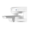
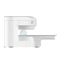
Anke MRI Openmark 5000 Permanent System
$0.00
Shipped from Abroad
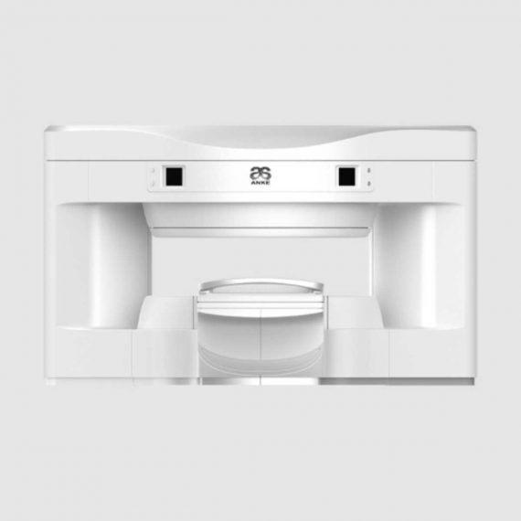
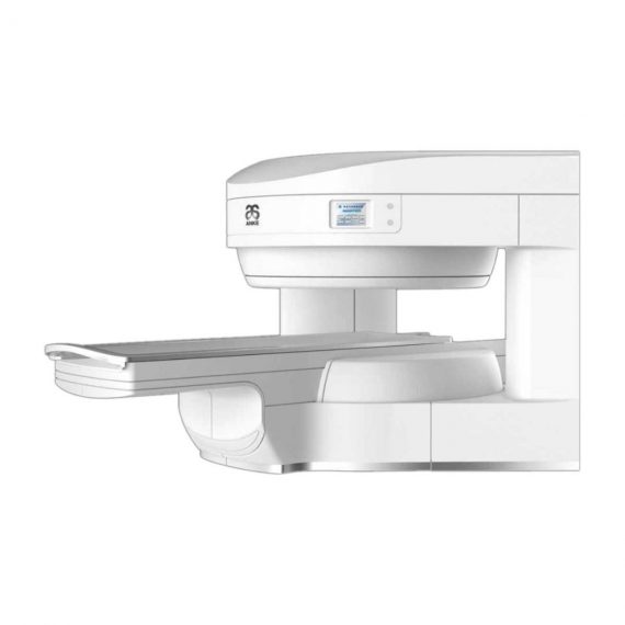
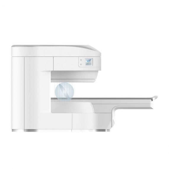
OPENMARK 5000 is 0.51T MRI. It’s approved by FDA and has CE mark. It adopts two-pillar magnet design with 280 degree openness and equipped with powerful
RF and gradient system, together with advanced imaging technology, making it as a high-end system which is comparable to high-field MRI.
Delivery & Availability:
Typically 90 working days – excluding furniture and heavy/bulky equipment. Please contact us for further information.
Description
OPENMARK 5000 is 0.51T MRI. It’s approved by FDA and has CE mark. It adopts two-pillar magnet design with 280 degree openness and equipped with powerful
RF and gradient system, together with advanced imaging technology, making it as a high-end system which is comparable to high-field MRI.
Features:
- With the highest system stability and the highest homogeneity of the
magnet field in permanent MRI - Screens on both sides facilitate positioning; 280 degree two-pillar magnet
design ensures stable magnet structure and facilitates interventional
treatment. - Active and passive shimming calibrate technology ensures the magnetic
field uniformity - Motor-driven patient couch makes it easier for patients to access and for
positioning - Powerful hardware and software platforms ensure the scan speed, image
quality and make it possible for advanced imaging functions - Fast scan speed eliminates motion artifact
- Rich scan sequences, advanced imaging technology and powerful postprocessing
technology ensure image quality, extend more applications,
which can fully satisfy the clinical needs - Intelligent user-friendly operating system ensures you easy operation
Technical Specifications:
| No. | Technique Description | Parameter |
| 1 | Magnet System | |
| 1.1 | Magnet Type | Permanent Magnet
Automatic constant temperature system |
| 1.2 | Field Strength | 0.51T |
| 1.3 | Magnet Shape | Dual-pillar shape |
| 1.4 | Homogeneity(40cm,DSV,VRMS) | ≤1.6ppm |
| 1.5 | Shim Method | Active/Passive |
| 1.6 | Magnet Vertical Gap (Cover) | 40cm |
| 1.7 | Magnetic Pole Dia. (Exclude Cover) | 145cm |
| 1.8 | Accessibility(Horizontal Opening Angle, | 280° |
| 1.9 | 5 Gauss fringe field | X-axis:horizontal ≤2.5m
Y-axis:Vertical ≤2.5m Z-axis:horizontal ≤2.5m |
| 2 | Patient Couch and Communication | |
| 2.1 | Patient Couch Driven mode | Motor-driven |
| 2.2 | Max. Patient Weight | ≥200kg(440lbs) |
| 2.3 | Patient Positioning Tools | Laser Light Localizer for positioning of center slice Motor-driven transfer to center of imaging volume |
| 2.4 | Position accuracy | ±1mm |
| 2.5 | Emergency Call Key | Yes |
| 2.6 | Intercom System | Yes |
| 3 | Gradient System | |
| 3.1 | Gradient Field Strength(Single Axis) | ≥30mT/m |
| 3.2 | Gradient Slew Rate (Single Axis) | ≥100mT/m/ms |
| 3.3 | Rise Time | ≤0.3ms |
| 3.4 | Gradient Cooling System ( Gradient coils
and Power electronics) |
Air Cooling |
| 4 | RF System | |
| 4.1 | RF System Type | Digital Transmit and
Receive signal |
| 4.2 | Number of RF Channels | 4 |
| 4.3 | Transmitter Amplifier Peak Power | 6kW |
| 4.4 | RF Bandwidth of Receiver | ≥1.25MHz |
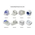
| 4.5 | Head Coil | Yes |
| 4.6 | Neck Coil | Yes |
| 4.7 | Body/Spine Coil (17 inch) | Yes |
| 4.8 | Body/Spine Coil (21 inch) | Yes |
| 4.9 | Knee Coil | Yes |
| 4.10 | Shoulder Coil | Yes |
| 4.11 | Flexible Coil | Optional |
| 4.12 | Breast Coil | Optional |
| 5 | Computer System | |
| 5.1 | Host Computer | DELL Computer (for MR) |
| 5.2 | System Software | Windows XP |
| 5.3 | Operation Software | APEX |
| 5.4 | CPU Clock rate | 3.0GHz |
| 5.5 | Main Memory | 4GB |
| 5.6 | Color LCD Monitor | 19” |
| 5.7 | Keyboard and Mouse | Standard |
| 5.8 | Image Reconstruction Speed(256 x 256
Matrix) |
200 frame/Sec. |
| 5.9 | Hard Disk | 500GB |
| 5.10 | Image Storage Capacity(256 x 256
Matrix) |
500,000 |
| 5.11 | Media Driver | DVD RW |
| 5.12 | DICOM 3.0 | Yes |
| 5.13 | Ethernet | Yes |
| 5.14 | Operation Console | Yes |
| 5.15 | Operation Chair | Yes |
| 6 | Scanning Parameter | |
| 6.1 | Max. FOV | 410mm |
| 6.2 | Min. FOV | 5mm |
| 6.3 | Min. TE(SE) | 5ms |
| 6.4 | Min. TR(SE) | 11ms |
| 6.5 | Min. TE(GR) | 1ms |
| 6.6 | Min. TR(GR) | 3ms |
| 6.7 | Min. 2D Thickness | 1.0mm |
| 6.8 | Min. 3D Thickness | 0.1mm |
| 6.9 | Max. Image Matrix | 512×512 |
| 7 | Scanning Sequence & Imaging Technique | |
| 7.1 | Spin Echo 2D/3D (SE 2D/3D) | Yes |
| 7.2 | DE/QE | Yes |
| 7.3 | Fast Spin Echo 2D/3D(FSE 2D/3D) | Yes |
| 7.4 | Single Shot FSE 2D/3D | Yes |
| 7.5 | Inversion Recovery(IR) | Yes |
| 7.6 | Fast Inversion Recovery(FIR) | Yes |
| 7.7 | Gradient Echo 2D/3D(GR 2D/3D) | Yes |
| 7.8 | Fast GR 2D/3D | Yes |
| 7.9 | SPGR | Yes |
| 7.10 | FLAIR | Yes |
| 7.11 | Fat Imaging | Yes |
| 7.12 | Fat Suppression imaging | Yes |
| 7.13 | Water-Fat Separation imaging | Yes |
| 7.14 | TOF MRA(2D/3D) | Yes |
| 7.15 | MRCP(2D/3D) | Yes |
| 7.16 | MRU (2D/3D) | Yes |
| 7.17 | MRM | Yes |
| 7.18 | Fast Hydrograph Imaging | Yes |
| 7.19 | Diffusion Weighted Imaging(DWI) | Yes |
| 7.20 | Max. b Value | 1000s/mm2 |
| 7.21 | Breath Hold Technology | Yes |
| 7.22 | Magnetization Transfer Contrast(MTC) | Yes |
| 7.23 | Multi-slice and Angle-free Presaturation | Yes |
| 7.24 | Saturation Tracking | Yes |
| 7.25 | Maximum Intensity Projection(MIP) | Yes |
| 7.26 | Multi-Angle Projection(MAP) | Yes |
| 7.27 | 3D Reconstruction | Yes |
| 7.28 | Multi-planar Reconstruction(MPR) | Yes |
| 7.29 | Multi-Artifacts Eliminating technology | Yes |
| 7.30 | Checking with Part Metal Implant | Yes |
| 7.31 | Online Image Filtration | Yes |
| 7.32 | Online Post Procession | Yes |
| 7.33 | 3D Scout | Yes |
| 7.34 | Scanning Protocol Preset | Yes |
| 7.35 | Scanning Protocol Queue Waiting | Yes |
| 7.36 | Advanced Image Post Processing | Yes |
| 7.37 | Image Fusion Technology of Vascular | Yes |
| 7.38 | Image Fusion Technology of Spine | Yes |
Click Here To Download Catalogue
Additional information
| Model | Advanced, Advanced Plus, Basic, Smart |
|---|
Quick Comparison
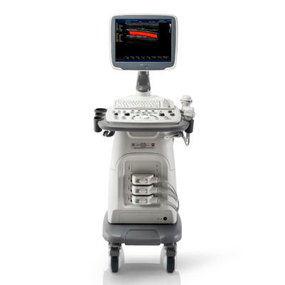
| Anke MRI Openmark 5000 Permanent System remove | ASPEL Ambulatory BP Machine remove | Jade Mobile X-ray machine (Analogue) remove | Sonoscape S11 Ultrasound Machine remove | Sonoscape P20 Ultrasound Machine remove | DrGem GXR-SD 400mA Floor Mounted Digital X-ray remove | ||||||||||||||||||||||||||||||||||||||||||||||||||||||||||||||||||||||||||||||||||||||||||||||||||||||||||||||||||||||||||||||||||||||||||||||||||||||||||||||||||||||||||||||||||||||||||||||||||||||||||||||||||||||||||||||||||||||||||||||||||||||||||||||||||||||||||||||||||||||||||||||||||||||||||||||||
|---|---|---|---|---|---|---|---|---|---|---|---|---|---|---|---|---|---|---|---|---|---|---|---|---|---|---|---|---|---|---|---|---|---|---|---|---|---|---|---|---|---|---|---|---|---|---|---|---|---|---|---|---|---|---|---|---|---|---|---|---|---|---|---|---|---|---|---|---|---|---|---|---|---|---|---|---|---|---|---|---|---|---|---|---|---|---|---|---|---|---|---|---|---|---|---|---|---|---|---|---|---|---|---|---|---|---|---|---|---|---|---|---|---|---|---|---|---|---|---|---|---|---|---|---|---|---|---|---|---|---|---|---|---|---|---|---|---|---|---|---|---|---|---|---|---|---|---|---|---|---|---|---|---|---|---|---|---|---|---|---|---|---|---|---|---|---|---|---|---|---|---|---|---|---|---|---|---|---|---|---|---|---|---|---|---|---|---|---|---|---|---|---|---|---|---|---|---|---|---|---|---|---|---|---|---|---|---|---|---|---|---|---|---|---|---|---|---|---|---|---|---|---|---|---|---|---|---|---|---|---|---|---|---|---|---|---|---|---|---|---|---|---|---|---|---|---|---|---|---|---|---|---|---|---|---|---|---|---|---|---|---|---|---|---|---|---|---|---|---|---|---|---|---|---|---|---|---|---|---|---|---|---|---|---|---|---|---|---|---|---|---|---|---|---|---|---|---|---|---|---|---|---|---|---|---|---|---|---|---|
| Name | Anke MRI Openmark 5000 Permanent System remove | ASPEL Ambulatory BP Machine remove | Jade Mobile X-ray machine (Analogue) remove | Sonoscape S11 Ultrasound Machine remove | Sonoscape P20 Ultrasound Machine remove | DrGem GXR-SD 400mA Floor Mounted Digital X-ray remove | |||||||||||||||||||||||||||||||||||||||||||||||||||||||||||||||||||||||||||||||||||||||||||||||||||||||||||||||||||||||||||||||||||||||||||||||||||||||||||||||||||||||||||||||||||||||||||||||||||||||||||||||||||||||||||||||||||||||||||||||||||||||||||||||||||||||||||||||||||||||||||||||||||||||||||||||
| Image |  | 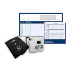 | 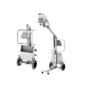 | 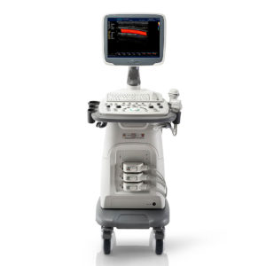 | 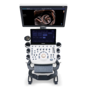 | 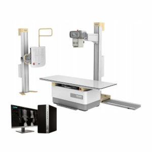 | |||||||||||||||||||||||||||||||||||||||||||||||||||||||||||||||||||||||||||||||||||||||||||||||||||||||||||||||||||||||||||||||||||||||||||||||||||||||||||||||||||||||||||||||||||||||||||||||||||||||||||||||||||||||||||||||||||||||||||||||||||||||||||||||||||||||||||||||||||||||||||||||||||||||||||||||
| SKU | SF1033560092-3 | SF1033560075-13 | SF1033560074-2 | SF1033560012-1 | SF1033560012-9 | SF1033560074-5 | |||||||||||||||||||||||||||||||||||||||||||||||||||||||||||||||||||||||||||||||||||||||||||||||||||||||||||||||||||||||||||||||||||||||||||||||||||||||||||||||||||||||||||||||||||||||||||||||||||||||||||||||||||||||||||||||||||||||||||||||||||||||||||||||||||||||||||||||||||||||||||||||||||||||||||||||
| Rating | |||||||||||||||||||||||||||||||||||||||||||||||||||||||||||||||||||||||||||||||||||||||||||||||||||||||||||||||||||||||||||||||||||||||||||||||||||||||||||||||||||||||||||||||||||||||||||||||||||||||||||||||||||||||||||||||||||||||||||||||||||||||||||||||||||||||||||||||||||||||||||||||||||||||||||||||||||||
| Price |
|
|
|
|
|
| |||||||||||||||||||||||||||||||||||||||||||||||||||||||||||||||||||||||||||||||||||||||||||||||||||||||||||||||||||||||||||||||||||||||||||||||||||||||||||||||||||||||||||||||||||||||||||||||||||||||||||||||||||||||||||||||||||||||||||||||||||||||||||||||||||||||||||||||||||||||||||||||||||||||||||||||
| Stock | |||||||||||||||||||||||||||||||||||||||||||||||||||||||||||||||||||||||||||||||||||||||||||||||||||||||||||||||||||||||||||||||||||||||||||||||||||||||||||||||||||||||||||||||||||||||||||||||||||||||||||||||||||||||||||||||||||||||||||||||||||||||||||||||||||||||||||||||||||||||||||||||||||||||||||||||||||||
| Availability | |||||||||||||||||||||||||||||||||||||||||||||||||||||||||||||||||||||||||||||||||||||||||||||||||||||||||||||||||||||||||||||||||||||||||||||||||||||||||||||||||||||||||||||||||||||||||||||||||||||||||||||||||||||||||||||||||||||||||||||||||||||||||||||||||||||||||||||||||||||||||||||||||||||||||||||||||||||
| Add to cart | |||||||||||||||||||||||||||||||||||||||||||||||||||||||||||||||||||||||||||||||||||||||||||||||||||||||||||||||||||||||||||||||||||||||||||||||||||||||||||||||||||||||||||||||||||||||||||||||||||||||||||||||||||||||||||||||||||||||||||||||||||||||||||||||||||||||||||||||||||||||||||||||||||||||||||||||||||||
| Description | Shipped from Abroad
OPENMARK 5000 is 0.51T MRI. It's approved by FDA and has CE mark. It adopts two-pillar magnet design with 280 degree openness and equipped with powerful
RF and gradient system, together with advanced imaging technology, making it as a high-end system which is comparable to high-field MRI.
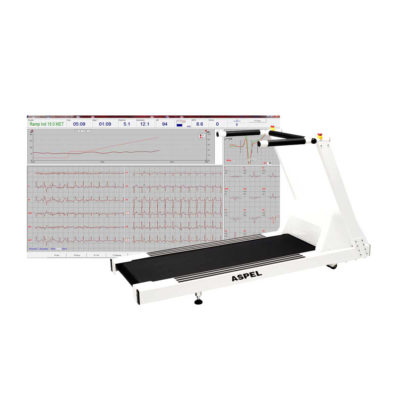
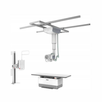
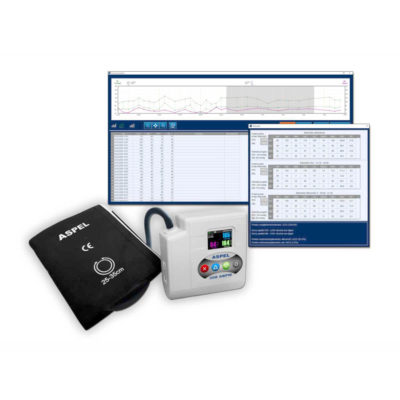
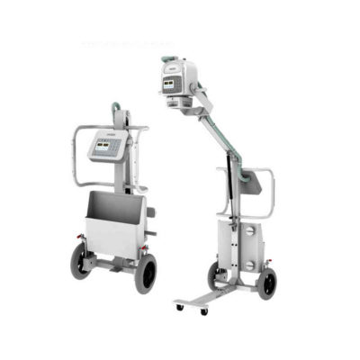
Delivery & Availability: Typically 90 working days – excluding furniture and heavy/bulky equipment. Please contact us for further information. | Shipped from Abroad ASPEL Ambulatory BP Machine - is a recorder of long-term records of non-invasive measurement of blood pressure intended for use in clinics, hospitals, outpatient centers and specialist surgeries. The recorder enables the assessment of blood pressure by the oscillometric method in adult patients, pregnant women, including preeclampsia and pediatric patients (from 3 years of age). Blood pressure is assessed by using an inflatable cuff, an accurate pressure transducer, and a deflation valve. Delivery & Availability: Typically 10 working days – excluding furniture and heavy/bulky equipment. Please contact us for further information. | In Stock JADE is one of the lightest portable X-ray systems on the market, allowing it to be used in any imaginable way including bedside, operating rooms, intensive care units and in veterinary fields. With a simple, easy-to-use operator console, three-way control, two-step foldable stand and auto lock system, JADE is a user-friendly portable X-ray system. Delivery & Availability: Typically 21 working days – excluding furniture and heavy/bulky equipment. Please contact us for further information. | In Stock A Value Choice beyond Your Expectation. SonoScape’s trolley color Doppler system S11 redefines price and performance with practical design. The S11 will go beyond your expectations but not your budget. Delivery & Availability: Typically 2 working days – excluding furniture and heavy/bulky equipment. Please contact us for further information. | Shipped from Abroad Incorporating innovative technologies, P20’s user-friendly design with a simple operation panel, intuitive user interface and a variety of intelligent auxiliary scanning tools, will significantly improve your daily examination experience. Besides general imaging applications, P20 has entitled with diagnostic 4D technology which has an extraordinary performance in obstetrics and gynecology applications. Delivery & Availability: Typically 5-7 working days – excluding furniture and heavy/bulky equipment. Please contact us for further information. | In Stock The GXR-SD Digital X-ray is a diagnostic digital radiography system that provides reliable high quality digital radiographic images with a reduced dose. The GXR-SD DR systems offer comprehensive digital solutions to all radiography needs, featuring ACQUIDR digital imaging system with stationary or portable digital flat-panel detectors as well as reliable high-frequency x-ray generators that are known worldwide for their excellent performance, lifetime and stability. Patient tables and wall stands are also offered. Delivery & Availability: Typically 21 working days – excluding furniture and heavy/bulky equipment. Please contact us for further information. | |||||||||||||||||||||||||||||||||||||||||||||||||||||||||||||||||||||||||||||||||||||||||||||||||||||||||||||||||||||||||||||||||||||||||||||||||||||||||||||||||||||||||||||||||||||||||||||||||||||||||||||||||||||||||||||||||||||||||||||||||||||||||||||||||||||||||||||||||||||||||||||||||||||||||||||||
| Content | OPENMARK 5000 is 0.51T MRI. It's approved by FDA and has CE mark. It adopts two-pillar magnet design with 280 degree openness and equipped with powerful
RF and gradient system, together with advanced imaging technology, making it as a high-end system which is comparable to high-field MRI.
Features:
Click Here To Download Catalogue | ASPEL Ambulatory BP Machine - is a recorder of long-term records of non-invasive measurement of blood pressure intended for use in clinics, hospitals, outpatient centers and specialist surgeries. The recorder enables the assessment of blood pressure by the oscillometric method in adult patients, pregnant women, including preeclampsia and pediatric patients (from 3 years of age). Blood pressure is assessed by using an inflatable cuff, an accurate pressure transducer, and a deflation valve.
Features:
Save-2-Safe: Double security system
Thanks to the use of two independent measuring systems with an additional valve, it meets the highest standards and takes care of patient safety even better.
Start-Easy: Quick start in two moves
The quick launch function allows you to use the device instantly, easily allows you to start recording in holter mode.
Memo-Care: Cuff pressure memory
Recorder remembers the pressure in the cuff. Thanks to the use of Intelligent Solutions, it adapts individually to the patient.
Power-Usb: USB connection
The device can work without batteries: by connecting to a computer via a USB cable.
Technical Specification:
Click Here To Download Catalogue | JADE Mobile X-ray machine is one of the lightest portable X-ray systems on the market, allowing it to be used in any imaginable way including bedside, operating rooms, intensive care units and veterinary fields. With a simple, easy-to-use operator console, three-way control, two-step foldable stand and auto-lock system, the JADE Mobile X-ray machine is a user-friendly portable X-ray system.
Convenient & Intuitive Operation:
JADE is one of the lightest portable X-ray systems on the market, allowing it to be used in any imaginable way including bedside, operating rooms, intensive care units and in veterinary fields. With a simple, easy-to-use operator console, three-way control, two-step foldable stand and auto-lock system, JADE is a user-friendly portable X-ray system.
Compact & Powerful Design:
JADE Mobile X-ray machine is an innovative, highly versatile portable X-ray system suitable for a variety of clinical uses. Utilizing the unique technology used in DRGEM’s universally recognized X-ray generators, JADE is a compact but powerful unit with a 4kW output and thoughtfully designed components to increase efficiency and maximize workflow. The core part of X-ray source adopts high-quality tube assembly, X-ray collimator and high frequency X-ray generator with excellent performance, lifetime and stability.
Features:
Click Here To Download Catalogue | DETAILS
SonoScape’s trolley colour Doppler system S11 redefines price and performance with practical design. The S11 will go beyond your expectations but not your budget. As an easy-to-use ultrasound system, the S11 is integrated with a new software platform, especially optimized for a smooth workflow and convenient operation. The system speeds up the exam process and makes file management easier.
SPECIFICATION
- 15-inch high definition LCD monitor with articulating arm
- Compact and agile trolley design
- 3 active transducer sockets available for a wide range of applications
- Duplex, Color Doppler, DPI, PW Doppler, tissue harmonic imaging, μ-scan speckle reduction imaging, compound imaging, trapezoidal imaging
- Customized settings based on your own working style
- Full patient database and image management solutions
Click Here To Download Catalogue | DETAILS
Upgraded Images with More Clarity
SonoScape never stops making progress in improving the image quality of its ultrasound products to enhance the confidence of diagnosis for doctors. With extraordinary images provided by P20, the anatomy structures are clearer than ever.
C-Xlasto Imaging
With C-xlasto Imaging, P20 enables comprehensive quantitative elastic analysis. Meanwhile, C-xlasto on P20 is supported by linear, convex and transvaginal probes, to ensure good reproducibility and highly consistent quantitative elastic results.
S-Live
S-Live allows for detailed visualization of subtle anatomical features, thereby enabling intuitive diagnosis with real-time 3D images and enriching patient communication.
Pelvic Floor 4D
Transperineal 4D pelvic floor ultrasound can provide useful clinical values in assessing the vaginal delivery impact on the female anterior compartment, judging whether the pelvic organs are prolapsed or not and the extent, determining if the pelvic muscles were torn accurately.
Anatomic M Mode
Anatomic M Mode helps you observe the myocardial motion at different phases by freely placing sample lines. It accurately measures the myocardial thickness and the heart size of even difficult patients and supports the myocardial function and LV wall-motion assessment.
Tissue Doppler Imaging
P20 is endowed with Tissue Doppler Imaging which provides velocities and other clinical information on myocardial functions, facilitating clinical doctors with the ability to analyze and compare the motions of different parts of the patient's heart.
Click Here To Download Catalogue | DrGem GXR-SD 400mA Floor Mounted Digital X-ray system matches with a radiographic room which perfectly fits your workow and can be easily upgraded to DR system with the help of DR interface and PC interface in GXR generator as well as Bucky suitable to Flat Panel Detector. GXR X-ray system is equipped with a high frequency X-ray generator which consistently produces high quality radiograph in favor of high quality X-ray output with a very small kV ripple and accurate mA and mAs. GXR X-ray system is designed to provide convenience to operator and comfort to patient
Features of DrGem GXR-SD 400mA Floor Mounted Digital X-ray:
Click Here To Download Catalogue | |||||||||||||||||||||||||||||||||||||||||||||||||||||||||||||||||||||||||||||||||||||||||||||||||||||||||||||||||||||||||||||||||||||||||||||||||||||||||||||||||||||||||||||||||||||||||||||||||||||||||||||||||||||||||||||||||||||||||||||||||||||||||||||||||||||||||||||||||||||||||||||||||||||||||||||||
| Weight | N/A | N/A | N/A | N/A | N/A | N/A | |||||||||||||||||||||||||||||||||||||||||||||||||||||||||||||||||||||||||||||||||||||||||||||||||||||||||||||||||||||||||||||||||||||||||||||||||||||||||||||||||||||||||||||||||||||||||||||||||||||||||||||||||||||||||||||||||||||||||||||||||||||||||||||||||||||||||||||||||||||||||||||||||||||||||||||||
| Dimensions | N/A | N/A | N/A | N/A | N/A | N/A | |||||||||||||||||||||||||||||||||||||||||||||||||||||||||||||||||||||||||||||||||||||||||||||||||||||||||||||||||||||||||||||||||||||||||||||||||||||||||||||||||||||||||||||||||||||||||||||||||||||||||||||||||||||||||||||||||||||||||||||||||||||||||||||||||||||||||||||||||||||||||||||||||||||||||||||||
| Additional information |
|

Reviews
There are no reviews yet.