
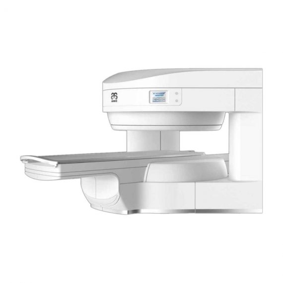
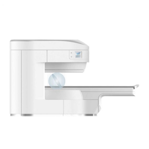
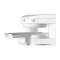
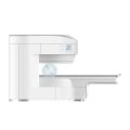
Anke MRI Openmark 5000 Permanent System
$0.00
Shipped from Abroad
OPENMARK 5000 is 0.51T MRI. It’s approved by FDA and has CE mark. It adopts two-pillar magnet design with 280 degree openness and equipped with powerful
RF and gradient system, together with advanced imaging technology, making it as a high-end system which is comparable to high-field MRI.
Delivery & Availability:
Typically 90 working days – excluding furniture and heavy/bulky equipment. Please contact us for further information.
Description
OPENMARK 5000 is 0.51T MRI. It’s approved by FDA and has CE mark. It adopts two-pillar magnet design with 280 degree openness and equipped with powerful
RF and gradient system, together with advanced imaging technology, making it as a high-end system which is comparable to high-field MRI.
Features:
- With the highest system stability and the highest homogeneity of the
magnet field in permanent MRI - Screens on both sides facilitate positioning; 280 degree two-pillar magnet
design ensures stable magnet structure and facilitates interventional
treatment. - Active and passive shimming calibrate technology ensures the magnetic
field uniformity - Motor-driven patient couch makes it easier for patients to access and for
positioning - Powerful hardware and software platforms ensure the scan speed, image
quality and make it possible for advanced imaging functions - Fast scan speed eliminates motion artifact
- Rich scan sequences, advanced imaging technology and powerful postprocessing
technology ensure image quality, extend more applications,
which can fully satisfy the clinical needs - Intelligent user-friendly operating system ensures you easy operation
Technical Specifications:
| No. | Technique Description | Parameter |
| 1 | Magnet System | |
| 1.1 | Magnet Type | Permanent Magnet
Automatic constant temperature system |
| 1.2 | Field Strength | 0.51T |
| 1.3 | Magnet Shape | Dual-pillar shape |
| 1.4 | Homogeneity(40cm,DSV,VRMS) | ≤1.6ppm |
| 1.5 | Shim Method | Active/Passive |
| 1.6 | Magnet Vertical Gap (Cover) | 40cm |
| 1.7 | Magnetic Pole Dia. (Exclude Cover) | 145cm |
| 1.8 | Accessibility(Horizontal Opening Angle, | 280° |
| 1.9 | 5 Gauss fringe field | X-axis:horizontal ≤2.5m
Y-axis:Vertical ≤2.5m Z-axis:horizontal ≤2.5m |
| 2 | Patient Couch and Communication | |
| 2.1 | Patient Couch Driven mode | Motor-driven |
| 2.2 | Max. Patient Weight | ≥200kg(440lbs) |
| 2.3 | Patient Positioning Tools | Laser Light Localizer for positioning of center slice Motor-driven transfer to center of imaging volume |
| 2.4 | Position accuracy | ±1mm |
| 2.5 | Emergency Call Key | Yes |
| 2.6 | Intercom System | Yes |
| 3 | Gradient System | |
| 3.1 | Gradient Field Strength(Single Axis) | ≥30mT/m |
| 3.2 | Gradient Slew Rate (Single Axis) | ≥100mT/m/ms |
| 3.3 | Rise Time | ≤0.3ms |
| 3.4 | Gradient Cooling System ( Gradient coils
and Power electronics) |
Air Cooling |
| 4 | RF System | |
| 4.1 | RF System Type | Digital Transmit and
Receive signal |
| 4.2 | Number of RF Channels | 4 |
| 4.3 | Transmitter Amplifier Peak Power | 6kW |
| 4.4 | RF Bandwidth of Receiver | ≥1.25MHz |
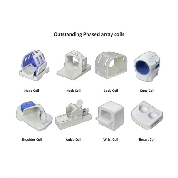
| 4.5 | Head Coil | Yes |
| 4.6 | Neck Coil | Yes |
| 4.7 | Body/Spine Coil (17 inch) | Yes |
| 4.8 | Body/Spine Coil (21 inch) | Yes |
| 4.9 | Knee Coil | Yes |
| 4.10 | Shoulder Coil | Yes |
| 4.11 | Flexible Coil | Optional |
| 4.12 | Breast Coil | Optional |
| 5 | Computer System | |
| 5.1 | Host Computer | DELL Computer (for MR) |
| 5.2 | System Software | Windows XP |
| 5.3 | Operation Software | APEX |
| 5.4 | CPU Clock rate | 3.0GHz |
| 5.5 | Main Memory | 4GB |
| 5.6 | Color LCD Monitor | 19” |
| 5.7 | Keyboard and Mouse | Standard |
| 5.8 | Image Reconstruction Speed(256 x 256
Matrix) |
200 frame/Sec. |
| 5.9 | Hard Disk | 500GB |
| 5.10 | Image Storage Capacity(256 x 256
Matrix) |
500,000 |
| 5.11 | Media Driver | DVD RW |
| 5.12 | DICOM 3.0 | Yes |
| 5.13 | Ethernet | Yes |
| 5.14 | Operation Console | Yes |
| 5.15 | Operation Chair | Yes |
| 6 | Scanning Parameter | |
| 6.1 | Max. FOV | 410mm |
| 6.2 | Min. FOV | 5mm |
| 6.3 | Min. TE(SE) | 5ms |
| 6.4 | Min. TR(SE) | 11ms |
| 6.5 | Min. TE(GR) | 1ms |
| 6.6 | Min. TR(GR) | 3ms |
| 6.7 | Min. 2D Thickness | 1.0mm |
| 6.8 | Min. 3D Thickness | 0.1mm |
| 6.9 | Max. Image Matrix | 512×512 |
| 7 | Scanning Sequence & Imaging Technique | |
| 7.1 | Spin Echo 2D/3D (SE 2D/3D) | Yes |
| 7.2 | DE/QE | Yes |
| 7.3 | Fast Spin Echo 2D/3D(FSE 2D/3D) | Yes |
| 7.4 | Single Shot FSE 2D/3D | Yes |
| 7.5 | Inversion Recovery(IR) | Yes |
| 7.6 | Fast Inversion Recovery(FIR) | Yes |
| 7.7 | Gradient Echo 2D/3D(GR 2D/3D) | Yes |
| 7.8 | Fast GR 2D/3D | Yes |
| 7.9 | SPGR | Yes |
| 7.10 | FLAIR | Yes |
| 7.11 | Fat Imaging | Yes |
| 7.12 | Fat Suppression imaging | Yes |
| 7.13 | Water-Fat Separation imaging | Yes |
| 7.14 | TOF MRA(2D/3D) | Yes |
| 7.15 | MRCP(2D/3D) | Yes |
| 7.16 | MRU (2D/3D) | Yes |
| 7.17 | MRM | Yes |
| 7.18 | Fast Hydrograph Imaging | Yes |
| 7.19 | Diffusion Weighted Imaging(DWI) | Yes |
| 7.20 | Max. b Value | 1000s/mm2 |
| 7.21 | Breath Hold Technology | Yes |
| 7.22 | Magnetization Transfer Contrast(MTC) | Yes |
| 7.23 | Multi-slice and Angle-free Presaturation | Yes |
| 7.24 | Saturation Tracking | Yes |
| 7.25 | Maximum Intensity Projection(MIP) | Yes |
| 7.26 | Multi-Angle Projection(MAP) | Yes |
| 7.27 | 3D Reconstruction | Yes |
| 7.28 | Multi-planar Reconstruction(MPR) | Yes |
| 7.29 | Multi-Artifacts Eliminating technology | Yes |
| 7.30 | Checking with Part Metal Implant | Yes |
| 7.31 | Online Image Filtration | Yes |
| 7.32 | Online Post Procession | Yes |
| 7.33 | 3D Scout | Yes |
| 7.34 | Scanning Protocol Preset | Yes |
| 7.35 | Scanning Protocol Queue Waiting | Yes |
| 7.36 | Advanced Image Post Processing | Yes |
| 7.37 | Image Fusion Technology of Vascular | Yes |
| 7.38 | Image Fusion Technology of Spine | Yes |
Click Here To Download Catalogue
Additional information
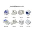
| Model | Advanced, Advanced Plus, Basic, Smart |
|---|
Quick Comparison
| Anke MRI Openmark 5000 Permanent System remove | DrGem Ceiling Analogue X-ray Machine remove | ASPEL Stress ECG with Ergometer and Software remove | Sonoscape S22 Ultrasound Machine remove | Sonoscape P10 Ultrasound Machine remove | DrGem Ceiling Mounted Digital X-ray remove | ||||||||||||||||||||||||||||||||||||||||||||||||||||||||||||||||||||||||||||||||||||||||||||||||||||||||||||||||||||||||||||||||||||||||||||||||||||||||||||||||||||||||||||||||||||||||||||||||||||||||||||||||||||||||||||||||||||||||||||||||||||||||||||||||||||||||||||||||||||||||||||||||||||||||||||||||
|---|---|---|---|---|---|---|---|---|---|---|---|---|---|---|---|---|---|---|---|---|---|---|---|---|---|---|---|---|---|---|---|---|---|---|---|---|---|---|---|---|---|---|---|---|---|---|---|---|---|---|---|---|---|---|---|---|---|---|---|---|---|---|---|---|---|---|---|---|---|---|---|---|---|---|---|---|---|---|---|---|---|---|---|---|---|---|---|---|---|---|---|---|---|---|---|---|---|---|---|---|---|---|---|---|---|---|---|---|---|---|---|---|---|---|---|---|---|---|---|---|---|---|---|---|---|---|---|---|---|---|---|---|---|---|---|---|---|---|---|---|---|---|---|---|---|---|---|---|---|---|---|---|---|---|---|---|---|---|---|---|---|---|---|---|---|---|---|---|---|---|---|---|---|---|---|---|---|---|---|---|---|---|---|---|---|---|---|---|---|---|---|---|---|---|---|---|---|---|---|---|---|---|---|---|---|---|---|---|---|---|---|---|---|---|---|---|---|---|---|---|---|---|---|---|---|---|---|---|---|---|---|---|---|---|---|---|---|---|---|---|---|---|---|---|---|---|---|---|---|---|---|---|---|---|---|---|---|---|---|---|---|---|---|---|---|---|---|---|---|---|---|---|---|---|---|---|---|---|---|---|---|---|---|---|---|---|---|---|---|---|---|---|---|---|---|---|---|---|---|---|---|---|---|---|---|---|---|---|---|
| Name | Anke MRI Openmark 5000 Permanent System remove | DrGem Ceiling Analogue X-ray Machine remove | ASPEL Stress ECG with Ergometer and Software remove | Sonoscape S22 Ultrasound Machine remove | Sonoscape P10 Ultrasound Machine remove | DrGem Ceiling Mounted Digital X-ray remove | |||||||||||||||||||||||||||||||||||||||||||||||||||||||||||||||||||||||||||||||||||||||||||||||||||||||||||||||||||||||||||||||||||||||||||||||||||||||||||||||||||||||||||||||||||||||||||||||||||||||||||||||||||||||||||||||||||||||||||||||||||||||||||||||||||||||||||||||||||||||||||||||||||||||||||||||
| Image |  |  | 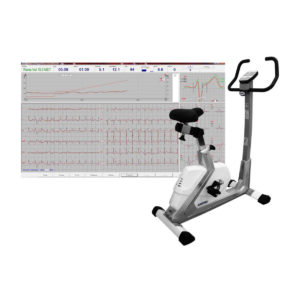 |  |  |  | |||||||||||||||||||||||||||||||||||||||||||||||||||||||||||||||||||||||||||||||||||||||||||||||||||||||||||||||||||||||||||||||||||||||||||||||||||||||||||||||||||||||||||||||||||||||||||||||||||||||||||||||||||||||||||||||||||||||||||||||||||||||||||||||||||||||||||||||||||||||||||||||||||||||||||||||
| SKU | SF1033560092-3 | SF1033560074-7 | SF1033560075-1 | SF1033560012-3 | SF1033560012-7 | SF1033560074-4 | |||||||||||||||||||||||||||||||||||||||||||||||||||||||||||||||||||||||||||||||||||||||||||||||||||||||||||||||||||||||||||||||||||||||||||||||||||||||||||||||||||||||||||||||||||||||||||||||||||||||||||||||||||||||||||||||||||||||||||||||||||||||||||||||||||||||||||||||||||||||||||||||||||||||||||||||
| Rating | |||||||||||||||||||||||||||||||||||||||||||||||||||||||||||||||||||||||||||||||||||||||||||||||||||||||||||||||||||||||||||||||||||||||||||||||||||||||||||||||||||||||||||||||||||||||||||||||||||||||||||||||||||||||||||||||||||||||||||||||||||||||||||||||||||||||||||||||||||||||||||||||||||||||||||||||||||||
| Price |
|
| $4,202.00 | $9,350.00 | $9,350.00 |
| |||||||||||||||||||||||||||||||||||||||||||||||||||||||||||||||||||||||||||||||||||||||||||||||||||||||||||||||||||||||||||||||||||||||||||||||||||||||||||||||||||||||||||||||||||||||||||||||||||||||||||||||||||||||||||||||||||||||||||||||||||||||||||||||||||||||||||||||||||||||||||||||||||||||||||||||
| Stock | |||||||||||||||||||||||||||||||||||||||||||||||||||||||||||||||||||||||||||||||||||||||||||||||||||||||||||||||||||||||||||||||||||||||||||||||||||||||||||||||||||||||||||||||||||||||||||||||||||||||||||||||||||||||||||||||||||||||||||||||||||||||||||||||||||||||||||||||||||||||||||||||||||||||||||||||||||||
| Availability | |||||||||||||||||||||||||||||||||||||||||||||||||||||||||||||||||||||||||||||||||||||||||||||||||||||||||||||||||||||||||||||||||||||||||||||||||||||||||||||||||||||||||||||||||||||||||||||||||||||||||||||||||||||||||||||||||||||||||||||||||||||||||||||||||||||||||||||||||||||||||||||||||||||||||||||||||||||
| Add to cart | |||||||||||||||||||||||||||||||||||||||||||||||||||||||||||||||||||||||||||||||||||||||||||||||||||||||||||||||||||||||||||||||||||||||||||||||||||||||||||||||||||||||||||||||||||||||||||||||||||||||||||||||||||||||||||||||||||||||||||||||||||||||||||||||||||||||||||||||||||||||||||||||||||||||||||||||||||||
| Description | Shipped from Abroad
OPENMARK 5000 is 0.51T MRI. It's approved by FDA and has CE mark. It adopts two-pillar magnet design with 280 degree openness and equipped with powerful
RF and gradient system, together with advanced imaging technology, making it as a high-end system which is comparable to high-field MRI.
Delivery & Availability: Typically 90 working days – excluding furniture and heavy/bulky equipment. Please contact us for further information. | Shipped from abroad The DrGem Ceiling Analogue X-ray Machine is a diagnostic radiography system that provides reliable high quality radiographic images with a reduced dose. The reliable high-frequency x-ray generators that are known worldwide for their excellent performance, lifetime and stability. Patient tables and wall stands are also offered. Delivery & Availability: Typically 21 working days – excluding furniture and heavy/bulky equipment. Please contact us for further information. | Shipped from Abroad Ergometer CRG 200 is dedicated for Exercise Stress Tests System CardioTEST. The Ergometer has been designed according to modern technologies. It is controlled from PC equipped in CardioTEST software. Load level is controlled by a microprocessor, therefore it does not depend on speed, which in turn can be adjusted according to patient’s individual needs. Ergometer is equipped with ECG mode recording 12 standard leads. Delivery & Availability: Typically 21 working days – excluding furniture and heavy/bulky equipment. Please contact us for further information. | Shipped from Abroad As SonoScape steps forward to add value and efficiency to ultrasound, the latest S22 was designed in a user-friendly platform to address current and future demanding needs. It represents an excellent mix in performance and price. Delivery & Availability: Typically 5-7 working days – excluding furniture and heavy/bulky equipment. Please contact us for further information. | Shipped from Abroad The P10 color Doppler ultrasound system is a new generation product from SonoScape. It is designed to give high quality images, rich probe configurations, various clinical tools and automatic analysis software to provide you with comprehensive solutions for your growing demand for clinical applications. Delivery & Availability: Typically 5-7 working days – excluding furniture and heavy/bulky equipment. Please contact us for further information. | In Stock The GXR-SD is a diagnostic digital radiography system that provides reliable high quality digital radiographic images with a reduced dose. The GXR-SD DR systems offer comprehensive digital solutions to all radiography needs, featuring ACQUIDR digital imaging system with stationary or portable digital flat-panel detectors as well as reliable high-frequency x-ray generators that are known worldwide for their excellent performance, lifetime and stability. Patient tables and wall stands are also offered. Delivery & Availability: Typically 21 working days – excluding furniture and heavy/bulky equipment. Please contact us for further information. | |||||||||||||||||||||||||||||||||||||||||||||||||||||||||||||||||||||||||||||||||||||||||||||||||||||||||||||||||||||||||||||||||||||||||||||||||||||||||||||||||||||||||||||||||||||||||||||||||||||||||||||||||||||||||||||||||||||||||||||||||||||||||||||||||||||||||||||||||||||||||||||||||||||||||||||||
| Content | OPENMARK 5000 is 0.51T MRI. It's approved by FDA and has CE mark. It adopts two-pillar magnet design with 280 degree openness and equipped with powerful
RF and gradient system, together with advanced imaging technology, making it as a high-end system which is comparable to high-field MRI.
Features:
Click Here To Download Catalogue | DrGem Ceiling Analogue X-ray Machine is a diagnostic radiography system X-ray Machine that provides reliable high quality radiographic images with a reduced dose. The reliable high-frequency x-ray generators that are known worldwide for their excellent performance, lifetime and stability. Patient tables and wall stands are also offered.
Features of DrGem Ceiling Analogue X-ray Machine
Click Here To Download Catalogue | Ergometer CRG 200 is dedicated for Exercise Stress Tests System CardioTEST. The Ergometer has been designed according to modern technologies. It is controlled from PC equipped in CardioTEST software. Load level is controlled by a microprocessor, therefore it does not depend on speed, which in turn can be adjusted according to patient’s individual needs. Ergometer is equipped with ECG mode recording 12 standard leads.
Technical Specifications:
Click Here To Download Catalogue | DETAILS
As SonoScape steps forward to add value and efficiency to ultrasound, the latest S22 was designed in a user-friendly platform to address current and future demanding needs. It represents an excellent mix in performance and price.
S22, is a shared service ultrasound system with a slim and elegant package that has combined mobility with utility to fit in specific clinical situations including emergency department, ICU, operating room and so on. Furthermore, its ergonomic design, easy operating and flexible data management will give you a memorable experience.
SPECIFICATION
• Large high-resolution widescreen LED
• Sensitive touch screen
• Four transducer sockets plus one socket for pencil probe
• A comprehensive selection of probes: linear, Convex, Micro-convex, Volumetric, Endocavity, Bi-plane, Phased Array, TEE, Intraoperative, Pencil
• Premium application technology: 4D, μ-scan speckle reduction, compound imaging, Pulse Inversion Harmonic Imaging, Color M-Mode, Steer M-Mode, PDI, TDI, Real-time Panoramic Imaging, Trapezoid Imaging, Auto-IMT…
• Full patient database and image management solutions: DICOM 3.0, AVI/JPG, USB 2.0, HDD, DVD, PDF report
• Multi-Language Input Keyboard
• Built-in battery
Click Here To Download Catalogue | DETAILS
B + Compound
B + Compound utilizes several lines of sight for optimal contrast resolution, speckle reduction and border detection, with which P10 is ideal for superficial and abdominal imaging with better clarity and improved continuity of structures.
μ-Scan
The new generation μ-Scan imaging technology gives you better image quality by reducing noise, improving signal strength and improving visualization.
P10 offers a comprehensive selection of electronic probes to maximize its capabilities to meet a wide range of applications including abdomen, pediatric, OB/GYN, cardiovascular, musculoskeletal, etc. The advanced probe technologies also effectively enhance the image quality and confidence in reaching clinical diagnoses, even in difficult patients.
Convex Probe 3C-A
Ideal for an abundant of application such as abdomen, gynecology, obstetrics, urology and even abdomen biopsy.
Linear Probe L741
This linear probe is designed to satisfy vascular, breast, thyroid, and other small parts diagnosis, and its adjustable parameters could also present users a clear view of MSK and deep vessels.
Phase Array Probe 3P-A
For the purpose of adult and pediatric cardiology and emergency, the phase array probe provides elaborate presets for different exam modes, even for difficult patients.
Intracavitary Probe 6V1
Intracavitary probe could face application of gynecology, urology, prostate, and its temperature detection technology not only protects the patient but also extends the service life.
Click Here To Download Catalogue | DrGem Ceiling Mounted Digital X-ray is a diagnostic digital radiography system that provides reliable high quality digital radiographic images with a reduced dose. The GXR-SD DR systems offer comprehensive digital solutions to all radiography needs, featuring ACQUIDR digital imaging system with stationary or portable digital flat-panel detectors as well as reliable high-frequency x-ray generators that are known worldwide for their excellent performance, lifetime and stability. Patient tables and wall stands are also offered.
Features:
Click Here To Download Catalogue | |||||||||||||||||||||||||||||||||||||||||||||||||||||||||||||||||||||||||||||||||||||||||||||||||||||||||||||||||||||||||||||||||||||||||||||||||||||||||||||||||||||||||||||||||||||||||||||||||||||||||||||||||||||||||||||||||||||||||||||||||||||||||||||||||||||||||||||||||||||||||||||||||||||||||||||||
| Weight | N/A | N/A | N/A | N/A | N/A | N/A | |||||||||||||||||||||||||||||||||||||||||||||||||||||||||||||||||||||||||||||||||||||||||||||||||||||||||||||||||||||||||||||||||||||||||||||||||||||||||||||||||||||||||||||||||||||||||||||||||||||||||||||||||||||||||||||||||||||||||||||||||||||||||||||||||||||||||||||||||||||||||||||||||||||||||||||||
| Dimensions | N/A | N/A | N/A | N/A | N/A | N/A | |||||||||||||||||||||||||||||||||||||||||||||||||||||||||||||||||||||||||||||||||||||||||||||||||||||||||||||||||||||||||||||||||||||||||||||||||||||||||||||||||||||||||||||||||||||||||||||||||||||||||||||||||||||||||||||||||||||||||||||||||||||||||||||||||||||||||||||||||||||||||||||||||||||||||||||||
| Additional information |
|


Reviews
There are no reviews yet.