
Anke Supermark 1.5T MRI Machine
$0.00
Shipped from Abroad
SuperMark 1.5T is a new generation superconducting MRI system based on years of experience in production and research. It’s applicable to whole body scan, such as, nervous system, spine, joint soft tissue, pelvic and abdominal cavity, etc
Delivery & Availability:
Typically 90 working days – excluding furniture and heavy/bulky equipment. Please contact us for further information.
Description
SuperMark 1.5T is a new generation superconducting MRI system based on years of experience in production and research. It’s applicable to whole body scan, such as, nervous system, spine, joint soft tissue, pelvic and abdominal cavity, etc. SuperMark 1.5T provides not only conventional pulse sequences and clinical diagnosis functions, but also provides advanced functional applications, for instance, 3D angiography and water imaging. It adopts brand new ANKE APEX operating system which ensures easy operation and fast diagnosis.
Technical Advantages:
- Reliable short cavity superconducting magnet system with zero liquid helium
consumption - New generation fully digitalized and extensible multichannel spectrometer
- Powerful high efficiency and high fidelity gradient system; Multi-channel PA RF
receiving coil with intelligent identification - English operating system and high extensible computer system
- High resolution conventional clinical images; Practical advanced functional
imaging
Superconducting MRI System:
- Highly open and humanization design -> Streamlined design
- Rich sequences and technology satisfy clinical needs -> Efficient service
Low Investment:
- High cost performance superconducting MRI system
- Zero liquid helium consumption, low running and maintenance cost
- Core technology by independent R & D supports full upgrade
- Low electric consumption
- Compact magnet design, minimum installation space: 35 square meters
High Return:
- High resolution thin slice images improve diagnosis
- Short cavity magnet design makes patients comfortable
- Fast scan speed improves work efficiency
Technical Specifications:
| No. | Technique Description | Parameter |
| 1 | Magnet System | |
| 1.1 | Magnet Type | Permanent Magnet
Automatic constant temperature system |
| 1.2 | Field Strength | 0.51T |
| 1.3 | Magnet Shape | Dual-pillar shape |
| 1.4 | Homogeneity(40cm,DSV,VRMS) | ≤1.6ppm |
| 1.5 | Shim Method | Active/Passive |
| 1.6 | Magnet Vertical Gap (Cover) | 40cm |
| 1.7 | Magnetic Pole Dia. (Exclude Cover) | 145cm |
| 1.8 | Accessibility(Horizontal Opening Angle, | 280° |
| 1.9 | 5 Gauss fringe field | X-axis:horizontal ≤2.5m
Y-axis:Vertical ≤2.5m Z-axis:horizontal ≤2.5m |
| 2 | Patient Couch and Communication | |
| 2.1 | Patient Couch Driven mode | Motor-driven |
| 2.2 | Max. Patient Weight | ≥200kg(440lbs) |
| 2.3 | Patient Positioning Tools | Laser Light Localizer for positioning of center slice Motor-driven transfer to center of imaging volume |
| 2.4 | Position accuracy | ±1mm |
| 2.5 | Emergency Call Key | Yes |
| 2.6 | Intercom System | Yes |
| 3 | Gradient System | |
| 3.1 | Gradient Field Strength(Single Axis) | ≥30mT/m |
| 3.2 | Gradient Slew Rate (Single Axis) | ≥100mT/m/ms |
| 3.3 | Rise Time | ≤0.3ms |
| 3.4 | Gradient Cooling System ( Gradient coils
and Power electronics) |
Air Cooling |
| 4 | RF System | |
| 4.1 | RF System Type | Digital Transmit and
Receive signal |
| 4.2 | Number of RF Channels | 4 |
| 4.3 | Transmitter Amplifier Peak Power | 6kW |
| 4.4 | RF Bandwidth of Receiver | ≥1.25MHz |
| 4.5 | Head Coil | Yes |
| 4.6 | Neck Coil | Yes |
| 4.7 | Body/Spine Coil (17 inch) | Yes |
| 4.8 | Body/Spine Coil (21 inch) | Yes |
| 4.9 | Knee Coil | Yes |
| 4.10 | Shoulder Coil | Yes |
| 4.11 | Flexible Coil | Optional |
| 4.12 | Breast Coil | Optional |
| 5 | Computer System | |
| 5.1 | Host Computer | DELL Computer (for MR) |
| 5.2 | System Software | Windows XP |
| 5.3 | Operation Software | APEX |
| 5.4 | CPU Clock rate | 3.0GHz |
| 5.5 | Main Memory | 4GB |
| 5.6 | Color LCD Monitor | 19” |
| 5.7 | Keyboard and Mouse | Standard |
| 5.8 | Image Reconstruction Speed(256 x 256
Matrix) |
200 frame/Sec. |
| 5.9 | Hard Disk | 500GB |
| 5.10 | Image Storage Capacity(256 x 256
Matrix) |
500,000 |
| 5.11 | Media Driver | DVD RW |
| 5.12 | DICOM 3.0 | Yes |
| 5.13 | Ethernet | Yes |
| 5.14 | Operation Console | Yes |
| 5.15 | Operation Chair | Yes |
| 6 | Scanning Parameter | |
| 6.1 | Max. FOV | 410mm |
| 6.2 | Min. FOV | 5mm |
| 6.3 | Min. TE(SE) | 5ms |
| 6.4 | Min. TR(SE) | 11ms |
| 6.5 | Min. TE(GR) | 1ms |
| 6.6 | Min. TR(GR) | 3ms |
| 6.7 | Min. 2D Thickness | 1.0mm |
| 6.8 | Min. 3D Thickness | 0.1mm |
| 6.9 | Max. Image Matrix | 512×512 |
| 7 | Scanning Sequence & Imaging Technique | |
| 7.1 | Spin Echo 2D/3D (SE 2D/3D) | Yes |
| 7.2 | DE/QE | Yes |
| 7.3 | Fast Spin Echo 2D/3D(FSE 2D/3D) | Yes |
| 7.4 | Single Shot FSE 2D/3D | Yes |
| 7.5 | Inversion Recovery(IR) | Yes |
| 7.6 | Fast Inversion Recovery(FIR) | Yes |
| 7.7 | Gradient Echo 2D/3D(GR 2D/3D) | Yes |
| 7.8 | Fast GR 2D/3D | Yes |
| 7.9 | SPGR | Yes |
| 7.10 | FLAIR | Yes |
| 7.11 | Fat Imaging | Yes |
| 7.12 | Fat Suppression imaging | Yes |
| 7.13 | Water-Fat Separation imaging | Yes |
| 7.14 | TOF MRA(2D/3D) | Yes |
| 7.15 | MRCP(2D/3D) | Yes |
| 7.16 | MRU (2D/3D) | Yes |
| 7.17 | MRM | Yes |
| 7.18 | Fast Hydrograph Imaging | Yes |
| 7.19 | Diffusion Weighted Imaging(DWI) | Yes |
| 7.20 | Max. b Value | 1000s/mm2 |
| 7.21 | Breath Hold Technology | Yes |
| 7.22 | Magnetization Transfer Contrast(MTC) | Yes |
| 7.23 | Multi-slice and Angle-free Presaturation | Yes |
| 7.24 | Saturation Tracking | Yes |
| 7.25 | Maximum Intensity Projection(MIP) | Yes |
| 7.26 | Multi-Angle Projection(MAP) | Yes |
| 7.27 | 3D Reconstruction | Yes |
| 7.28 | Multi-planar Reconstruction(MPR) | Yes |
| 7.29 | Multi-Artifacts Eliminating technology | Yes |
| 7.30 | Checking with Part Metal Implant | Yes |
| 7.31 | Online Image Filtration | Yes |
| 7.32 | Online Post Procession | Yes |
| 7.33 | 3D Scout | Yes |
| 7.34 | Scanning Protocol Preset | Yes |
| 7.35 | Scanning Protocol Queue Waiting | Yes |
| 7.36 | Advanced Image Post Processing | Yes |
| 7.37 | Image Fusion Technology of Vascular | Yes |
| 7.38 | Image Fusion Technology of Spine | Yes |
Click Here To Download Catalogue
Additional information
| Model | Advanced, Advanced Plus, Basic, Smart |
|---|
Quick Comparison
| Settings | Anke Supermark 1.5T MRI Machine remove | Sonoscape S22 Ultrasound Machine remove | Sonoscape P20 Ultrasound Machine remove | ASPEL AsCARD Grey ECG Machine remove | ASPEL AsCARD Green B/W ECG Machine remove | Bistos BT-770-12.1" Touchscreen Patient Monitor remove | |||||||||||||||||||||||||||||||||||||||||||||||||||||||||||||||||||||||||||||||||||||||||||||||||||||||||||||||||||||||||||||||||||||||||||||||||||||||||||||||||||||||||||||||||||||||||||||||||||||||||||||||||||||||||||||||||||||||||||||||||||||||||||||||||||||||||||||||||||||||||||||||||||||||||||||||
|---|---|---|---|---|---|---|---|---|---|---|---|---|---|---|---|---|---|---|---|---|---|---|---|---|---|---|---|---|---|---|---|---|---|---|---|---|---|---|---|---|---|---|---|---|---|---|---|---|---|---|---|---|---|---|---|---|---|---|---|---|---|---|---|---|---|---|---|---|---|---|---|---|---|---|---|---|---|---|---|---|---|---|---|---|---|---|---|---|---|---|---|---|---|---|---|---|---|---|---|---|---|---|---|---|---|---|---|---|---|---|---|---|---|---|---|---|---|---|---|---|---|---|---|---|---|---|---|---|---|---|---|---|---|---|---|---|---|---|---|---|---|---|---|---|---|---|---|---|---|---|---|---|---|---|---|---|---|---|---|---|---|---|---|---|---|---|---|---|---|---|---|---|---|---|---|---|---|---|---|---|---|---|---|---|---|---|---|---|---|---|---|---|---|---|---|---|---|---|---|---|---|---|---|---|---|---|---|---|---|---|---|---|---|---|---|---|---|---|---|---|---|---|---|---|---|---|---|---|---|---|---|---|---|---|---|---|---|---|---|---|---|---|---|---|---|---|---|---|---|---|---|---|---|---|---|---|---|---|---|---|---|---|---|---|---|---|---|---|---|---|---|---|---|---|---|---|---|---|---|---|---|---|---|---|---|---|---|---|---|---|---|---|---|---|---|---|---|---|---|---|---|---|---|---|---|---|---|---|---|
| Name | Anke Supermark 1.5T MRI Machine remove | Sonoscape S22 Ultrasound Machine remove | Sonoscape P20 Ultrasound Machine remove | ASPEL AsCARD Grey ECG Machine remove | ASPEL AsCARD Green B/W ECG Machine remove | Bistos BT-770-12.1" Touchscreen Patient Monitor remove | |||||||||||||||||||||||||||||||||||||||||||||||||||||||||||||||||||||||||||||||||||||||||||||||||||||||||||||||||||||||||||||||||||||||||||||||||||||||||||||||||||||||||||||||||||||||||||||||||||||||||||||||||||||||||||||||||||||||||||||||||||||||||||||||||||||||||||||||||||||||||||||||||||||||||||||||
| Image |  |  |  | 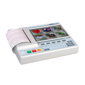 |  | 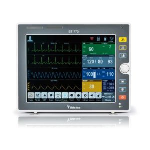 | |||||||||||||||||||||||||||||||||||||||||||||||||||||||||||||||||||||||||||||||||||||||||||||||||||||||||||||||||||||||||||||||||||||||||||||||||||||||||||||||||||||||||||||||||||||||||||||||||||||||||||||||||||||||||||||||||||||||||||||||||||||||||||||||||||||||||||||||||||||||||||||||||||||||||||||||
| SKU | SF1033560092-4 | SF1033560012-3 | SF1033560012-9 | SF1033560075-5 | SF1033560075-8 | SF1033560059-1 | |||||||||||||||||||||||||||||||||||||||||||||||||||||||||||||||||||||||||||||||||||||||||||||||||||||||||||||||||||||||||||||||||||||||||||||||||||||||||||||||||||||||||||||||||||||||||||||||||||||||||||||||||||||||||||||||||||||||||||||||||||||||||||||||||||||||||||||||||||||||||||||||||||||||||||||||
| Rating | |||||||||||||||||||||||||||||||||||||||||||||||||||||||||||||||||||||||||||||||||||||||||||||||||||||||||||||||||||||||||||||||||||||||||||||||||||||||||||||||||||||||||||||||||||||||||||||||||||||||||||||||||||||||||||||||||||||||||||||||||||||||||||||||||||||||||||||||||||||||||||||||||||||||||||||||||||||
| Price |
| $9,350.00 |
| $1,166.00 |
| $902.00 | |||||||||||||||||||||||||||||||||||||||||||||||||||||||||||||||||||||||||||||||||||||||||||||||||||||||||||||||||||||||||||||||||||||||||||||||||||||||||||||||||||||||||||||||||||||||||||||||||||||||||||||||||||||||||||||||||||||||||||||||||||||||||||||||||||||||||||||||||||||||||||||||||||||||||||||||
| Stock | |||||||||||||||||||||||||||||||||||||||||||||||||||||||||||||||||||||||||||||||||||||||||||||||||||||||||||||||||||||||||||||||||||||||||||||||||||||||||||||||||||||||||||||||||||||||||||||||||||||||||||||||||||||||||||||||||||||||||||||||||||||||||||||||||||||||||||||||||||||||||||||||||||||||||||||||||||||
| Availability | |||||||||||||||||||||||||||||||||||||||||||||||||||||||||||||||||||||||||||||||||||||||||||||||||||||||||||||||||||||||||||||||||||||||||||||||||||||||||||||||||||||||||||||||||||||||||||||||||||||||||||||||||||||||||||||||||||||||||||||||||||||||||||||||||||||||||||||||||||||||||||||||||||||||||||||||||||||
| Add to cart | |||||||||||||||||||||||||||||||||||||||||||||||||||||||||||||||||||||||||||||||||||||||||||||||||||||||||||||||||||||||||||||||||||||||||||||||||||||||||||||||||||||||||||||||||||||||||||||||||||||||||||||||||||||||||||||||||||||||||||||||||||||||||||||||||||||||||||||||||||||||||||||||||||||||||||||||||||||
| Description | Shipped from Abroad
SuperMark 1.5T is a new generation superconducting MRI system based on years of experience in production and research. It's applicable to whole body scan, such as, nervous system, spine, joint soft tissue, pelvic and abdominal cavity, etc
Delivery & Availability: Typically 90 working days – excluding furniture and heavy/bulky equipment. Please contact us for further information. | Shipped from Abroad As SonoScape steps forward to add value and efficiency to ultrasound, the latest S22 was designed in a user-friendly platform to address current and future demanding needs. It represents an excellent mix in performance and price. Delivery & Availability: Typically 5-7 working days – excluding furniture and heavy/bulky equipment. Please contact us for further information. | Shipped from Abroad Incorporating innovative technologies, P20’s user-friendly design with a simple operation panel, intuitive user interface and a variety of intelligent auxiliary scanning tools, will significantly improve your daily examination experience. Besides general imaging applications, P20 has entitled with diagnostic 4D technology which has an extraordinary performance in obstetrics and gynecology applications. Delivery & Availability: Typically 5-7 working days – excluding furniture and heavy/bulky equipment. Please contact us for further information. | Shipped from Abroad Electrocardiograph AsCARD Grey v.07.225 - is a 1, 3, 6, 12 channel ECG unit which enables to make electrocardiogram in full 12 leads. It is intended to conduct ECG examinations of adults and paediatric patients in all types of health care centres. ECG examination may be recorded in manual or automatic mode, with the possibility of analysis and interpretation. The device can be powered from 100 V ÷ 240 V mains supply or by an internal battery. Delivery & Availability: Typically 10 working days – excluding furniture and heavy/bulky equipment. Please contact us for further information. | Shipped from Abroad AsCARD Green electrocardiograph is a 1- and 3-channel ECG unit which enables to make electrocardiogram in full 12 leads. Intended for ECG examinations of adult and paediatric patients aimed at identification of cardiological abnormalities, myocardial ischaemia or infarction. The device is intended for use in healthcare facilities by duly trained personnel. ECG examination may be recorded in manual or automatic mode with the ability to perform the analysis and interpretation. Delivery & Availability: Typically 10 working days – excluding furniture and heavy/bulky equipment. Please contact us for further information. | Shipped from Abroad The Bistos BT-770 patient monitor is equipped with a 12.1" touchscreen display, which allows for an easy operation and readability with a powerful rechargeable battery guaranteeing a continuous operation of 5 hours to monitor ECG, SpO2, NIBP, temperature and respiration Delivery & Availability: Typically 14 working days – excluding furniture and heavy/bulky equipment. Please contact us for further information. | |||||||||||||||||||||||||||||||||||||||||||||||||||||||||||||||||||||||||||||||||||||||||||||||||||||||||||||||||||||||||||||||||||||||||||||||||||||||||||||||||||||||||||||||||||||||||||||||||||||||||||||||||||||||||||||||||||||||||||||||||||||||||||||||||||||||||||||||||||||||||||||||||||||||||||||||
| Content | SuperMark 1.5T is a new generation superconducting MRI system based on years of experience in production and research. It's applicable to whole body scan, such as, nervous system, spine, joint soft tissue, pelvic and abdominal cavity, etc. SuperMark 1.5T provides not only conventional pulse sequences and clinical diagnosis functions, but also provides advanced functional applications, for instance, 3D angiography and water imaging. It adopts brand new ANKE APEX operating system which ensures easy operation and fast diagnosis.
Technical Advantages:
Click Here To Download Catalogue | DETAILS
As SonoScape steps forward to add value and efficiency to ultrasound, the latest S22 was designed in a user-friendly platform to address current and future demanding needs. It represents an excellent mix in performance and price.
S22, is a shared service ultrasound system with a slim and elegant package that has combined mobility with utility to fit in specific clinical situations including emergency department, ICU, operating room and so on. Furthermore, its ergonomic design, easy operating and flexible data management will give you a memorable experience.
SPECIFICATION
• Large high-resolution widescreen LED
• Sensitive touch screen
• Four transducer sockets plus one socket for pencil probe
• A comprehensive selection of probes: linear, Convex, Micro-convex, Volumetric, Endocavity, Bi-plane, Phased Array, TEE, Intraoperative, Pencil
• Premium application technology: 4D, μ-scan speckle reduction, compound imaging, Pulse Inversion Harmonic Imaging, Color M-Mode, Steer M-Mode, PDI, TDI, Real-time Panoramic Imaging, Trapezoid Imaging, Auto-IMT…
• Full patient database and image management solutions: DICOM 3.0, AVI/JPG, USB 2.0, HDD, DVD, PDF report
• Multi-Language Input Keyboard
• Built-in battery
Click Here To Download Catalogue | DETAILS
Upgraded Images with More Clarity
SonoScape never stops making progress in improving the image quality of its ultrasound products to enhance the confidence of diagnosis for doctors. With extraordinary images provided by P20, the anatomy structures are clearer than ever.
C-Xlasto Imaging
With C-xlasto Imaging, P20 enables comprehensive quantitative elastic analysis. Meanwhile, C-xlasto on P20 is supported by linear, convex and transvaginal probes, to ensure good reproducibility and highly consistent quantitative elastic results.
S-Live
S-Live allows for detailed visualization of subtle anatomical features, thereby enabling intuitive diagnosis with real-time 3D images and enriching patient communication.
Pelvic Floor 4D
Transperineal 4D pelvic floor ultrasound can provide useful clinical values in assessing the vaginal delivery impact on the female anterior compartment, judging whether the pelvic organs are prolapsed or not and the extent, determining if the pelvic muscles were torn accurately.
Anatomic M Mode
Anatomic M Mode helps you observe the myocardial motion at different phases by freely placing sample lines. It accurately measures the myocardial thickness and the heart size of even difficult patients and supports the myocardial function and LV wall-motion assessment.
Tissue Doppler Imaging
P20 is endowed with Tissue Doppler Imaging which provides velocities and other clinical information on myocardial functions, facilitating clinical doctors with the ability to analyze and compare the motions of different parts of the patient's heart.
Click Here To Download Catalogue |
Electrocardiograph AsCARD Grey v.07.225 - is a 1, 3, 6, 12 channel ECG unit which enables to make electrocardiogram in full 12 leads. It is intended to conduct ECG examinations of adults and paediatric patients in all types of health care centres. ECG examination may be recorded in manual or automatic mode, with the possibility of analysis and interpretation. The device can be powered from 100 V ÷ 240 V mains supply or by an internal battery.
Technical Specification:1. Visualisation of 1, 3, 6 or 12 ECG waveforms, analysis results and interpretations, examinations stored in memory.
2. Recording of 12 standard leads.
3. Print out in 1, 3, 6 or 12 ECG waveforms mode. Printing of a selected group:
Click Here To Download Catalogue | AsCARD Green electrocardiograph is a 1- and 3-channel ECG unit which enables to make electrocardiogram in full 12 leads. Intended for ECG examinations of adult and paediatric patients aimed at identification of cardiological abnormalities, myocardial ischaemia or infarction. The device is intended for use in healthcare facilities by duly trained personnel. ECG examination may be recorded in manual or automatic mode with the ability to perform the analysis and interpretation.
Electrocardiograph is based on advanced microprocessor technology. It is equipped with a thermal printer with high-resolution head and graphical LCD display. A hightech membrane keyboard makes the AsCARD Green device operation intuitive, and its menu navigation exceptionally easy. This light-weight, small-footprint and battery powered cause that device can be easily transported to any location. With plastic casing and foil covered keyboard, the device is neat and easy to clean.
Technical Specifications:
Click Here To Download Catalogue |
Bistos BT-770 is a 12.1" touchscreen patient monitor designed for easy operations.
SPECIFICATIONS
Click Here To Download Catalogue | |||||||||||||||||||||||||||||||||||||||||||||||||||||||||||||||||||||||||||||||||||||||||||||||||||||||||||||||||||||||||||||||||||||||||||||||||||||||||||||||||||||||||||||||||||||||||||||||||||||||||||||||||||||||||||||||||||||||||||||||||||||||||||||||||||||||||||||||||||||||||||||||||||||||||||||||
| Weight | N/A | N/A | N/A | N/A | N/A | N/A | |||||||||||||||||||||||||||||||||||||||||||||||||||||||||||||||||||||||||||||||||||||||||||||||||||||||||||||||||||||||||||||||||||||||||||||||||||||||||||||||||||||||||||||||||||||||||||||||||||||||||||||||||||||||||||||||||||||||||||||||||||||||||||||||||||||||||||||||||||||||||||||||||||||||||||||||
| Dimensions | N/A | N/A | N/A | N/A | N/A | N/A | |||||||||||||||||||||||||||||||||||||||||||||||||||||||||||||||||||||||||||||||||||||||||||||||||||||||||||||||||||||||||||||||||||||||||||||||||||||||||||||||||||||||||||||||||||||||||||||||||||||||||||||||||||||||||||||||||||||||||||||||||||||||||||||||||||||||||||||||||||||||||||||||||||||||||||||||
| Additional information |
|


Reviews
There are no reviews yet.