- Sorry, this product cannot be purchased.

ASPEL AsCARD Green ECG Machine
Ask for Price$0.00
Shipped from Abroad
AsCARD Green v.06.101 is a 1-, 3-, 6- and 12-channel ECG unit which enables to make electrocardiogram in full 12 leads. Intended for ECG examinations of adult and paediatric patients aimed at identification of cardiological abnormalities, myocardial ischaemia or infarction. The device is intended for use in healthcare facilities by duly trained personnel. ECG examination may be recorded in manual or automatic mode with the ability to perform the analysis and interpretation.
Delivery & Availability:
Typically 10 working days – excluding furniture and heavy/bulky equipment. Please contact us for further information.

Description
AsCARD Green v.06.101 is a 1-, 3-, 6- and 12-channel ECG unit which enables to make electrocardiogram in full 12 leads. Intended for ECG examinations of adult and paediatric patients aimed at identification of cardiological abnormalities, myocardial ischaemia or infarction. The device is intended for use in healthcare facilities by duly trained personnel. ECG examination may be recorded in manual or automatic mode with the ability to perform the analysis and interpretation.
Electrocardiograph is based on advanced microprocessor technology .It is equipped with a thermal printer with high-resolution head and 4,3″ LCD display. A touch panel and high-tech membrane keyboard makes this device intuitive in usage and its menu navigation exceptionally easy. This light-weight, small-footprint and battery powered cause that device can be easily transported to any location. With plastic casing and foil covered keyboard, the device is neat and easy to clean.
Technical Specifications:
- dimensions: 220x153x55 mm (LxWxH)
- weight: < 0,6 kg
- recording of standard 12 ECG leads
- saving the ECG signal simultaneously from all the 12 leads during the automatic recording, together with the date and time of the examination, filter settings, examination recording time and, optionally, patient’s data, in the internal memory
- printout of the automatic ECG recording from internal memory, in groups of three leads
- printout of the analysis, interpretation of automatic ECG examination
- display of 1, 3, 6 or 12 lead ECG recordings
- ECG printout in 1-, 3-, 6- or 12-channel modes
- attaching the name of the patient to the ECG printout
- LCD TFT (4,3″, 480×282) touch screen
- membrane alphanumeric keyboard with functional keys
- graphical menu displayed on the screen for easy operation using keyboard
- automatic analysis and interpretation in compliance with EN 60601-2-25 (CSE database) – interpretation dependable on age & sex of a patient
- memory of the last automatic examinations, with a limit from 5 to 1000
- up to 130 automatic examinations in battery mode
- continuous heart rate (HR) measurement and display
- automatic detection of QRS complex
- adapted to direct operation on an open heart
- power line interference filter: 50 Hz, 60 Hz
- muscle interference filter; filters available: 25 Hz, 35 Hz, 45 Hz
- contour line filters; filters available: 0.15 Hz, 0.45 Hz, 0.75 Hz, 1.5 Hz
- detection of electrode detachment, independent for each channel
- selectable channel to detect heart rate
- thickness of the printing ECG waveforms line to choose from: normal or bold
- presentation of the curves in the standard layout or Cabrera layout
- multilingual menu
- external USB communication port to connect a PC and the CardioTEKA software, for real-time transmission of ECG signal
- acoustic signalling of detected beats
- protection against defibrillation pulse
- detection and presentation of stimulating pulses on printout
- automatic examination with the printout of patient data and clinic data
- battery power saving functions
Standard Equipment:
- clamp electrodes (set of 4)
- chest electrodes (set of 6)
- KEKG-30R ECG cable
- medical power connector M12-15
- ECG paper RB1 58 mm wide (1 roll)
- ECG gel
- operation manual
Click Here To Catalogue Download
Review(1)
Quick Comparison
| Settings | ASPEL AsCARD Green ECG Machine remove | Sonoscape P15 Ultrasound Machine With Four Probes remove | Sonoscape S8 Exp Portable Ultrasound remove | DrGem Ceiling Mounted Digital X-ray remove | Jade Mobile X-ray machine (Analogue) remove | Sonoscape P20 Ultrasound Machine remove |
|---|---|---|---|---|---|---|
| Name | ASPEL AsCARD Green ECG Machine remove | Sonoscape P15 Ultrasound Machine With Four Probes remove | Sonoscape S8 Exp Portable Ultrasound remove | DrGem Ceiling Mounted Digital X-ray remove | Jade Mobile X-ray machine (Analogue) remove | Sonoscape P20 Ultrasound Machine remove |
| Image | 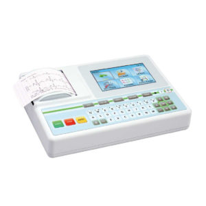 |  |  |  |  |  |
| SKU | SF1033560075-9 | SF1033560012-8 | SF1033560012-15 | SF1033560074-4 | SF1033560074-2 | SF1033560012-9 |
| Rating | ||||||
| Price | Ask for Price | $13,900.00 | $7,700.00 | $68,468.00 | $6,930.00 | Ask for Price |
| Stock | ||||||
| Availability | ||||||
| Add to cart | ||||||
| Description | Shipped from Abroad AsCARD Green v.06.101 is a 1-, 3-, 6- and 12-channel ECG unit which enables to make electrocardiogram in full 12 leads. Intended for ECG examinations of adult and paediatric patients aimed at identification of cardiological abnormalities, myocardial ischaemia or infarction. The device is intended for use in healthcare facilities by duly trained personnel. ECG examination may be recorded in manual or automatic mode with the ability to perform the analysis and interpretation. Delivery & Availability: Typically 10 working days – excluding furniture and heavy/bulky equipment. Please contact us for further information. | In Stock A feature-rich system inheriting the Wi-Sono high-end platform, the P15 uses an array of advanced tools to help enhance the image quality. It's a cost-effective, simplified console with an intuitive user interface and multiple intelligent functions. Delivery & Availability: Typically 2 working days – excluding furniture and heavy/bulky equipment. Please contact us for further information. | Shipped from Abroad With ultra-modern innovative design and the clinically-proven technologies, S8 Exp is portable ultrasound scanner well equipped as a low-physical-effort and enhanced-image-quality ultrasound scanner, which not only provides optimized solutions for versatile applications, but does help to improve the user-experience for both routine and non-traditional challenges. Delivery & Availability: Typically 5-7 working days – excluding furniture and heavy/bulky equipment. Please contact us for further information. | In Stock The GXR-SD is a diagnostic digital radiography system that provides reliable high quality digital radiographic images with a reduced dose. The GXR-SD DR systems offer comprehensive digital solutions to all radiography needs, featuring ACQUIDR digital imaging system with stationary or portable digital flat-panel detectors as well as reliable high-frequency x-ray generators that are known worldwide for their excellent performance, lifetime and stability. Patient tables and wall stands are also offered. Delivery & Availability: Typically 21 working days – excluding furniture and heavy/bulky equipment. Please contact us for further information. | In Stock JADE is one of the lightest portable X-ray systems on the market, allowing it to be used in any imaginable way including bedside, operating rooms, intensive care units and in veterinary fields. With a simple, easy-to-use operator console, three-way control, two-step foldable stand and auto lock system, JADE is a user-friendly portable X-ray system. Delivery & Availability: Typically 21 working days – excluding furniture and heavy/bulky equipment. Please contact us for further information. | Shipped from Abroad Incorporating innovative technologies, P20’s user-friendly design with a simple operation panel, intuitive user interface and a variety of intelligent auxiliary scanning tools, will significantly improve your daily examination experience. Besides general imaging applications, P20 has entitled with diagnostic 4D technology which has an extraordinary performance in obstetrics and gynecology applications. Delivery & Availability: Typically 5-7 working days – excluding furniture and heavy/bulky equipment. Please contact us for further information. |
| Content | AsCARD Green v.06.101 is a 1-, 3-, 6- and 12-channel ECG unit which enables to make electrocardiogram in full 12 leads. Intended for ECG examinations of adult and paediatric patients aimed at identification of cardiological abnormalities, myocardial ischaemia or infarction. The device is intended for use in healthcare facilities by duly trained personnel. ECG examination may be recorded in manual or automatic mode with the ability to perform the analysis and interpretation.
Electrocardiograph is based on advanced microprocessor technology .It is equipped with a thermal printer with high-resolution head and 4,3" LCD display. A touch panel and high-tech membrane keyboard makes this device intuitive in usage and its menu navigation exceptionally easy. This light-weight, small-footprint and battery powered cause that device can be easily transported to any location. With plastic casing and foil covered keyboard, the device is neat and easy to clean.
Technical Specifications:
Click Here To Catalogue Download | DETAILS
Super Wide-bandwidth Platform
Inheriting Wi-sono's ultra-wide system platform and with the advanced probe technology, high-resolution and deep penetration images are provided for precision medicine.
Spatial Compound Imaging
Spatial Compound Imaging utilizes several lines of sight for optimal contrast resolution, speckle reduction and border detection, with which P15 is ideal for superficial and abdominal imaging with better clarity and improved continuity of structures.
μ-Scan+
The new generation μ-Scan imaging technology gives you better image quality by reducing noise, improving signal strength and improving visualization.
Dynamic Color
Dynamic color improves upon already existing color Doppler technologies for a clearer capture of color flow and detailed visualization of even tiny veins with lower velocities.
Real-time Panoramic
With real-time panoramic, you can acquire an extended field of view for large organs or long vessels for easy measurement and diagnostic efficiency. Accomplished in real-time for the convenience of the sonographers, any mistake can also be easily back tracked and corrected without interrupting the scan.
3D/4D
Outstanding volume performance with speed and convenience makes P15 outshine others on volume imaging.
Tissue Doppler Imaging
Tissue Doppler Imaging allows clinical doctors to quantitatively evaluate local myocardial movements and functions, facilitating them with the ability to analyze and compare the motions of the different parts of the patient's heart.
Auto IMT
Quick measurement of intra-media vessel thickness ensures good reproducibility and high diagnostic efficiency.
Click Here To Download Catalogue | Sonoscape S8 Exp Portable Ultrasound scannerDETAILS Agile and Versatile With ultra-modern innovative design and the clinically-proven technologies, S8 Exp Portable Ultrasound scanner is well equipped as a low-physical-effort and enhanced-image-quality ultrasound scanner, which not only provides optimized solutions for versatile applications but does help to improve the user experience for both routine and non-traditional challenges. Working with S8 Exp, it will trigger your unlimited reverie and endow you with endless charm. Carrying forward the classical design of SonoScape's portable ultrasound products, S8 Exp successfully combines the best ergonomics, attractive design and ease of use. This charismatic identity is also enhanced by a sophisticated color palette—with sedate grey as its interior paint color and pearl white as exterior cover, S8 Exp reveals a style of aristocrat and strong character among SonoScape's ultrasound systems. Workflow The S8 Exp is a portable ultrasound scanner that adapts to your workflow, whether you are in the consulting room, at the bedside, or at a remote location. With easy-to-use new platform designed for sonographers' needs and full connection interfaces for easy connectivity and data sharing, S8 Exp leads to improved user comfort and clinical outcome as well as patient throughput and working efficiency. Powerful Platform Embedded with SonoScape's core imaging technologies such as μ-scan, PHI and Spatial Compound, S8 Exp boasts exceptional 2D image, sensitive spectral, Color and Power Doppler, displaying well-defined anatomy and pathology and facilitating a highly optimized diagnostic user environment for conclusive diagnoses. Besides, S8 Exp offers a comprehensive selection of electronic probes to maximally extend its capabilities to meet a wide range of applications including the abdomen, pediatric, OB/GYN, cardiovascular, musculoskeletal, etc. The advanced probe technologies also effectively enhance the image quality and confidence in reaching clinical diagnoses even in difficult patients.Click Here To Download Catalogue | DrGem Ceiling Mounted Digital X-ray is a diagnostic digital radiography system that provides reliable high quality digital radiographic images with a reduced dose. The GXR-SD DR systems offer comprehensive digital solutions to all radiography needs, featuring ACQUIDR digital imaging system with stationary or portable digital flat-panel detectors as well as reliable high-frequency x-ray generators that are known worldwide for their excellent performance, lifetime and stability. Patient tables and wall stands are also offered.
Features:
Click Here To Download Catalogue | JADE Mobile X-ray machine is one of the lightest portable X-ray systems on the market, allowing it to be used in any imaginable way including bedside, operating rooms, intensive care units and veterinary fields. With a simple, easy-to-use operator console, three-way control, two-step foldable stand and auto-lock system, the JADE Mobile X-ray machine is a user-friendly portable X-ray system.
Convenient & Intuitive Operation:
JADE is one of the lightest portable X-ray systems on the market, allowing it to be used in any imaginable way including bedside, operating rooms, intensive care units and in veterinary fields. With a simple, easy-to-use operator console, three-way control, two-step foldable stand and auto-lock system, JADE is a user-friendly portable X-ray system.
Compact & Powerful Design:
JADE Mobile X-ray machine is an innovative, highly versatile portable X-ray system suitable for a variety of clinical uses. Utilizing the unique technology used in DRGEM’s universally recognized X-ray generators, JADE is a compact but powerful unit with a 4kW output and thoughtfully designed components to increase efficiency and maximize workflow. The core part of X-ray source adopts high-quality tube assembly, X-ray collimator and high frequency X-ray generator with excellent performance, lifetime and stability.
Features:
Click Here To Download Catalogue | DETAILS
Upgraded Images with More Clarity
SonoScape never stops making progress in improving the image quality of its ultrasound products to enhance the confidence of diagnosis for doctors. With extraordinary images provided by P20, the anatomy structures are clearer than ever.
C-Xlasto Imaging
With C-xlasto Imaging, P20 enables comprehensive quantitative elastic analysis. Meanwhile, C-xlasto on P20 is supported by linear, convex and transvaginal probes, to ensure good reproducibility and highly consistent quantitative elastic results.
S-Live
S-Live allows for detailed visualization of subtle anatomical features, thereby enabling intuitive diagnosis with real-time 3D images and enriching patient communication.
Pelvic Floor 4D
Transperineal 4D pelvic floor ultrasound can provide useful clinical values in assessing the vaginal delivery impact on the female anterior compartment, judging whether the pelvic organs are prolapsed or not and the extent, determining if the pelvic muscles were torn accurately.
Anatomic M Mode
Anatomic M Mode helps you observe the myocardial motion at different phases by freely placing sample lines. It accurately measures the myocardial thickness and the heart size of even difficult patients and supports the myocardial function and LV wall-motion assessment.
Tissue Doppler Imaging
P20 is endowed with Tissue Doppler Imaging which provides velocities and other clinical information on myocardial functions, facilitating clinical doctors with the ability to analyze and compare the motions of different parts of the patient's heart.
Click Here To Download Catalogue |
| Weight | N/A | N/A | N/A | N/A | N/A | N/A |
| Dimensions | N/A | N/A | N/A | N/A | N/A | N/A |
| Additional information |


WAHID
Dear
I have ASPEL AsCARD Green ECG Machine v. 04.202
I need the manual because I have some difficulties
thanks