- Sorry, this product cannot be purchased.

ASPEL AsCARD Grey ECG Machine
$0.00
Shipped from Abroad
Electrocardiograph AsCARD Grey v.07.225 – is a 1, 3, 6, 12 channel ECG unit which enables to make electrocardiogram in full 12 leads. It is intended to conduct ECG examinations of adults and paediatric patients in all types of health care centres. ECG examination may be recorded in manual or automatic mode, with the possibility of analysis and interpretation. The device can be powered from 100 V ÷ 240 V mains supply or by an internal battery.
Delivery & Availability:
Typically 10 working days – excluding furniture and heavy/bulky equipment. Please contact us for further information.

Description
Electrocardiograph AsCARD Grey v.07.225 – is a 1, 3, 6, 12 channel ECG unit which enables to make electrocardiogram in full 12 leads. It is intended to conduct ECG examinations of adults and paediatric patients in all types of health care centres. ECG examination may be recorded in manual or automatic mode, with the possibility of analysis and interpretation. The device can be powered from 100 V ÷ 240 V mains supply or by an internal battery.
Technical Specification:1. Visualisation of 1, 3, 6 or 12 ECG waveforms, analysis results and interpretations, examinations stored in memory.
2. Recording of 12 standard leads.
3. Print out in 1, 3, 6 or 12 ECG waveforms mode. Printing of a selected group:
- 1 channel – (I, II, III, aVR, aVL, aVF, V1, V2, V3, V4, V5, V6),
- 3 channels in standard format – (I-II-III, aVR-aVL-aVF, V1-V2-V3, V4-V5-V6),
- 3 channels in Cabrera format – (aVL-I-aVR, II-aVF-III, V1-V2-V3, V4-V5-V6),
- 6 channels in standard format – (I-II-III-aVR-aVL-aVF, V1-V2-V3-V4-V5-V6),
- 6 channels in Cabrera format – (aVL-I-aVR-II-aVF-III, V1-V2-V3-V4-V5-V6),
- 12 channels in standard format – (I-II-III-aVR-aVL-aVF-V1-V2-V3-V4-V5-V6),
- 12 channels in Cabrera format – (aVL-I-aVR-II-aVF-III-V1-V2-V3-V4-V5-V6).
4. Available types of examinations: manual, AUTO, SPIRO, automatic to clipboard, AUTOMANUAL, LONG (v.07.xx5).
5. Automatic record ECG signal from all 12 leads simultaneously, with save to temporary memory function, then depending on the settings:
print out examination’s result, analysis, interpretation or save to database.
6. Adjustable length of automatic examination recording — interval of 6–30 seconds.
7. Reverse recording in automatic examination to clipboard and in manual examination (v.07.xx5).
8. Rhythm print out in AUTO examination and automatic examination to clipboard.
9. Definable stages of examination according to set up parameters in AUTOMANUAL examination.
10. Storing an examination in memory, 1–15 minutes in LONG mode (v.07.xx5).
11. ECG printed directly from the electrocardiograph using PCL5/6 printer.
12. Printout from database. Possibility to printout additional information about examination or patient.
13. Alpha-numerical soft-key keyboard with functional buttons.
14. Possibility to set parameters: speed, sensitive, intensity of printing.
15. Menu displayed on the screen for easy operation using touch panel.
16. Database of patients and examinations. Internal memory up to 1000 patients, 1000 examinations.
17. Viewing of the memory-stored examinations on display, with the possibility of changing the number of leads, augmentation and speed.
18. Automatic analysis and interpretation in compliance with EN 60601-2-51 (CSE database) – Analysis and interpretation dependable on age & sex of a patient.
19. Up to 130 automatic examinations in battery mode.
20. Continuous heart rate (HR) measurement and display.
21. Adapted to direct operation on an open heart.
22. Device allows to enable and disable the following filters:
- power line interference filter; filters available: 50 Hz, 60 Hz,
- muscle interference filter; filters available: 25 Hz, 35 Hz, 45 Hz,
- contour line filters; filters available: 0.15 Hz, 0.45 Hz, 0.75 Hz, 1.5 Hz,
- the low-pass filter (v.07.224, v.07.324, v.07.225, v.07.325): 75 Hz, 100 Hz, 125 Hz, 150 Hz,
- the auto-adaptive filter (v.07.224, v.07.324, v.07.225, v.07.325).
23. Detection of electrode detachment, independent for each channel (INOP control).
24. Detection of cardiac stimulator.
25. Sound signalling of detected stimulations of heart stimulator.
26. Protection against defibrillation pulse.
27. ECG recordings optionally saved on PenDrive, send on email or to other AsCARD via EKG-MAIL.
28. Wireless communication with the LAN or the Internet (v.07.3xx).
29. Wired communication with the LAN or the Internet.
30. Cooperation with CardioTEKA and CardioTEL.
31. Possibility of accepting orders for examination and sending back the results in HL7 standard via Internet (v.07.3xx).
32. Possibility of conducting preliminary spirometry examinations by using SPIRO-31 attachment.
33. EMR – keep performed examinations in specified period of time on external data storage (USB memory).
Accessories
1. Limb electrodes, 4 pieces (EKK type).
2. Chest electrodes, 6 pieces (EPP type).
3. KEKG 30R ECG cable.
4. Main cable.
5. R-A4 paper, 112 mm wide (1 reel).
6. ECG gel.
7. Operation manual.
Click Here To Download Catalogue
Quick Comparison
| ASPEL AsCARD Grey ECG Machine remove | ASPEL Ambulatory BP Machine remove | ASPEL AsCARD Green ECG Machine remove | Sonoscape P15 Ultrasound Machine With Four Probes remove | ASPEL Stress ECG with Treadmill and Software remove | DrGem Ceiling Analogue X-ray Machine remove | |
|---|---|---|---|---|---|---|
| Name | ASPEL AsCARD Grey ECG Machine remove | ASPEL Ambulatory BP Machine remove | ASPEL AsCARD Green ECG Machine remove | Sonoscape P15 Ultrasound Machine With Four Probes remove | ASPEL Stress ECG with Treadmill and Software remove | DrGem Ceiling Analogue X-ray Machine remove |
| Image | 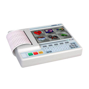 | 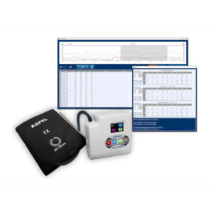 | 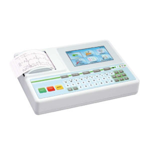 | 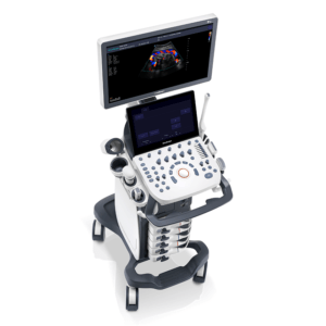 | 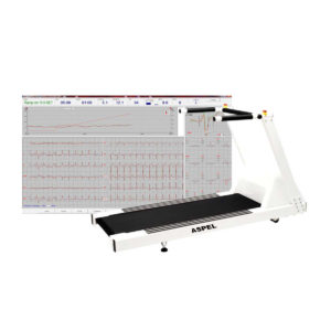 | 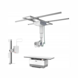 |
| SKU | SF1033560075-5 | SF1033560075-13 | SF1033560075-9 | SF1033560012-8 | SF1033560075-2 | SF1033560074-7 |
| Rating | ||||||
| Price |
|
|
|
|
|
|
| Stock | ||||||
| Availability | ||||||
| Add to cart | ||||||
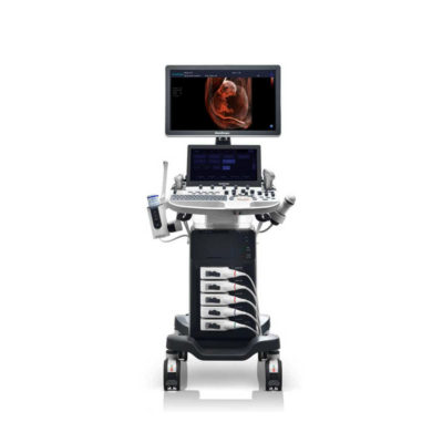
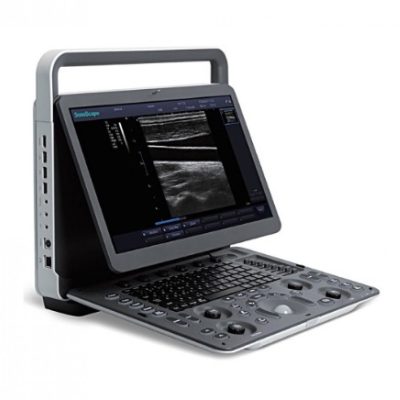
| Description | Shipped from Abroad Electrocardiograph AsCARD Grey v.07.225 - is a 1, 3, 6, 12 channel ECG unit which enables to make electrocardiogram in full 12 leads. It is intended to conduct ECG examinations of adults and paediatric patients in all types of health care centres. ECG examination may be recorded in manual or automatic mode, with the possibility of analysis and interpretation. The device can be powered from 100 V ÷ 240 V mains supply or by an internal battery. Delivery & Availability: Typically 10 working days – excluding furniture and heavy/bulky equipment. Please contact us for further information. | Shipped from Abroad ASPEL Ambulatory BP Machine - is a recorder of long-term records of non-invasive measurement of blood pressure intended for use in clinics, hospitals, outpatient centers and specialist surgeries. The recorder enables the assessment of blood pressure by the oscillometric method in adult patients, pregnant women, including preeclampsia and pediatric patients (from 3 years of age). Blood pressure is assessed by using an inflatable cuff, an accurate pressure transducer, and a deflation valve. Delivery & Availability: Typically 10 working days – excluding furniture and heavy/bulky equipment. Please contact us for further information. | Shipped from Abroad AsCARD Green v.06.101 is a 1-, 3-, 6- and 12-channel ECG unit which enables to make electrocardiogram in full 12 leads. Intended for ECG examinations of adult and paediatric patients aimed at identification of cardiological abnormalities, myocardial ischaemia or infarction. The device is intended for use in healthcare facilities by duly trained personnel. ECG examination may be recorded in manual or automatic mode with the ability to perform the analysis and interpretation. Delivery & Availability: Typically 10 working days – excluding furniture and heavy/bulky equipment. Please contact us for further information. | In Stock A feature-rich system inheriting the Wi-Sono high-end platform, the P15 uses an array of advanced tools to help enhance the image quality. It's a cost-effective, simplified console with an intuitive user interface and multiple intelligent functions. Delivery & Availability: Typically 2 working days – excluding furniture and heavy/bulky equipment. Please contact us for further information. | Shipped from Abroad It is a system with professional tool dedicated to exercise and resting ECG examination. Treadmill has 12 lead ECG modules. With ECG Analyzing Software. Delivery & Availability: Typically 21 working days – excluding furniture and heavy/bulky equipment. Please contact us for further information. | Shipped from abroad The DrGem Ceiling Analogue X-ray Machine is a diagnostic radiography system that provides reliable high quality radiographic images with a reduced dose. The reliable high-frequency x-ray generators that are known worldwide for their excellent performance, lifetime and stability. Patient tables and wall stands are also offered. Delivery & Availability: Typically 21 working days – excluding furniture and heavy/bulky equipment. Please contact us for further information. |
| Content |
Electrocardiograph AsCARD Grey v.07.225 - is a 1, 3, 6, 12 channel ECG unit which enables to make electrocardiogram in full 12 leads. It is intended to conduct ECG examinations of adults and paediatric patients in all types of health care centres. ECG examination may be recorded in manual or automatic mode, with the possibility of analysis and interpretation. The device can be powered from 100 V ÷ 240 V mains supply or by an internal battery.
Technical Specification:1. Visualisation of 1, 3, 6 or 12 ECG waveforms, analysis results and interpretations, examinations stored in memory.
2. Recording of 12 standard leads.
3. Print out in 1, 3, 6 or 12 ECG waveforms mode. Printing of a selected group:
Click Here To Download Catalogue | ASPEL Ambulatory BP Machine - is a recorder of long-term records of non-invasive measurement of blood pressure intended for use in clinics, hospitals, outpatient centers and specialist surgeries. The recorder enables the assessment of blood pressure by the oscillometric method in adult patients, pregnant women, including preeclampsia and pediatric patients (from 3 years of age). Blood pressure is assessed by using an inflatable cuff, an accurate pressure transducer, and a deflation valve.
Features:
Save-2-Safe: Double security system
Thanks to the use of two independent measuring systems with an additional valve, it meets the highest standards and takes care of patient safety even better.
Start-Easy: Quick start in two moves
The quick launch function allows you to use the device instantly, easily allows you to start recording in holter mode.
Memo-Care: Cuff pressure memory
Recorder remembers the pressure in the cuff. Thanks to the use of Intelligent Solutions, it adapts individually to the patient.
Power-Usb: USB connection
The device can work without batteries: by connecting to a computer via a USB cable.
Technical Specification:
Click Here To Download Catalogue | AsCARD Green v.06.101 is a 1-, 3-, 6- and 12-channel ECG unit which enables to make electrocardiogram in full 12 leads. Intended for ECG examinations of adult and paediatric patients aimed at identification of cardiological abnormalities, myocardial ischaemia or infarction. The device is intended for use in healthcare facilities by duly trained personnel. ECG examination may be recorded in manual or automatic mode with the ability to perform the analysis and interpretation.
Electrocardiograph is based on advanced microprocessor technology .It is equipped with a thermal printer with high-resolution head and 4,3" LCD display. A touch panel and high-tech membrane keyboard makes this device intuitive in usage and its menu navigation exceptionally easy. This light-weight, small-footprint and battery powered cause that device can be easily transported to any location. With plastic casing and foil covered keyboard, the device is neat and easy to clean.
Technical Specifications:
Click Here To Catalogue Download | DETAILS
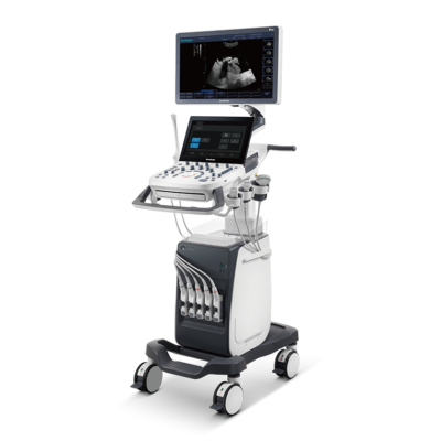
Super Wide-bandwidth Platform
Inheriting Wi-sono's ultra-wide system platform and with the advanced probe technology, high-resolution and deep penetration images are provided for precision medicine.
Spatial Compound Imaging
Spatial Compound Imaging utilizes several lines of sight for optimal contrast resolution, speckle reduction and border detection, with which P15 is ideal for superficial and abdominal imaging with better clarity and improved continuity of structures.
μ-Scan+
The new generation μ-Scan imaging technology gives you better image quality by reducing noise, improving signal strength and improving visualization.
Dynamic Color
Dynamic color improves upon already existing color Doppler technologies for a clearer capture of color flow and detailed visualization of even tiny veins with lower velocities.
Real-time Panoramic
With real-time panoramic, you can acquire an extended field of view for large organs or long vessels for easy measurement and diagnostic efficiency. Accomplished in real-time for the convenience of the sonographers, any mistake can also be easily back tracked and corrected without interrupting the scan.
3D/4D
Outstanding volume performance with speed and convenience makes P15 outshine others on volume imaging.
Tissue Doppler Imaging
Tissue Doppler Imaging allows clinical doctors to quantitatively evaluate local myocardial movements and functions, facilitating them with the ability to analyze and compare the motions of the different parts of the patient's heart.
Auto IMT
Quick measurement of intra-media vessel thickness ensures good reproducibility and high diagnostic efficiency.
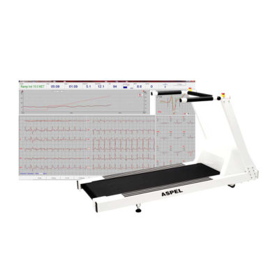
Click Here To Download Catalogue | It is a system with professional tool dedicated to exercise and resting ECG examination. Treadmill has 12 lead ECG modules. With ECG Analyzing Software.
Technical Specification:
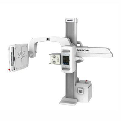
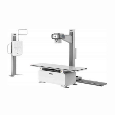
Click Here To Download Catalogue | DrGem Ceiling Analogue X-ray Machine is a diagnostic radiography system X-ray Machine that provides reliable high quality radiographic images with a reduced dose. The reliable high-frequency x-ray generators that are known worldwide for their excellent performance, lifetime and stability. Patient tables and wall stands are also offered.
Features of DrGem Ceiling Analogue X-ray Machine
Click Here To Download Catalogue |
| Weight | N/A | N/A | N/A | N/A | N/A | N/A |
| Dimensions | N/A | N/A | N/A | N/A | N/A | N/A |
| Additional information |

Reviews
There are no reviews yet.