| Description | | Shipped from Abroad
The ANATOM 64 CT scanner is the latest innovation for cardiac imaging based on Precision Platform system. The excellent design of Ahart technology which innovatively combined single spiral scan + gated imaging + mA modulation for easy heart imaging at extremely low radiation dose. We provide you ANATOM 64 Clarity/Precision of two models which are low/high configurations for preferences. It also offers you conventional clinical applications of low dose, better image quality and faster exams.
Delivery & Availability:
Typically 90 working days – excluding furniture and heavy/bulky equipment. Please contact us for further information. | Shipped from Abroad
SonoScape has developed a new probe and function for the E1 Exp. With these additions the E1 Exp will bring users a more efficient examination experience with satisfying image quality and a smooth workflow.
Delivery & Availability:
Typically 5-7 working days – excluding furniture and heavy/bulky equipment. Please contact us for further information. | In Stock
DRGEM’s TOPAZ X-ray machine is a state-of-the-art mobile digital radiography system, designed with maximum comfort for patients and users in mind. From its user-friendly software to smooth movements, TOPAZ is made to improve your workflow and provide you with high-quality images.
Delivery & Availability:
Typically 21 working days – excluding furniture and heavy/bulky equipment. Please contact us for further information. | In Stock
A feature-rich system inheriting the Wi-Sono high-end platform, the P15 uses an array of advanced tools to help enhance the image quality. It's a cost-effective, simplified console with an intuitive user interface and multiple intelligent functions.
Delivery & Availability:
Typically 2 working days – excluding furniture and heavy/bulky equipment. Please contact us for further information. | Shipped from Abroad
This Machine gives a possibility to perform computed tomography without any problems and on high quality level. This device is used to conduct exams of internal organs and their functioning. With its help, a physician has a possibility to assess the condition of the human body as a whole.
Delivery & Availability:
Typically 90 working days – excluding furniture and heavy/bulky equipment. Please contact us for further information. |
| Content |
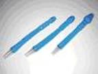
For gastroenterology
Rectal cannulas are made of PVC for medical use.
They are flexible and have a smooth surface. The balloon can be filled either with air than water (about 100 ml). Upon request, all cannulas can be provided with a foam pad
| The ANATOM 64 CT scanner is the latest innovation for cardiac imaging based on Precision Platform system. The excellent design of Ahart technology which innovatively combined single spiral scan + gated imaging + mA modulation for easy heart imaging at extremely low radiation dose. We provide you ANATOM 64 Clarity/Precision of two models which are low/high configurations for preferences. It also offers you conventional clinical applications of low dose, better image quality and faster exams.
Features:
- Modularized OptiWave HD detector features low-cost & easy maintenance, high spatial resolution and long lifetime
- Admir3D iterative technology delivers optimal dose efficiency and noise reduction without compromising image quality
- High configurations of main components ensure the best results and maximum patient throughput
- Uniquely and creatively uses 140kV and 80kV dual energy scan mode for brain imaging on 16-slice CT to offers you extraordinary image quality both in low and high density resolutions
- AdoseTM mA modulation ensures you low dose imaging without compromising image quality particularly useful in pediatric applications
- Equipped with dedicated Abast and Amast for bone and metal artifacts
- The brilliant Ahart technology enables you to experience so easy and low-dose cardiac imaging applications
Technical Specifications:
| Model |
ANATOM 64 Precision |
ANATOM 64 Fit |
| Rack type |
Low pressure slip ring |
Low pressure slip ring |
| Scan aperture |
70cm |
70cm |
| Rack Physical inclination |
± 30 ° |
N.A |
| Rack digital inclination |
± 50 ° |
± 50 ° |
| cooling method |
Air-cooled |
Air-cooled |
| Focus to the center distance |
56 cm |
53 cm |
| |
|
|
| Maximum power (non-equivalent) |
80kW |
42kW |
| Votage (kV) |
80kV / 100kV / 120kV / 140kV |
70kV / 80kV / 100kV / 120kV /
140kV |
| |
|
|
| Heat capacity |
8MHU |
5.0MHU |
| Heat dissipation rate |
931 kHU / min |
748kHU / min |
| cooling method |
Oil cool |
Oil cool |
| Large focus size |
1.1mm × 1.2mm |
1.2mm × 1.4mm |
| Small focus size |
0.6mm × 1.2mm |
0.7mm × 0.8mm |
| Tube current range |
10-670mA |
10-350mA |
| |
|
|
| Detector type |
Optiwave detectors |
Optiwave detectors |
| Number of Z-axis |
32 |
32 |
| The width of the Z-axis |
20mm |
20mm |
| The number of elements per row |
912 |
848 |
| Total number of detectors |
29184 |
27136 |
| Acquisition mode |
64x0.625, 32x0.625,
16x0.625 |
64x0.625, 32x0.625,
16x0.625 |
| |
|
|
| Scanning range |
1800mm |
1800mm |
| Horizontal positioning accuracy |
± 0.25mm |
± 0.25mm |
| weight capacity |
205kg |
205kg |
| Minimum height |
43cm |
43cm |
| Anti - collision protection device |
Yes |
Yes |
| Foot control switch |
Yes |
Yes |
| IV rack |
Yes |
Yes |
| |
|
|
| CPU |
3.5GHz |
3.5GHz |
| RAM |
16 GB × 4 |
16 GB × 4 |
| Hard drive capacity |
1T × 2 |
1T × 2 |
| Display size |
24 inch LCD monitor |
24 inch LCD monitor |
| Display resolution |
1920 × 1200 |
1920 × 1200 |
| Computer operating system |
Windows 7 |
Windows 7 |
| Image reconstruction speed |
65 frames/ second |
65 frames/ second |
| Number of image store |
1000000 |
1000000 |
| Data external storage mode |
CD / DVD / USB |
CD / DVD / USB |
| |
|
|
| Minimum Scan Time of 360 degree |
0.39sec |
0.75sec |
| Sub-millimeter acquisition layers |
64 |
64 |
| Double sub-millimeter acquisition
layers |
64 |
64 |
| Thinnest acquisition thickness |
0.625mm |
0.625mm |
| The thinnest reconstruction
thickness |
0.3125mm |
0.625mm |
| Conventional reconstruction
thickness (mm) |
0.3125 mm, 0.625 mm, 1.25 mm, 2.5
mm, 5.0 mm, 7.5 mm, 10 mm |
0.625 mm, 1.25 mm, 2.5 mm, 5.0
mm, 7.5 mm, 10 mm |
| The reconstruction matrix |
512 x 512, 1024 x 1024 |
512 x 512, 1024 x 1024 |
| Display matrix |
1024 × 1024 |
1024 × 1024 |
| Max FOV |
52cm |
50cm |
| The maximum display field of view |
70cm |
50cm |
| Maximum scan length |
1800mm |
1800mm |
| Maximum continuous helix scan
time |
120s |
120s |
| Pitch range |
0.5-1.5 |
0.5, 1.0, 1.5 |
| |
|
|
| High contrast resolution |
21 Lp / cm @ 0% MTF |
21 Lp / cm @ 0% MTF |
| Low contrast resolution |
2mm @ 0.3% |
2mm @ 0.3% |
| Image noise |
≤ 0.25 |
≤ 0.29 |
| |
|
|
| MPR |
Yes |
Yes |
| CPR |
Yes |
Yes |
| SSD |
Yes |
Yes |
| VR |
Yes |
Yes |
| MIP |
Yes |
Yes |
| MinIP |
Yes |
Yes |
| Virtual endoscopy |
Yes |
Yes |
| CT angiography |
Yes |
Yes |
| Tissue segmentation |
Yes |
Yes |
| One-key bone removal |
Yes |
Yes |
| Automatically patient table removal |
Yes |
Yes |
| Contrast Agent Automatic Tracking
Technology- bolus tracking |
Yes |
Yes |
| Automatic linkage trigger
technology |
Yes |
Yes |
| Cine mode display |
Yes |
Yes |
| Bone artifact suppression technique |
AbastTM |
AbastTM |
| Metal artifact suppression technique |
AbastTM |
AbastTM |
| Iterative reconstruction technique |
Admir3D global iteration |
Admir3D full-domain iteration |
| Low - dose children 's scanning
technology |
Yes |
Yes |
| Low - dose lung scan |
Yes |
Yes |
| Gray matter enhancement
technology |
AccuHead |
AccuHead |
| High resolution imaging of the lung |
AccuLung |
AccuLung |
| Inner ear high resolution imaging |
AccuOtica |
AccuOtica |
| Body high resolution imaging |
AccuBody |
AccuBody |
| Bone high resolution imaging |
AccuBone |
AccuBone |
| Head dual-energy imaging |
Ahead |
Ahead |
| CT perfusion imaging |
Optional |
Optional |
| Quantitative analysis of blood
vessels |
Optional |
Optional |
| Heart coronary artery imaging |
Aheart |
N.A |
| ECG gated |
Yes |
N.A |
| Low dose cardiac scan |
Yes |
N.A |
| |
|
|
| Green energy saving technology |
AccuSaving |
AccuSaving |
| Dual-energy scan technology |
Optional |
Optional |
| DETAILS
Efficient Diagnosis
μ-Scan, Speckle Reduction & Edge Enhancement
Spatial Compound Imaging
PIH - Pure Inversion Harmonic
Wide Scan - Enlarged Image Area
Tissue-Specific Imaging
SR Flow
Ergonomic Designs
Up to 2 Transducer Ports
Light Weight and Compact
15.6 inch Anti-flickering HD LED Screen
Tilting Monitor Angle Adjustment
Backlit Keyboard and Intelligent Panel
Long-lasting Battery for 90 mins
Ease of Use
Quick Boot Up
Auto-Brightness Adjustment
Auto Image Optimization
Auto IMT
Auto Trace
Equipped Accessories
Wi-Fi and Bluetooth Available
DICOM
500GB Hard Disk
Height Adjustable Trolley
Durable, Carry-on Site Suitcase
| TOPAZ X-ray machine is among the high end X-ray machine manufactured by DRGEM, a digital X-ray system that provides quality images with little or no effort.
It begins with Advanced Technology
Integrating high technology and over a decade of experience in conventional and digital radiography systems, DRGEM’s TOPAZ X-ray machine is a state-of-the-art mobile digital radiography system, designed with maximum comfort for patients and users. From its user-friendly software to smooth movements, TOPAZ X-ray machine is made to improve your workflow and provide you with high-quality images.
Full Featured Imaging Software & Excellent Digital Image Processing
With a high-performance, built-in touchscreen, TOPAZ X-ray machine offers a user-friendly interface and powerful software for easy operation and increased workflow. The anatomical view-based digital image processing, automatically optimizes and enhances the quality of the image. it also comes with automatic image storage and print with DICOM 3.0 networking capability. additionally, the system offers increasing exam throughput while decreasing examination time.
- Provides convenient user interface and easy operation
- Anatomical view-based digital image processing
- Radiographic stand and automatic collimator control function
- DICOM 3.0 networking interface features include: work-list, print, store, and query for
- integration with any PACS or RIS.
Features of Topaz X-ray Machine:
- Outstanding image quality by optimized digital image processing
- Easy driving and maneuverable with ergonomic and compact design
- Convenient and enough space for detector, battery and other necessary stuff
- Swift mobility with 5km/h speed allows you to save time, cost and satisfy your patient with quick processing
- Accurate positioning and precise movement provided with 4 direction buttons on this control panel
- Longer arm stroke and high column provide wider coverage and patient-friendly operation service
- A safety function with front safety bumper & brake, spring loaded front wheel and status LED indicator
- Provide best satisfaction and convenience for your patient and operator. It will prevent any unexpected and secondary accident
Technical Specification:
Topaz X-ray Machine
- Output Rating - 32kW/40kW
- kVRange - 40 to 125V/150kV
- mA Range - 10 to 400mA/500mA
- mAs Range - 0.1 to 400mA/500mAs
Digital Flat-panel Detector
- Size – 14X17inch
- Scintillator - Csl/GOS
- Type - Wireless
| DETAILS
Super Wide-bandwidth Platform
Inheriting Wi-sono's ultra-wide system platform and with the advanced probe technology, high-resolution and deep penetration images are provided for precision medicine.
Spatial Compound Imaging
Spatial Compound Imaging utilizes several lines of sight for optimal contrast resolution, speckle reduction and border detection, with which P15 is ideal for superficial and abdominal imaging with better clarity and improved continuity of structures.
μ-Scan+
The new generation μ-Scan imaging technology gives you better image quality by reducing noise, improving signal strength and improving visualization.
Dynamic Color
Dynamic color improves upon already existing color Doppler technologies for a clearer capture of color flow and detailed visualization of even tiny veins with lower velocities.
Real-time Panoramic
With real-time panoramic, you can acquire an extended field of view for large organs or long vessels for easy measurement and diagnostic efficiency. Accomplished in real-time for the convenience of the sonographers, any mistake can also be easily back tracked and corrected without interrupting the scan.
3D/4D
Outstanding volume performance with speed and convenience makes P15 outshine others on volume imaging.
Tissue Doppler Imaging
Tissue Doppler Imaging allows clinical doctors to quantitatively evaluate local myocardial movements and functions, facilitating them with the ability to analyze and compare the motions of the different parts of the patient's heart.
Auto IMT
Quick measurement of intra-media vessel thickness ensures good reproducibility and high diagnostic efficiency.
| This Machine gives a possibility to perform computed tomography without any problems and on high quality level. This device is used to conduct exams of internal organs and their functioning. With its help, a physician has a possibility to assess the condition of the human body as a whole.
Features:
- It is easy to use;
- Convenience;
- Multi functionality;
- Obtained images are of high definition;
- High-definition 3D images of the area under study;
- The procedure is pain-free;
- The data is processed fast;
- The image can be stored in the computer memory;
- The diagnostics does not take a lot of time;
- Acceptable radiation dose.
Technical Specifications:
| No. |
Technical Features |
Descriptions |
| 1 |
Gantry |
|
| 1.01 |
Gantry type |
Low voltage slip-ring |
| 1.02 |
Gantry driven type |
Strap-driven |
| 1.03 |
Patient opening |
70cm |
| 1.04 |
Gantry tilt mode |
Digital gantry tilt |
| 1.05 |
Digital tilt capability |
±50° |
| 1.06 |
Detector type |
OptiWave rare-earth ceramic detector |
| 1.07 |
Numbers of detector rows |
16 |
| 1.08 |
Width of Z-axle detector |
20mm |
| 1.09 |
Detector columns of channels per row |
848 |
| 1.10 |
Numbers of detector columns |
13568 |
| 1.11 |
Data-transfer type |
RF, optical fiber communication |
| 1.12 |
Distance of focus-ISO-center |
53cm |
| 1.13 |
Distance of focus-detector |
94cm |
| 1.14 |
3D laser orientation |
Provided |
| 1.15 |
13" integrated display panel |
Provided |
| 1.16 |
Adose automatic exposure control (mA
Modulation) |
Provided |
| 1.17 |
Auto-voice manager |
Breath Graphical Display
Hold Message (Record/Playback)
Breath Message (Record/Playback) |
| 1.18 |
AccuSaving energy conservation management |
Provided |
| 2 |
HVPS and X-ray tube |
|
| 2.01 |
Maximum continuous output of HVgenerator |
42kW |
| 2.02 |
Tube kV selections |
70kV, 80kV, 100 kV, 120 kV, 140 kV |
| 2.03 |
Tube mA range |
10~350mA |
| 2.04 |
Tube anode heat capacity |
3.5MHU |
| 2.05 |
Max. anode cooling rate |
735kHU/min |
| 2.06 |
Type of cooling |
Oil cooling + Air cooling |
| 2.07 |
Tube focus |
Large: 1.2mm×1.4mm
Small: 0.7mm×0.8mm |
| 2.08 |
Collimator width selection |
4-level election |
| 2.09 |
Focus spot tracking technology |
Provided |
| 3 |
Patient table |
|
| 3.01 |
Maximum horizontal-movable range |
1850mm |
| 3.02 |
Table horizontal-scannablerange |
1800mm |
| 3.03 |
Table horizontal-position repeatability |
±0.25mm |
| 3.04 |
Minimum height above floor |
430mm |
| 3.05 |
Maximum vertical-movable range |
500mm |
| 3.06 |
Maximum speed of vertical movement |
35mm |
| 3.07 |
Maximum speed of horizontal movement |
150mm/s |
| 3.08 |
Maximum patient weight |
205kg |
| 3.09 |
Foot pedal of patient table control |
Provided |
| 4 |
Computer |
|
| 4.01 |
CPU |
3.5GHz |
| 4.02 |
Memory |
32GB |
| 4.03 |
Storage of hard-disk |
1TB×2 |
| 4.04 |
Monitor |
24’’ LCD Monitor |
| 4.05 |
Resolution of monitor |
1920×1200 |
| 4.06 |
Image-data external storage type |
CD/DVD/USB |
| 4.07 |
Time of image reconstruction (512×512) |
33.3ms/image |
| 4.08 |
Speed of image reconstruction (512×12) |
30fps |
| 4.09 |
DICOM 3.0 interface |
Provided |
| 4.10 |
Printer DICOM 3.0 interface |
Provided |
| 4.11 |
Auto filming |
Provided |
| 4.12 |
Worklist function |
Provided |
| 5 |
Scan parameters |
|
| 5.01 |
Shortest 360 degree rotation time |
0.75s |
| 5.02 |
Allowed rotation times |
0.75s, 1.0s, 1.5s, 2.0s, 3.0s, 4.0s |
| 5.03 |
Maximum slice numbers per rotation |
32 |
| 5.04 |
Minimum slice thickness of scan |
1.25mm |
| 5.05 |
Minimum slice thickness of reconstruction |
0.625mm |
| 5.06 |
Maximum slice thickness of scan |
20mm |
| 5.07 |
Nominal reconstruction slice thickness |
0.625mm, 1.25mm, 2.5mm, 5.0mm, 7.5mm,
10mm, 20mm |
| 5.08 |
Speed of image reconstruction (512×512) |
30 frames/s |
| 5.09 |
Scan FOV |
50cm |
| 5.10 |
Image reconstruction matrix |
512×512, 1024×1024 (Optional) |
| 5.11 |
Image reconstruction matrix |
512×512, 1024×1024 (Optional) |
| 5.12 |
Image display matrix |
512×512, 1024×1024 (Optional) |
| 5.13 |
Maximum continuous scan duration |
120s |
| 5.14 |
Maximum continuous scan length |
180cm |
| 5.15 |
Direction of TOPO |
Front-back, Left-right |
| 5.16 |
Max. length of TOPO |
180cm |
| 5.17 |
Range of pitch |
0.5~1.5 |
| 5.18 |
Scan mode |
Scout scan
Axial scan
Helical scan
Cine scan |
| 6 |
Image Quality |
|
| 6.01 |
High contrast resolution |
21lp/cm@0%MTF |
| 6.02 |
Low contrast resolution |
2.0mm@0.30% |
| 6.03 |
Isotropic imaging resolution |
0.24mm |
| 6.04 |
Range of CT numbers |
-32767~32768 |
| 6.05 |
Image noise |
≤0.29@28mGy |
| 7 |
Advanced application |
|
| 7.01 |
Multi-Planar Reconstruction (MPR) |
Provided |
| 7.02 |
Curve Multi-Planar Reconstruction (CPR) |
Provided |
| 7.03 |
Surface Shaded Display (SSD) |
Provided |
| 7.04 |
Volume Rendering (VR) |
Provided |
| 7.05 |
Maximum Intensity Projection (MIP) |
Provided |
| 7.06 |
Minimum Intensity Projection (MinIP) |
Provided |
| 7.07 |
Virtual Endoscopy (VE) |
Provided |
| 7.08 |
CT angiography (CTA) |
Provided |
| 7.09 |
Tissue segmentation |
Provided |
| 7.10 |
One click bone remove |
Provided |
| 7.11 |
One click patient table remove |
Provided |
| 7.12 |
Bolus-tracking Technology |
Provided |
| 7.13 |
Spiral auto start |
Provided |
| 7.14 |
Cine display |
Provided |
| 7.15 |
AbastTM bone artifact suppression technology |
Provided |
| 7.16 |
AmastTM metal artifact suppression technology |
Provided |
| 7.17 |
Admir3D all-domain iterative reconstruction |
Provided |
| 7.18 |
Low-dose pediatric scan technology |
Provided |
| 7.19 |
Low-dose lung scan technology |
Provided |
| 7.20 |
AccuHead grey-white matter enhanced
technology |
Provided |
| 7.21 |
AccuOrgan lung high resolution scan technology |
Provided |
| 7.22 |
AccuOrgan inner-ear high resolution scan
technology |
Provided |
| 7.23 |
AccuOrgan body high resolution scan technology |
Provided |
| 7.24 |
AccuOrgan bone high resolution scan technology |
Provided |
| 7.25 |
AccuMatter dual-energy with Admir3D for new
application |
Provided |
|


Reviews
There are no reviews yet.