| Description | | Shipped from Abroad
OPENMARK 5000 is 0.51T MRI. It's approved by FDA and has CE mark. It adopts two-pillar magnet design with 280 degree openness and equipped with powerful
RF and gradient system, together with advanced imaging technology, making it as a high-end system which is comparable to high-field MRI.
Delivery & Availability:
Typically 90 working days – excluding furniture and heavy/bulky equipment. Please contact us for further information. | Shipped from Abroad
Sonoscape E2 portable ultrasound machine is a color Doppler ultrasound system that reaches beyond your expectations due to its compact and fashionable appearance. It fulfills GI, OB/GYN, Cardiac and POC applications to fit your routine scanning needs while its color mode will help you for more accurate and efficient diagnosis of lesions. E2 provides a wide range of applications to assist users with routine scanning. E2 provides automatic calculations to enhance your diagnostic confidence and save you time for patient communication.
Delivery & Availability:
Typically 14 working days – excluding furniture and heavy/bulky equipment. Please contact us for further information. | Shipped from Abroad
SuperMark 1.5T is a new generation superconducting MRI system based on years of experience in production and research. It's applicable to whole body scan, such as, nervous system, spine, joint soft tissue, pelvic and abdominal cavity, etc
Delivery & Availability:
Typically 90 working days – excluding furniture and heavy/bulky equipment. Please contact us for further information. |
Shipped from Abroad
SUPiA made by Signers offers such a better clinic environment with no chemicals, ideal space, high-resolution image quality, and affordability.
Delivery & Availability:
Typically 14 working days – excluding furniture and heavy/bulky equipment. Please contact us for further information. | Shipped from Abroad
As SonoScape steps forward to add value and efficiency to ultrasound, the latest S22 was designed in a user-friendly platform to address current and future demanding needs. It represents an excellent mix in performance and price.
Delivery & Availability:
Typically 5-7 working days – excluding furniture and heavy/bulky equipment. Please contact us for further information. |
| Content |
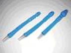
For gastroenterology
Rectal cannulas are made of PVC for medical use.
They are flexible and have a smooth surface. The balloon can be filled either with air than water (about 100 ml). Upon request, all cannulas can be provided with a foam pad
| OPENMARK 5000 is 0.51T MRI. It's approved by FDA and has CE mark. It adopts two-pillar magnet design with 280 degree openness and equipped with powerful
RF and gradient system, together with advanced imaging technology, making it as a high-end system which is comparable to high-field MRI.
Features:
- With the highest system stability and the highest homogeneity of the
magnet field in permanent MRI
- Screens on both sides facilitate positioning; 280 degree two-pillar magnet
design ensures stable magnet structure and facilitates interventional
treatment.
- Active and passive shimming calibrate technology ensures the magnetic
field uniformity
- Motor-driven patient couch makes it easier for patients to access and for
positioning
- Powerful hardware and software platforms ensure the scan speed, image
quality and make it possible for advanced imaging functions
- Fast scan speed eliminates motion artifact
- Rich scan sequences, advanced imaging technology and powerful postprocessing
technology ensure image quality, extend more applications,
which can fully satisfy the clinical needs
- Intelligent user-friendly operating system ensures you easy operation
Technical Specifications:
| No. |
Technique Description |
Parameter |
| 1 |
Magnet System |
|
| 1.1 |
Magnet Type |
Permanent Magnet
Automatic constant temperature
system |
| 1.2 |
Field Strength |
0.51T |
| 1.3 |
Magnet Shape |
Dual-pillar shape |
| 1.4 |
Homogeneity(40cm,DSV,VRMS) |
≤1.6ppm |
| 1.5 |
Shim Method |
Active/Passive |
| 1.6 |
Magnet Vertical Gap (Cover) |
40cm |
| 1.7 |
Magnetic Pole Dia. (Exclude Cover) |
145cm |
| 1.8 |
Accessibility(Horizontal Opening Angle, |
280° |
| 1.9 |
5 Gauss fringe field |
X-axis:horizontal ≤2.5m
Y-axis:Vertical ≤2.5m
Z-axis:horizontal ≤2.5m |
| 2 |
Patient Couch and Communication |
|
| 2.1 |
Patient Couch Driven mode |
Motor-driven |
| 2.2 |
Max. Patient Weight |
≥200kg(440lbs) |
| 2.3 |
Patient Positioning Tools |
Laser Light Localizer for positioning of center slice Motor-driven transfer to center of imaging volume |
| 2.4 |
Position accuracy |
±1mm |
| 2.5 |
Emergency Call Key |
Yes |
| 2.6 |
Intercom System |
Yes |
| 3 |
Gradient System |
|
| 3.1 |
Gradient Field Strength(Single Axis) |
≥30mT/m |
| 3.2 |
Gradient Slew Rate (Single Axis) |
≥100mT/m/ms |
| 3.3 |
Rise Time |
≤0.3ms |
| 3.4 |
Gradient Cooling System ( Gradient coils
and Power electronics) |
Air Cooling |
| 4 |
RF System |
|
| 4.1 |
RF System Type |
Digital Transmit and
Receive signal |
| 4.2 |
Number of RF Channels |
4 |
| 4.3 |
Transmitter Amplifier Peak Power |
6kW |
| 4.4 |
RF Bandwidth of Receiver |
≥1.25MHz |
| 4.5 |
Head Coil |
Yes |
| 4.6 |
Neck Coil |
Yes |
| 4.7 |
Body/Spine Coil (17 inch) |
Yes |
| 4.8 |
Body/Spine Coil (21 inch) |
Yes |
| 4.9 |
Knee Coil |
Yes |
| 4.10 |
Shoulder Coil |
Yes |
| 4.11 |
Flexible Coil |
Optional |
| 4.12 |
Breast Coil |
Optional |
| 5 |
Computer System |
|
| 5.1 |
Host Computer |
DELL Computer (for MR) |
| 5.2 |
System Software |
Windows XP |
| 5.3 |
Operation Software |
APEX |
| 5.4 |
CPU Clock rate |
3.0GHz |
| 5.5 |
Main Memory |
4GB |
| 5.6 |
Color LCD Monitor |
19” |
| 5.7 |
Keyboard and Mouse |
Standard |
| 5.8 |
Image Reconstruction Speed(256 x 256
Matrix) |
200 frame/Sec. |
| 5.9 |
Hard Disk |
500GB |
| 5.10 |
Image Storage Capacity(256 x 256
Matrix) |
500,000 |
| 5.11 |
Media Driver |
DVD RW |
| 5.12 |
DICOM 3.0 |
Yes |
| 5.13 |
Ethernet |
Yes |
| 5.14 |
Operation Console |
Yes |
| 5.15 |
Operation Chair |
Yes |
| 6 |
Scanning Parameter |
|
| 6.1 |
Max. FOV |
410mm |
| 6.2 |
Min. FOV |
5mm |
| 6.3 |
Min. TE(SE) |
5ms |
| 6.4 |
Min. TR(SE) |
11ms |
| 6.5 |
Min. TE(GR) |
1ms |
| 6.6 |
Min. TR(GR) |
3ms |
| 6.7 |
Min. 2D Thickness |
1.0mm |
| 6.8 |
Min. 3D Thickness |
0.1mm |
| 6.9 |
Max. Image Matrix |
512x512 |
| 7 |
Scanning Sequence & Imaging Technique |
|
| 7.1 |
Spin Echo 2D/3D (SE 2D/3D) |
Yes |
| 7.2 |
DE/QE |
Yes |
| 7.3 |
Fast Spin Echo 2D/3D(FSE 2D/3D) |
Yes |
| 7.4 |
Single Shot FSE 2D/3D |
Yes |
| 7.5 |
Inversion Recovery(IR) |
Yes |
| 7.6 |
Fast Inversion Recovery(FIR) |
Yes |
| 7.7 |
Gradient Echo 2D/3D(GR 2D/3D) |
Yes |
| 7.8 |
Fast GR 2D/3D |
Yes |
| 7.9 |
SPGR |
Yes |
| 7.10 |
FLAIR |
Yes |
| 7.11 |
Fat Imaging |
Yes |
| 7.12 |
Fat Suppression imaging |
Yes |
| 7.13 |
Water-Fat Separation imaging |
Yes |
| 7.14 |
TOF MRA(2D/3D) |
Yes |
| 7.15 |
MRCP(2D/3D) |
Yes |
| 7.16 |
MRU (2D/3D) |
Yes |
| 7.17 |
MRM |
Yes |
| 7.18 |
Fast Hydrograph Imaging |
Yes |
| 7.19 |
Diffusion Weighted Imaging(DWI) |
Yes |
| 7.20 |
Max. b Value |
1000s/mm2 |
| 7.21 |
Breath Hold Technology |
Yes |
| 7.22 |
Magnetization Transfer Contrast(MTC) |
Yes |
| 7.23 |
Multi-slice and Angle-free Presaturation |
Yes |
| 7.24 |
Saturation Tracking |
Yes |
| 7.25 |
Maximum Intensity Projection(MIP) |
Yes |
| 7.26 |
Multi-Angle Projection(MAP) |
Yes |
| 7.27 |
3D Reconstruction |
Yes |
| 7.28 |
Multi-planar Reconstruction(MPR) |
Yes |
| 7.29 |
Multi-Artifacts Eliminating technology |
Yes |
| 7.30 |
Checking with Part Metal Implant |
Yes |
| 7.31 |
Online Image Filtration |
Yes |
| 7.32 |
Online Post Procession |
Yes |
| 7.33 |
3D Scout |
Yes |
| 7.34 |
Scanning Protocol Preset |
Yes |
| 7.35 |
Scanning Protocol Queue Waiting |
Yes |
| 7.36 |
Advanced Image Post Processing |
Yes |
| 7.37 |
Image Fusion Technology of Vascular |
Yes |
| 7.38 |
Image Fusion Technology of Spine |
Yes |
| SONOSCAPE E2 DETAILS
Auto Image Optimization
A portable ultrasound machine with the press of a button, the image is automatically adjusted and optimized, saving you time with parameter adjustments. Additionally, with Auto Focus on, the focus area follows the depth of the ROI box as it is moved in the scanning field, providing users with excellent image quality in the desired area of interest.
Automated Calculation
Auto IMT is used when determining the level of vascular sclerosis present in the patient by automatically tracing the thickness of the carotid vessels.
Auto trace provides users sensitive and accurate wave tracing, avoiding the error of manual trace and giving out calculation result in no time
In-Build Battery pack
This portable ultrasound machine was equipped with an in-build battery pack which enable the user to perform image scanning when AC power is not available.
| SuperMark 1.5T is a new generation superconducting MRI system based on years of experience in production and research. It's applicable to whole body scan, such as, nervous system, spine, joint soft tissue, pelvic and abdominal cavity, etc. SuperMark 1.5T provides not only conventional pulse sequences and clinical diagnosis functions, but also provides advanced functional applications, for instance, 3D angiography and water imaging. It adopts brand new ANKE APEX operating system which ensures easy operation and fast diagnosis.
Technical Advantages:
- Reliable short cavity superconducting magnet system with zero liquid helium
consumption
- New generation fully digitalized and extensible multichannel spectrometer
- Powerful high efficiency and high fidelity gradient system; Multi-channel PA RF
receiving coil with intelligent identification
- English operating system and high extensible computer system
- High resolution conventional clinical images; Practical advanced functional
imaging
Superconducting MRI System:
- Highly open and humanization design -> Streamlined design
- Rich sequences and technology satisfy clinical needs -> Efficient service
Low Investment:
- High cost performance superconducting MRI system
- Zero liquid helium consumption, low running and maintenance cost
- Core technology by independent R & D supports full upgrade
- Low electric consumption
- Compact magnet design, minimum installation space: 35 square meters
High Return:
- High resolution thin slice images improve diagnosis
- Short cavity magnet design makes patients comfortable
- Fast scan speed improves work efficiency
Technical Specifications:
| No. |
Technique Description |
Parameter |
| 1 |
Magnet System |
|
| 1.1 |
Magnet Type |
Permanent Magnet
Automatic constant temperature
system |
| 1.2 |
Field Strength |
0.51T |
| 1.3 |
Magnet Shape |
Dual-pillar shape |
| 1.4 |
Homogeneity(40cm,DSV,VRMS) |
≤1.6ppm |
| 1.5 |
Shim Method |
Active/Passive |
| 1.6 |
Magnet Vertical Gap (Cover) |
40cm |
| 1.7 |
Magnetic Pole Dia. (Exclude Cover) |
145cm |
| 1.8 |
Accessibility(Horizontal Opening Angle, |
280° |
| 1.9 |
5 Gauss fringe field |
X-axis:horizontal ≤2.5m
Y-axis:Vertical ≤2.5m
Z-axis:horizontal ≤2.5m |
| 2 |
Patient Couch and Communication |
|
| 2.1 |
Patient Couch Driven mode |
Motor-driven |
| 2.2 |
Max. Patient Weight |
≥200kg(440lbs) |
| 2.3 |
Patient Positioning Tools |
Laser Light Localizer for positioning of center slice Motor-driven transfer to center of imaging volume |
| 2.4 |
Position accuracy |
±1mm |
| 2.5 |
Emergency Call Key |
Yes |
| 2.6 |
Intercom System |
Yes |
| 3 |
Gradient System |
|
| 3.1 |
Gradient Field Strength(Single Axis) |
≥30mT/m |
| 3.2 |
Gradient Slew Rate (Single Axis) |
≥100mT/m/ms |
| 3.3 |
Rise Time |
≤0.3ms |
| 3.4 |
Gradient Cooling System ( Gradient coils
and Power electronics) |
Air Cooling |
| 4 |
RF System |
|
| 4.1 |
RF System Type |
Digital Transmit and
Receive signal |
| 4.2 |
Number of RF Channels |
4 |
| 4.3 |
Transmitter Amplifier Peak Power |
6kW |
| 4.4 |
RF Bandwidth of Receiver |
≥1.25MHz |
| 4.5 |
Head Coil |
Yes |
| 4.6 |
Neck Coil |
Yes |
| 4.7 |
Body/Spine Coil (17 inch) |
Yes |
| 4.8 |
Body/Spine Coil (21 inch) |
Yes |
| 4.9 |
Knee Coil |
Yes |
| 4.10 |
Shoulder Coil |
Yes |
| 4.11 |
Flexible Coil |
Optional |
| 4.12 |
Breast Coil |
Optional |
| 5 |
Computer System |
|
| 5.1 |
Host Computer |
DELL Computer (for MR) |
| 5.2 |
System Software |
Windows XP |
| 5.3 |
Operation Software |
APEX |
| 5.4 |
CPU Clock rate |
3.0GHz |
| 5.5 |
Main Memory |
4GB |
| 5.6 |
Color LCD Monitor |
19” |
| 5.7 |
Keyboard and Mouse |
Standard |
| 5.8 |
Image Reconstruction Speed(256 x 256
Matrix) |
200 frame/Sec. |
| 5.9 |
Hard Disk |
500GB |
| 5.10 |
Image Storage Capacity(256 x 256
Matrix) |
500,000 |
| 5.11 |
Media Driver |
DVD RW |
| 5.12 |
DICOM 3.0 |
Yes |
| 5.13 |
Ethernet |
Yes |
| 5.14 |
Operation Console |
Yes |
| 5.15 |
Operation Chair |
Yes |
| 6 |
Scanning Parameter |
|
| 6.1 |
Max. FOV |
410mm |
| 6.2 |
Min. FOV |
5mm |
| 6.3 |
Min. TE(SE) |
5ms |
| 6.4 |
Min. TR(SE) |
11ms |
| 6.5 |
Min. TE(GR) |
1ms |
| 6.6 |
Min. TR(GR) |
3ms |
| 6.7 |
Min. 2D Thickness |
1.0mm |
| 6.8 |
Min. 3D Thickness |
0.1mm |
| 6.9 |
Max. Image Matrix |
512x512 |
| 7 |
Scanning Sequence & Imaging Technique |
|
| 7.1 |
Spin Echo 2D/3D (SE 2D/3D) |
Yes |
| 7.2 |
DE/QE |
Yes |
| 7.3 |
Fast Spin Echo 2D/3D(FSE 2D/3D) |
Yes |
| 7.4 |
Single Shot FSE 2D/3D |
Yes |
| 7.5 |
Inversion Recovery(IR) |
Yes |
| 7.6 |
Fast Inversion Recovery(FIR) |
Yes |
| 7.7 |
Gradient Echo 2D/3D(GR 2D/3D) |
Yes |
| 7.8 |
Fast GR 2D/3D |
Yes |
| 7.9 |
SPGR |
Yes |
| 7.10 |
FLAIR |
Yes |
| 7.11 |
Fat Imaging |
Yes |
| 7.12 |
Fat Suppression imaging |
Yes |
| 7.13 |
Water-Fat Separation imaging |
Yes |
| 7.14 |
TOF MRA(2D/3D) |
Yes |
| 7.15 |
MRCP(2D/3D) |
Yes |
| 7.16 |
MRU (2D/3D) |
Yes |
| 7.17 |
MRM |
Yes |
| 7.18 |
Fast Hydrograph Imaging |
Yes |
| 7.19 |
Diffusion Weighted Imaging(DWI) |
Yes |
| 7.20 |
Max. b Value |
1000s/mm2 |
| 7.21 |
Breath Hold Technology |
Yes |
| 7.22 |
Magnetization Transfer Contrast(MTC) |
Yes |
| 7.23 |
Multi-slice and Angle-free Presaturation |
Yes |
| 7.24 |
Saturation Tracking |
Yes |
| 7.25 |
Maximum Intensity Projection(MIP) |
Yes |
| 7.26 |
Multi-Angle Projection(MAP) |
Yes |
| 7.27 |
3D Reconstruction |
Yes |
| 7.28 |
Multi-planar Reconstruction(MPR) |
Yes |
| 7.29 |
Multi-Artifacts Eliminating technology |
Yes |
| 7.30 |
Checking with Part Metal Implant |
Yes |
| 7.31 |
Online Image Filtration |
Yes |
| 7.32 |
Online Post Procession |
Yes |
| 7.33 |
3D Scout |
Yes |
| 7.34 |
Scanning Protocol Preset |
Yes |
| 7.35 |
Scanning Protocol Queue Waiting |
Yes |
| 7.36 |
Advanced Image Post Processing |
Yes |
| 7.37 |
Image Fusion Technology of Vascular |
Yes |
| 7.38 |
Image Fusion Technology of Spine |
Yes |
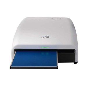
| SUPiA X-ray Digitizer made by Signers offers such a better clinic environment with no chemicals, ideal space, high-resolution image quality, and affordability
FEATURE
Rigid Type
• No damage or scratch on image plates during scanning & erasing
• Scanning & Erasing without a roller
• No cut-off image during winter and cold period
Durability
• Extremely simple structure design
• Strong aluminum base plate
• Flip covers preventing dust from inside scanner
Barcode System
• Automatically recognising cassette sizes(14x17", 10x12", 18x24cm) by barcode reader
Compact & lightweight design
- Very small and compatible CR on desktop (only 63.5cm)
- Only 21.5kg (47.4lbs)
Cassette
Strong structure
• Strong enough against external impact
• Totally metal frame
• Enduring under 150kg on cassette
Featherlight
• 14x17" : 2.05kg 10x12" : 0.99kg 18x24cm : 0.75kg
Dust free & Easy cleaning
• Easy to clean up dust on IPs
• Prevent dust from outside
User friendly design
• Various colors 14x17"(Green),
10x12"(Blue), 18x24cm(Pink))
• Barcode label
TECHNICAL SPECIFICATION
|
SUPiA Specifications |
|
| Cassette |
SUPiA CR Cassette 14x17 (inch)
SUPiA CR Cassette 10x12 (inch)
SUPiA CR Cassette 18x24 (cm) |
| Throughput |
Up to 94 IPs/hour (14x17"/160μm) |
| Slots |
Single Cassette feed |
| Dimensions (W x D x H) |
436 x 636 x 196mm |
| Weight |
21.5kg (47.4lbs) |
| Grayscale Resolution |
Acquisition : 16 bits per pixel
Display : 12 bits per pixel |
| Power Supply Conditions |
Single Phase 50 ~ 60Hz
AC 90 ~ 264V |
| Network |
100 MBit |
| PC Connection |
USB 2.0 |
| Computer Min.
requirements |
OS : Win 7 or 10
CPU : Intel i5
RAM : 4GB
Graphic Card : Intel HD 2500
HDD : 500GB
Monitor : FHD 1920 x 1080
|
| Operating conditions |
Temperature : 10 ~ 30˚C
Humidity : 15 ~ 85% RH |
| DETAILS
As SonoScape steps forward to add value and efficiency to ultrasound, the latest S22 was designed in a user-friendly platform to address current and future demanding needs. It represents an excellent mix in performance and price.
S22, is a shared service ultrasound system with a slim and elegant package that has combined mobility with utility to fit in specific clinical situations including emergency department, ICU, operating room and so on. Furthermore, its ergonomic design, easy operating and flexible data management will give you a memorable experience.
SPECIFICATION
• Large high-resolution widescreen LED
• Sensitive touch screen
• Four transducer sockets plus one socket for pencil probe
• A comprehensive selection of probes: linear, Convex, Micro-convex, Volumetric, Endocavity, Bi-plane, Phased Array, TEE, Intraoperative, Pencil
• Premium application technology: 4D, μ-scan speckle reduction, compound imaging, Pulse Inversion Harmonic Imaging, Color M-Mode, Steer M-Mode, PDI, TDI, Real-time Panoramic Imaging, Trapezoid Imaging, Auto-IMT…
• Full patient database and image management solutions: DICOM 3.0, AVI/JPG, USB 2.0, HDD, DVD, PDF report
• Multi-Language Input Keyboard
• Built-in battery
|


Reviews
There are no reviews yet.