Description
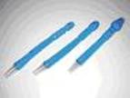
For gastroenterology
Rectal cannulas are made of PVC for medical use.
They are flexible and have a smooth surface. The balloon can be filled either with air than water (about 100 ml). Upon request, all cannulas can be provided with a foam pad
Quick Comparison
| Cannulla disposable rectal remove | SIGNERS SUPiA X-ray Digitizer ( CR Scanner) remove | DrGem Diamond All-In-One Digital X-ray Machine remove | Anke Anatom 32 Fit Multi-Slice Spiral CT Scan remove | Sonoscape S22 Ultrasound Machine remove | DrGem Floor Mounted Analogue X-ray remove | |||||||||||||||||||||||||||||||||||||||||||||||||||||||||||||||||||||||||||||||||||||||||||||||||||||||||||||||||||||||||||||||||||||||||||||||||||||||||||||||||||||||||||||||||||||||||||||||||||||||||||||||||||||||||||||||||||||||||||||||||||||||||||||||||||||||||||||||||||||||||||||||||||||||||||||||||||||||||||||||||||||||||||||||||
|---|---|---|---|---|---|---|---|---|---|---|---|---|---|---|---|---|---|---|---|---|---|---|---|---|---|---|---|---|---|---|---|---|---|---|---|---|---|---|---|---|---|---|---|---|---|---|---|---|---|---|---|---|---|---|---|---|---|---|---|---|---|---|---|---|---|---|---|---|---|---|---|---|---|---|---|---|---|---|---|---|---|---|---|---|---|---|---|---|---|---|---|---|---|---|---|---|---|---|---|---|---|---|---|---|---|---|---|---|---|---|---|---|---|---|---|---|---|---|---|---|---|---|---|---|---|---|---|---|---|---|---|---|---|---|---|---|---|---|---|---|---|---|---|---|---|---|---|---|---|---|---|---|---|---|---|---|---|---|---|---|---|---|---|---|---|---|---|---|---|---|---|---|---|---|---|---|---|---|---|---|---|---|---|---|---|---|---|---|---|---|---|---|---|---|---|---|---|---|---|---|---|---|---|---|---|---|---|---|---|---|---|---|---|---|---|---|---|---|---|---|---|---|---|---|---|---|---|---|---|---|---|---|---|---|---|---|---|---|---|---|---|---|---|---|---|---|---|---|---|---|---|---|---|---|---|---|---|---|---|---|---|---|---|---|---|---|---|---|---|---|---|---|---|---|---|---|---|---|---|---|---|---|---|---|---|---|---|---|---|---|---|---|---|---|---|---|---|---|---|---|---|---|---|---|---|---|---|---|---|---|---|---|---|---|---|---|---|---|---|---|---|---|---|---|---|---|---|---|---|---|---|---|---|---|---|---|---|---|---|---|---|---|
| Name | Cannulla disposable rectal remove | SIGNERS SUPiA X-ray Digitizer ( CR Scanner) remove | DrGem Diamond All-In-One Digital X-ray Machine remove | Anke Anatom 32 Fit Multi-Slice Spiral CT Scan remove | Sonoscape S22 Ultrasound Machine remove | DrGem Floor Mounted Analogue X-ray remove | ||||||||||||||||||||||||||||||||||||||||||||||||||||||||||||||||||||||||||||||||||||||||||||||||||||||||||||||||||||||||||||||||||||||||||||||||||||||||||||||||||||||||||||||||||||||||||||||||||||||||||||||||||||||||||||||||||||||||||||||||||||||||||||||||||||||||||||||||||||||||||||||||||||||||||||||||||||||||||||||||||||||||||||||||
| Image | | 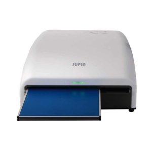 |  |  |  |  | ||||||||||||||||||||||||||||||||||||||||||||||||||||||||||||||||||||||||||||||||||||||||||||||||||||||||||||||||||||||||||||||||||||||||||||||||||||||||||||||||||||||||||||||||||||||||||||||||||||||||||||||||||||||||||||||||||||||||||||||||||||||||||||||||||||||||||||||||||||||||||||||||||||||||||||||||||||||||||||||||||||||||||||||||
| SKU | SF103356013086-27 | SF1033560050-01 | SF1033560074-3 | SF1033560092-1 | SF1033560012-3 | SF1033560074-6 | ||||||||||||||||||||||||||||||||||||||||||||||||||||||||||||||||||||||||||||||||||||||||||||||||||||||||||||||||||||||||||||||||||||||||||||||||||||||||||||||||||||||||||||||||||||||||||||||||||||||||||||||||||||||||||||||||||||||||||||||||||||||||||||||||||||||||||||||||||||||||||||||||||||||||||||||||||||||||||||||||||||||||||||||||
| Rating | ||||||||||||||||||||||||||||||||||||||||||||||||||||||||||||||||||||||||||||||||||||||||||||||||||||||||||||||||||||||||||||||||||||||||||||||||||||||||||||||||||||||||||||||||||||||||||||||||||||||||||||||||||||||||||||||||||||||||||||||||||||||||||||||||||||||||||||||||||||||||||||||||||||||||||||||||||||||||||||||||||||||||||||||||||||||
| Price |
| $6,930.00 |
|
| $9,350.00 |
| ||||||||||||||||||||||||||||||||||||||||||||||||||||||||||||||||||||||||||||||||||||||||||||||||||||||||||||||||||||||||||||||||||||||||||||||||||||||||||||||||||||||||||||||||||||||||||||||||||||||||||||||||||||||||||||||||||||||||||||||||||||||||||||||||||||||||||||||||||||||||||||||||||||||||||||||||||||||||||||||||||||||||||||||||
| Stock | ||||||||||||||||||||||||||||||||||||||||||||||||||||||||||||||||||||||||||||||||||||||||||||||||||||||||||||||||||||||||||||||||||||||||||||||||||||||||||||||||||||||||||||||||||||||||||||||||||||||||||||||||||||||||||||||||||||||||||||||||||||||||||||||||||||||||||||||||||||||||||||||||||||||||||||||||||||||||||||||||||||||||||||||||||||||
| Availability | ||||||||||||||||||||||||||||||||||||||||||||||||||||||||||||||||||||||||||||||||||||||||||||||||||||||||||||||||||||||||||||||||||||||||||||||||||||||||||||||||||||||||||||||||||||||||||||||||||||||||||||||||||||||||||||||||||||||||||||||||||||||||||||||||||||||||||||||||||||||||||||||||||||||||||||||||||||||||||||||||||||||||||||||||||||||
| Add to cart | ||||||||||||||||||||||||||||||||||||||||||||||||||||||||||||||||||||||||||||||||||||||||||||||||||||||||||||||||||||||||||||||||||||||||||||||||||||||||||||||||||||||||||||||||||||||||||||||||||||||||||||||||||||||||||||||||||||||||||||||||||||||||||||||||||||||||||||||||||||||||||||||||||||||||||||||||||||||||||||||||||||||||||||||||||||||
| Description | Shipped from Abroad SUPiA made by Signers offers such a better clinic environment with no chemicals, ideal space, high-resolution image quality, and affordability. Delivery & Availability: Typically 14 working days – excluding furniture and heavy/bulky equipment. Please contact us for further information. | Shipped from Abroad DrGem Diamond All-In-One Digital X-ray Machine is a fully automatic digital radiography system providing state-of-the-art image quality, image processing and user interface. With a wide selection of anatomical studies on the imaging software, DIAMOND automatically sets up the x-ray generator’s preprogrammed exposure technique settings, motorized radiographic stand positioning, x-ray collimation and post-image processing for the selected study. Specifically designed to increase workflow, this fully digital system offers convenient auto-positioning and advanced image processing to achieve big performance with little effort. Delivery & Availability: Typically 21 working days – excluding furniture and heavy/bulky equipment. Please contact us for further information. | Shipped from Abroad
This Machine gives a possibility to perform computed tomography without any problems and on high quality level. This device is used to conduct exams of internal organs and their functioning. With its help, a physician has a possibility to assess the condition of the human body as a whole.
Delivery & Availability: Typically 90 working days – excluding furniture and heavy/bulky equipment. Please contact us for further information. | Shipped from Abroad As SonoScape steps forward to add value and efficiency to ultrasound, the latest S22 was designed in a user-friendly platform to address current and future demanding needs. It represents an excellent mix in performance and price. Delivery & Availability: Typically 5-7 working days – excluding furniture and heavy/bulky equipment. Please contact us for further information. | In Stock GXR Analogue X-ray system matches with a radiographic room which perfectly fits your workow and can be easily upgraded to DR system with the help of DR interface and PC interface in GXR generator as well as Bucky suitable to Flat Panel Detector. GXR X-ray system is equipped with a high frequency X-ray generator which consistently produces high quality radiograph in favor of high quality X-ray output with a very small kV ripple and accurate mA and mAs. GXR X-ray system is designed to provide convenience to operator and comfort to patient. Delivery & Availability: Typically 21 working days – excluding furniture and heavy/bulky equipment. Please contact us for further information. | |||||||||||||||||||||||||||||||||||||||||||||||||||||||||||||||||||||||||||||||||||||||||||||||||||||||||||||||||||||||||||||||||||||||||||||||||||||||||||||||||||||||||||||||||||||||||||||||||||||||||||||||||||||||||||||||||||||||||||||||||||||||||||||||||||||||||||||||||||||||||||||||||||||||||||||||||||||||||||||||||||||||||||||||||
| Content | For gastroenterology
Rectal cannulas are made of PVC for medical use.
They are flexible and have a smooth surface. The balloon can be filled either with air than water (about 100 ml). Upon request, all cannulas can be provided with a foam pad
| SUPiA X-ray Digitizer made by Signers offers such a better clinic environment with no chemicals, ideal space, high-resolution image quality, and affordability
FEATURE
Rigid Type
• No damage or scratch on image plates during scanning & erasing
• Scanning & Erasing without a roller
• No cut-off image during winter and cold period
Durability
• Extremely simple structure design
• Strong aluminum base plate
• Flip covers preventing dust from inside scanner
Barcode System
• Automatically recognising cassette sizes(14x17", 10x12", 18x24cm) by barcode reader
Compact & lightweight design
Click Here To Download Catalogue | DrGem Diamond All-In-One Digital X-ray Machine is a fully automatic digital radiography system providing state-of-the-art image quality, image processing and user interface. With a wide selection of anatomical studies on the imaging software, DIAMOND automatically sets up the x-ray generator’s pre-programmed exposure technique settings, motorized radiographic stand positioning, x-ray collimation and post-image processing for the selected study. Specifically designed to increase workflow, this fully digital system offers convenient auto-positioning and advanced image processing to achieve big performance with little effort.
Features of DrGem Diamond All-In-One Digital X-ray Machine:
Outstanding Image Quality -
Digital radiography via at panel detector improves your workflow, exam speed and comfort with efficiency. Digital at panel detector with Csl screen provides excellent spatial resolution, MTF, DQE and stability based on ne pixel pitch. A 3-field ion-chamber is provided for AEC function.
Automatic Collimation –
Automatic x-ray eld size control of the motorized collimator corresponds to dierent SIDs. Includes user adjustable lamp timer with on/oswitch.
Automatic Positioning –
Click Here To Download Catalogue | This Machine gives a possibility to perform computed tomography without any problems and on high quality level. This device is used to conduct exams of internal organs and their functioning. With its help, a physician has a possibility to assess the condition of the human body as a whole.
Features:
Click Here To Download Catalogue | DETAILS
As SonoScape steps forward to add value and efficiency to ultrasound, the latest S22 was designed in a user-friendly platform to address current and future demanding needs. It represents an excellent mix in performance and price.
S22, is a shared service ultrasound system with a slim and elegant package that has combined mobility with utility to fit in specific clinical situations including emergency department, ICU, operating room and so on. Furthermore, its ergonomic design, easy operating and flexible data management will give you a memorable experience.
SPECIFICATION
• Large high-resolution widescreen LED
• Sensitive touch screen
• Four transducer sockets plus one socket for pencil probe
• A comprehensive selection of probes: linear, Convex, Micro-convex, Volumetric, Endocavity, Bi-plane, Phased Array, TEE, Intraoperative, Pencil
• Premium application technology: 4D, μ-scan speckle reduction, compound imaging, Pulse Inversion Harmonic Imaging, Color M-Mode, Steer M-Mode, PDI, TDI, Real-time Panoramic Imaging, Trapezoid Imaging, Auto-IMT…
• Full patient database and image management solutions: DICOM 3.0, AVI/JPG, USB 2.0, HDD, DVD, PDF report
• Multi-Language Input Keyboard
• Built-in battery
Click Here To Download Catalogue | DrGem GXR Floor Mounted Analogue X-ray system matches with a radiographic room which perfectly fits your workflow and can be easily upgraded to DR system with the help of DR interface and PC interface in GXR generator as well as Bucky suitable to Flat Panel Detector. GXR (Analogue X-ray)system is equipped with a high frequency X-ray generator which consistently produces high quality radiograph in favor of high quality X-ray output with a very small kV ripple and accurate mA and mAs. GXR (Analogue X-ray) system is designed to provide convenience to operator and comfort to patient.
Features of DrGem GXR Floor Mounted Analogue X-ray:
Click Here To Download Catalogue | ||||||||||||||||||||||||||||||||||||||||||||||||||||||||||||||||||||||||||||||||||||||||||||||||||||||||||||||||||||||||||||||||||||||||||||||||||||||||||||||||||||||||||||||||||||||||||||||||||||||||||||||||||||||||||||||||||||||||||||||||||||||||||||||||||||||||||||||||||||||||||||||||||||||||||||||||||||||||||||||||||||||||||||||||
| Weight | N/A | N/A | N/A | N/A | N/A | N/A | ||||||||||||||||||||||||||||||||||||||||||||||||||||||||||||||||||||||||||||||||||||||||||||||||||||||||||||||||||||||||||||||||||||||||||||||||||||||||||||||||||||||||||||||||||||||||||||||||||||||||||||||||||||||||||||||||||||||||||||||||||||||||||||||||||||||||||||||||||||||||||||||||||||||||||||||||||||||||||||||||||||||||||||||||
| Dimensions | N/A | N/A | N/A | N/A | N/A | N/A | ||||||||||||||||||||||||||||||||||||||||||||||||||||||||||||||||||||||||||||||||||||||||||||||||||||||||||||||||||||||||||||||||||||||||||||||||||||||||||||||||||||||||||||||||||||||||||||||||||||||||||||||||||||||||||||||||||||||||||||||||||||||||||||||||||||||||||||||||||||||||||||||||||||||||||||||||||||||||||||||||||||||||||||||||
| Additional information |
|


Reviews
There are no reviews yet.