| Description | Shipped From Abroad
11mm Diameter, Round Plate
Delivery & Availability:
Typically 10-21 working days – excluding furniture and heavy/bulky equipment. Please contact us for further information.
| In Stock
- Durable and light weight
- Supplied in a soft pouch
- Long life bright white Xenon bulb
- Macular beam dioptres lenses from 0 to +20 and 0 to -20
- 3 year guarantee (excludes consumables)
Delivery & Availability:
Typically 5-7 working days – excluding furniture and heavy/bulky equipment. Please contact us for further information.
| Shipped from abroad
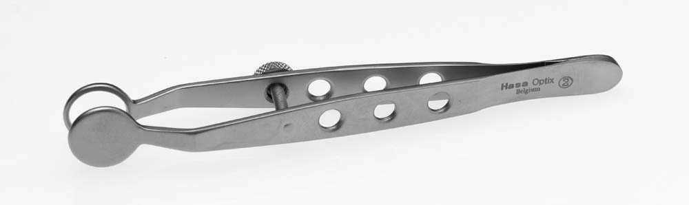
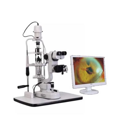
The product is designed on the principle basis of Goldman tonometer. It can be connected with slit lamp(Carl Zeiss type).
Delivery & Availability:
Typically 14 working days – excluding furniture and heavy/bulky equipment. Please contact us for further information.
| Shipped from abroad
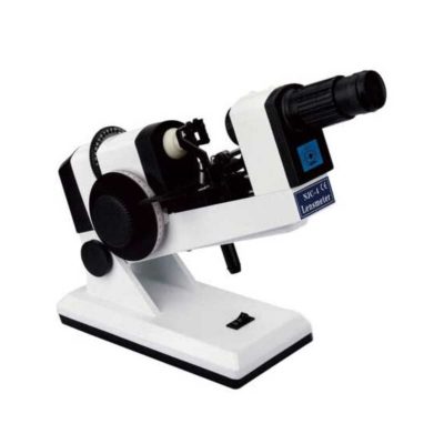
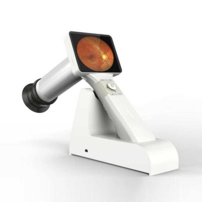
Ultra-small corneal curvature tester, is mainly used to measure the corneal curvature radius and diopter, wireless output print data.
Delivery & Availability:
Typically 14 working days – excluding furniture and heavy/bulky equipment. Please contact us for further information.
| Shipped from abroad
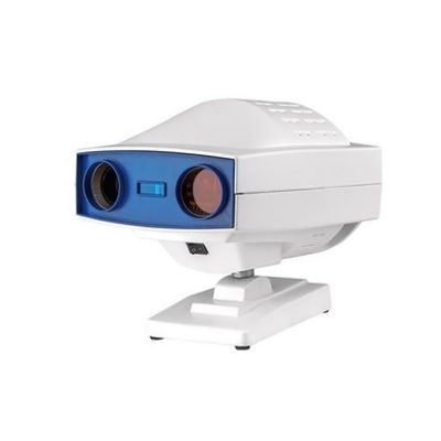
- 7.0-inch color LCD touch panel.
- Hartman sensor with 108 multiple measure points.
- Green measurement light beam.
- Dyeing lens, sunglasses measured easily.
Delivery & Availability:
Typically 14 working days – excluding furniture and heavy/bulky equipment. Please contact us for further information.
| Shipped from abroad
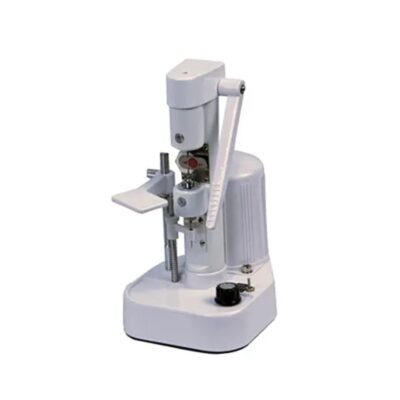
Super lightweight design, reduce fatigue, operation is very convenient.
Delivery & Availability:
Typically 14 working days – excluding furniture and heavy/bulky equipment. Please contact us for further information.
|
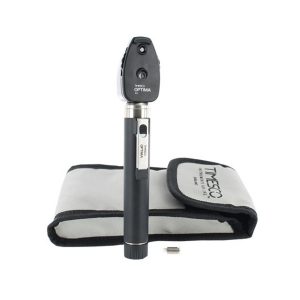
| Content | | Timesco Ophthalmoscope features a head made from lightweight hermetically sealed durable plastic, precision optics and a latex free rubber eyebrow rest. A bright white light from long life standard bulbs provides crystal clear illumination in ophthalmic diagnostic procedures.
- Durable and lightweight
- Supplied in a soft pouch
- Long life bright white Standard bulb
- Macular beam dioptres lenses from 0 to +20 and 0 to -20
- 3-year guarantee (excludes consumables)
| Applanation Tonometer is designed on the principle basis of Goldman tonometer. It can be connected with slit lamp(Carl Zeiss type).
Features of Applanation Tonometer:
- When used with slit lamp, it can be used to examine the eyes and measure ocular pressure.
- Accurate measurement and total tolerance are less than 0.066KPa(0.55Hg).
- Directly get the ocular pressure and do not need to look up the conversion table.
- The measured ocular pressure is not affected by hardness of eye.
- The affected?ocular volume is just 0.56 cubic millimeter.
- Adjustable measuring pressure ensures the long-term stability and reliability.
Technical Specifications Applanation Tonometer:
- Measuring Range: 0~1064 Kpa
- Light Ring Displacement: 1.53×2=3.06mm
- Diameter of Prism Head: 7mm
- Moving Range of Prism Head: 3mm
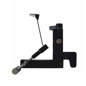
| Portable Keratometer Features:
Ultra-small corneal curvature tester, is mainly used to measure the corneal curvature radius and diopter, wireless output print data.
Technical Specifications:
| Model |
SW-100 |
| Measuring Range |
6.5mm~9.5mm: ±0.05mm: 0.01mm |
| Precision |
± 0.05mm |
| Resolution of Curvature Radius of Cornea |
0.01mm |
| Single Measuring Time |
0.03s |
| Output |
Wireless Infrared Thermal Printer |
| Can observe the eye directly through the screen. |
| Weight |
0.5Kg(with batteries) |
| Dimension |
240mmx90mmx60mm |
| Power |
500mW+15% |
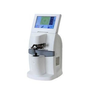
| Features:
- 7.0-inch color LCD touch panel.
- Hartman sensor with 108 multiple measure points.
- Green measurement light beam.
- Dyeing lens, sunglasses measured easily.
Technical Specifications:
- Sphere lenses: 0~±25D
- Cylinder lenses: 0~±10D
- Cylinder Axis Angle: 0°~180°
- Add.:0~10D
- Power supply: 100~240V, 50/60HZ, 30W
- Weight: 5kg
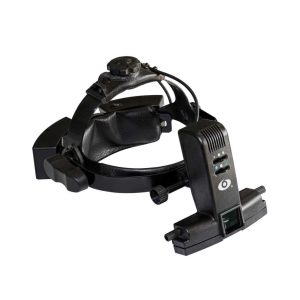
| Ophthalmoscope Features:
- LED bulb(low-temperature,low-pressure,high-brightness) with long lifetime to make the illumination uniformly and bright. The illumination brightness can be adjusted continuously.
- Imaging clearly,strong dimensional sense and large field of view range.The main body part of this device adopts engineering plastics and is designed to be compact and exquisite.
- Super lightweight design, reduce fatigue,operation is very convenient.
- The portable power supply is suitable for surgery, ward round and consultation.
- This device is equipped with color filter,teaching mirror and sclera depressor to be more handy in operation.
Technical Specifications:
| Pupilary Adjustment |
52mm~74mm adjustable |
| Intensity of Illumination |
continuous adjustment with the maximum intensity no less than 500Lx |
| Headband |
520~640mm in circumference, 85~125mm in depth |
| Light Spots |
Big spot, middle spot and small spot |
| Filters |
Red-free, white and cobalt blue |
| Light Source |
LED light |
| Source of Power |
Li+ battery, DC 7.4V |
| AC 100~240V, 50/60Hz |
| Input Power |
Battery charger: 15 VA |
| AC charger: 50VA |
| Dimension of Main Part |
142mm × 48mm × 128mm(long×breadth×high) |
| Weight of Main Part |
230g |
|


Reviews
There are no reviews yet.