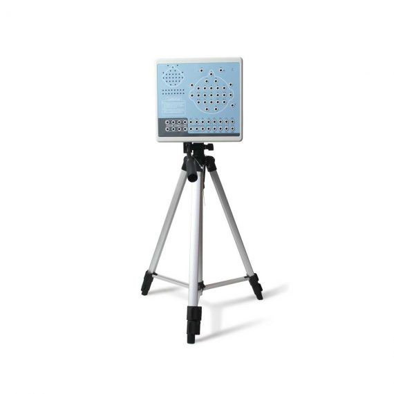
Contec KT88-3200 Electroencephalogram (EEG) Machine
$1,320.00
Shipped From Abroad
KT88-3200 Digital Brain Electric Activity Mapping collects EEG signal with electrodes, via integrated amplification, A/D transformation, PC auto-analysis, FFT, to form an electroencephalogram that displays with color depth. The product is applicable for checking such diseases as epilepsy, intracranial inflammation, cerebrovascular diseases, and brain tumors.
Delivery & Availability:
Typically 5-7 working days – excluding furniture and heavy/bulky equipment. Please contact us for further information.
Description
KT88-3200 Digital Brain Electric Activity Mapping collects EEG signal with electrodes, via integrated amplification, A/D transformation, PC auto-analysis, FFT, to form electroencephalogram that displays with color depth. The product is applicable for checking such diseases as epilepsy, intracranial inflammation, cerebrovascular diseases and brain tumors.
Features:
- Method of 10/20 electrodes placement under international standard system, leads can be changed during replaying. Support different types of combined leads during sampling.
- Adopt bioelectricity amplifier for distilling brainwave, continuous recording time can be up to 24 hours, integrated full-automatic calibration system.
- Powerful playback function: amplitude and display speed are adjustable. The special subdividing time line divides the waveform in one second into 5 parts, which is easy for doctors to look over the waveform.
- Digital filter system can be set as required, providing different window types
- EEG signal clipping function, analyze and store any section of EEG wave, and select several waveform segments for automatically analyzing and distilling to different parameters.
- Electronic frequency ruler, convenient to measure the basic information of any appointed EEG waveform. With partial enlarging window, accurate measurement of EEG period, amplitude and frequency, which can be adjusted according to personnel judgment.
- Mark EEG wave under the events of opening eyes, closing eyes and flashing with different colors, and user-defined events can be added, waveform color for evoked event can also be set freely, ensures that the waveform in corresponding time can be rapidly found by event name during case playback.
- Powerful automatic analysis function, can carry through the power spectrum analysis and pathologic wave detection for appointed waveform. Many graphs can be displayed in the same screen, including kinds of BEAM, numerical BEAM, compressed spectrum graph, trend graph, and so on.
- Professional isolation transformer, dual power supply isolation system and optoelectronic data transmission to ensure security. Use USB interface to transmit data which just need to be inserted.
- Multifunctional flash stimulator of USB interface, and flashing can be controlled manually or automatically. A flash stimulation scheme can be set and performed in the process of sampling.
- Perfect case management function, provides many means for research and quick statistic information; convenient case export and import function, and stores with MO or CD-RW disk, which is easy for data research.
- Integrative image and character report, report can be edited in mode and switched to Word document.
- Case files can be transformed into EDF and BDF data format, convenient for data exchange, academic exchange and further analysis.
- System parameters and display modes can be set as required, which meets different user’s requirement.
- Add marks and annotations to the waveform designated, which can rapidly find the waveform in that time by marks.
- Optional video function: USB camera is easy to install, convenient to use and exact to record. With flexible playback function, which can browse the waveform of any time along with the corresponding isochronous sampled image.
- SpO2 function is optional.
Technical Specifications:
- 32 channels of EEG
- Sampling rate: 200 dots/s
- Accuracy: 12bit
- Input impedance: ≥10MΩ
- Patient leak current: < 10µA
- Noise level: ≤5µVp-p
- CMRR: ≥90dB
- Magnification multiple: 10000
- Filter constant: all digital and free enactment
- Display speed(paper speed): 5, 10, 15, 30, 60, 120 mm/s
- Amplitude: 1, 1.5, 2, 3, 5, 7.5, 10, 12, 15, 20, 30, 50 mm/50µV
- Playback speed:1 time, 2 times, 3 times, 10 times, 20 times, 40 times, 60 times
- 50Hz interference suppression: ≥30dB
- Safety type:Class II, type BF applied part
Accesories:
- EEG lead
- EEG electrode
- Data line
- Headgear
- Strobe light
- Strobe light power supply
- PC software CD
- User Manual
- Earth wire
- EEG bracket
- Aluminum block
Review(1)
Quick Comparison
| Contec KT88-3200 Electroencephalogram (EEG) Machine remove | DrGem Diamond All-In-One Digital X-ray Machine remove | ASPEL AsCARD Coral PC Based ECG Machine remove | DrGem Floor Mounted Analogue X-ray remove | DrGem GXR-SD 400mA Floor Mounted Digital X-ray remove | Sonoscape S22 Ultrasound Machine remove | |
|---|---|---|---|---|---|---|
| Name | Contec KT88-3200 Electroencephalogram (EEG) Machine remove | DrGem Diamond All-In-One Digital X-ray Machine remove | ASPEL AsCARD Coral PC Based ECG Machine remove | DrGem Floor Mounted Analogue X-ray remove | DrGem GXR-SD 400mA Floor Mounted Digital X-ray remove | Sonoscape S22 Ultrasound Machine remove |
| Image |  |  |  |  |  |  |
| SKU | SF1033560084-249 | SF1033560074-3 | SF1033560075-11 | SF1033560074-6 | SF1033560074-5 | SF1033560012-3 |
| Rating | ||||||
| Price | $1,320.00 |
| $486.00 |
|
| $9,350.00 |
| Stock | ||||||
| Availability | ||||||
| Add to cart | ||||||
| Description | Shipped From Abroad
KT88-3200 Digital Brain Electric Activity Mapping collects EEG signal with electrodes, via integrated amplification, A/D transformation, PC auto-analysis, FFT, to form an electroencephalogram that displays with color depth. The product is applicable for checking such diseases as epilepsy, intracranial inflammation, cerebrovascular diseases, and brain tumors.
| Shipped from Abroad DrGem Diamond All-In-One Digital X-ray Machine is a fully automatic digital radiography system providing state-of-the-art image quality, image processing and user interface. With a wide selection of anatomical studies on the imaging software, DIAMOND automatically sets up the x-ray generator’s preprogrammed exposure technique settings, motorized radiographic stand positioning, x-ray collimation and post-image processing for the selected study. Specifically designed to increase workflow, this fully digital system offers convenient auto-positioning and advanced image processing to achieve big performance with little effort. Delivery & Availability: Typically 21 working days – excluding furniture and heavy/bulky equipment. Please contact us for further information. | Shipped from Abroad AsCARD Coral electrocardiograph is a 3-, 6-, 12-channel ECG equipped with CardioTEKA software allows transmission of full 12 ECG leads to the user PC through USB interface. It is intended for carrying out ECG examinations in adults and pediatric patients in all types of health care centres. ECG procedures can be performed by qualified personnel only. AsCARD Coral can cooperate also with CardioTEST system as 12-channel ECG device allows transmission of full 12 ECG leads to the user PC through USB interface. Delivery & Availability: Typically 10 working days – excluding furniture and heavy/bulky equipment. Please contact us for further information. | In Stock GXR Analogue X-ray system matches with a radiographic room which perfectly fits your workow and can be easily upgraded to DR system with the help of DR interface and PC interface in GXR generator as well as Bucky suitable to Flat Panel Detector. GXR X-ray system is equipped with a high frequency X-ray generator which consistently produces high quality radiograph in favor of high quality X-ray output with a very small kV ripple and accurate mA and mAs. GXR X-ray system is designed to provide convenience to operator and comfort to patient. Delivery & Availability: Typically 21 working days – excluding furniture and heavy/bulky equipment. Please contact us for further information. | In Stock The GXR-SD Digital X-ray is a diagnostic digital radiography system that provides reliable high quality digital radiographic images with a reduced dose. The GXR-SD DR systems offer comprehensive digital solutions to all radiography needs, featuring ACQUIDR digital imaging system with stationary or portable digital flat-panel detectors as well as reliable high-frequency x-ray generators that are known worldwide for their excellent performance, lifetime and stability. Patient tables and wall stands are also offered. Delivery & Availability: Typically 21 working days – excluding furniture and heavy/bulky equipment. Please contact us for further information. | Shipped from Abroad As SonoScape steps forward to add value and efficiency to ultrasound, the latest S22 was designed in a user-friendly platform to address current and future demanding needs. It represents an excellent mix in performance and price. Delivery & Availability: Typically 5-7 working days – excluding furniture and heavy/bulky equipment. Please contact us for further information. |
| Content | KT88-3200 Digital Brain Electric Activity Mapping collects EEG signal with electrodes, via integrated amplification, A/D transformation, PC auto-analysis, FFT, to form electroencephalogram that displays with color depth. The product is applicable for checking such diseases as epilepsy, intracranial inflammation, cerebrovascular diseases and brain tumors.
Features:
Technical Specifications:
| DrGem Diamond All-In-One Digital X-ray Machine is a fully automatic digital radiography system providing state-of-the-art image quality, image processing and user interface. With a wide selection of anatomical studies on the imaging software, DIAMOND automatically sets up the x-ray generator’s pre-programmed exposure technique settings, motorized radiographic stand positioning, x-ray collimation and post-image processing for the selected study. Specifically designed to increase workflow, this fully digital system offers convenient auto-positioning and advanced image processing to achieve big performance with little effort.
Features of DrGem Diamond All-In-One Digital X-ray Machine:
Outstanding Image Quality -
Digital radiography via at panel detector improves your workflow, exam speed and comfort with efficiency. Digital at panel detector with Csl screen provides excellent spatial resolution, MTF, DQE and stability based on ne pixel pitch. A 3-field ion-chamber is provided for AEC function.
Automatic Collimation –
Automatic x-ray eld size control of the motorized collimator corresponds to dierent SIDs. Includes user adjustable lamp timer with on/oswitch.
Automatic Positioning –
Click Here To Download Catalogue |
AsCARD Coral electrocardiograph is a 3-, 6-, 12-channel ECG equipped with CardioTEKA software allows transmission of full 12 ECG leads to the user PC through USB interface. It is intended for carrying out ECG examinations in adults and pediatric patients in all types of health care centres. ECG procedures can be performed by qualified personnel only. AsCARD Coral can cooperate also with CardioTEST system as 12-channel ECG device allows transmission of full 12 ECG leads to the user PC through USB interface.
Technical Specification:
Click Here To Download Catalogue | DrGem GXR Floor Mounted Analogue X-ray system matches with a radiographic room which perfectly fits your workflow and can be easily upgraded to DR system with the help of DR interface and PC interface in GXR generator as well as Bucky suitable to Flat Panel Detector. GXR (Analogue X-ray)system is equipped with a high frequency X-ray generator which consistently produces high quality radiograph in favor of high quality X-ray output with a very small kV ripple and accurate mA and mAs. GXR (Analogue X-ray) system is designed to provide convenience to operator and comfort to patient.
Features of DrGem GXR Floor Mounted Analogue X-ray:
Click Here To Download Catalogue | DrGem GXR-SD 400mA Floor Mounted Digital X-ray system matches with a radiographic room which perfectly fits your workow and can be easily upgraded to DR system with the help of DR interface and PC interface in GXR generator as well as Bucky suitable to Flat Panel Detector. GXR X-ray system is equipped with a high frequency X-ray generator which consistently produces high quality radiograph in favor of high quality X-ray output with a very small kV ripple and accurate mA and mAs. GXR X-ray system is designed to provide convenience to operator and comfort to patient
Features of DrGem GXR-SD 400mA Floor Mounted Digital X-ray:
Click Here To Download Catalogue | DETAILS
As SonoScape steps forward to add value and efficiency to ultrasound, the latest S22 was designed in a user-friendly platform to address current and future demanding needs. It represents an excellent mix in performance and price.
S22, is a shared service ultrasound system with a slim and elegant package that has combined mobility with utility to fit in specific clinical situations including emergency department, ICU, operating room and so on. Furthermore, its ergonomic design, easy operating and flexible data management will give you a memorable experience.
SPECIFICATION
• Large high-resolution widescreen LED
• Sensitive touch screen
• Four transducer sockets plus one socket for pencil probe
• A comprehensive selection of probes: linear, Convex, Micro-convex, Volumetric, Endocavity, Bi-plane, Phased Array, TEE, Intraoperative, Pencil
• Premium application technology: 4D, μ-scan speckle reduction, compound imaging, Pulse Inversion Harmonic Imaging, Color M-Mode, Steer M-Mode, PDI, TDI, Real-time Panoramic Imaging, Trapezoid Imaging, Auto-IMT…
• Full patient database and image management solutions: DICOM 3.0, AVI/JPG, USB 2.0, HDD, DVD, PDF report
• Multi-Language Input Keyboard
• Built-in battery
Click Here To Download Catalogue |
| Weight | N/A | N/A | N/A | N/A | N/A | N/A |
| Dimensions | N/A | N/A | N/A | N/A | N/A | N/A |
| Additional information |


Mbah Prince
Cost of KT88-3200