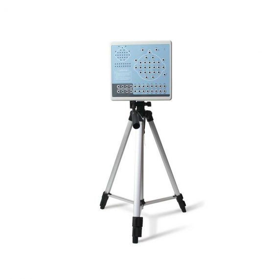
Contec KT88-3200 Electroencephalogram (EEG) Machine
Ask for Price$0.00
Shipped From Abroad
KT88-3200 Digital Brain Electric Activity Mapping collects EEG signal with electrodes, via integrated amplification, A/D transformation, PC auto-analysis, FFT, to form an electroencephalogram that displays with color depth. The product is applicable for checking such diseases as epilepsy, intracranial inflammation, cerebrovascular diseases, and brain tumors.
Delivery & Availability:
Typically 5-7 working days – excluding furniture and heavy/bulky equipment. Please contact us for further information.
Description
KT88-3200 Digital Brain Electric Activity Mapping collects EEG signal with electrodes, via integrated amplification, A/D transformation, PC auto-analysis, FFT, to form electroencephalogram that displays with color depth. The product is applicable for checking such diseases as epilepsy, intracranial inflammation, cerebrovascular diseases and brain tumors.
Features:
- Method of 10/20 electrodes placement under international standard system, leads can be changed during replaying. Support different types of combined leads during sampling.
- Adopt bioelectricity amplifier for distilling brainwave, continuous recording time can be up to 24 hours, integrated full-automatic calibration system.
- Powerful playback function: amplitude and display speed are adjustable. The special subdividing time line divides the waveform in one second into 5 parts, which is easy for doctors to look over the waveform.
- Digital filter system can be set as required, providing different window types
- EEG signal clipping function, analyze and store any section of EEG wave, and select several waveform segments for automatically analyzing and distilling to different parameters.
- Electronic frequency ruler, convenient to measure the basic information of any appointed EEG waveform. With partial enlarging window, accurate measurement of EEG period, amplitude and frequency, which can be adjusted according to personnel judgment.
- Mark EEG wave under the events of opening eyes, closing eyes and flashing with different colors, and user-defined events can be added, waveform color for evoked event can also be set freely, ensures that the waveform in corresponding time can be rapidly found by event name during case playback.
- Powerful automatic analysis function, can carry through the power spectrum analysis and pathologic wave detection for appointed waveform. Many graphs can be displayed in the same screen, including kinds of BEAM, numerical BEAM, compressed spectrum graph, trend graph, and so on.
- Professional isolation transformer, dual power supply isolation system and optoelectronic data transmission to ensure security. Use USB interface to transmit data which just need to be inserted.
- Multifunctional flash stimulator of USB interface, and flashing can be controlled manually or automatically. A flash stimulation scheme can be set and performed in the process of sampling.
- Perfect case management function, provides many means for research and quick statistic information; convenient case export and import function, and stores with MO or CD-RW disk, which is easy for data research.
- Integrative image and character report, report can be edited in mode and switched to Word document.
- Case files can be transformed into EDF and BDF data format, convenient for data exchange, academic exchange and further analysis.
- System parameters and display modes can be set as required, which meets different user’s requirement.
- Add marks and annotations to the waveform designated, which can rapidly find the waveform in that time by marks.
- Optional video function: USB camera is easy to install, convenient to use and exact to record. With flexible playback function, which can browse the waveform of any time along with the corresponding isochronous sampled image.
- SpO2 function is optional.
Technical Specifications:
- 32 channels of EEG
- Sampling rate: 200 dots/s
- Accuracy: 12bit
- Input impedance: ≥10MΩ
- Patient leak current: < 10µA
- Noise level: ≤5µVp-p
- CMRR: ≥90dB
- Magnification multiple: 10000
- Filter constant: all digital and free enactment
- Display speed(paper speed): 5, 10, 15, 30, 60, 120 mm/s
- Amplitude: 1, 1.5, 2, 3, 5, 7.5, 10, 12, 15, 20, 30, 50 mm/50µV
- Playback speed:1 time, 2 times, 3 times, 10 times, 20 times, 40 times, 60 times
- 50Hz interference suppression: ≥30dB
- Safety type:Class II, type BF applied part
Accesories:
- EEG lead
- EEG electrode
- Data line
- Headgear
- Strobe light
- Strobe light power supply
- PC software CD
- User Manual
- Earth wire
- EEG bracket
- Aluminum block
Review(1)
Quick Comparison
| Settings | Contec KT88-3200 Electroencephalogram (EEG) Machine remove | Sonoscape S11 Ultrasound Machine remove | Sonoscape P15 Ultrasound Machine With Four Probes remove | Sonoscape E2 Ultrasound Machine remove | Sonoscape P20 Ultrasound Machine remove | Sonoscape P10 Ultrasound Machine remove |
|---|---|---|---|---|---|---|
| Name | Contec KT88-3200 Electroencephalogram (EEG) Machine remove | Sonoscape S11 Ultrasound Machine remove | Sonoscape P15 Ultrasound Machine With Four Probes remove | Sonoscape E2 Ultrasound Machine remove | Sonoscape P20 Ultrasound Machine remove | Sonoscape P10 Ultrasound Machine remove |
| Image | 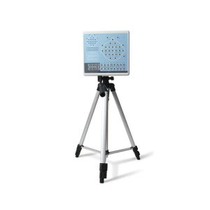 |  |  |  |  |  |
| SKU | SF1033560084-249 | SF1033560012-1 | SF1033560012-8 | SF1033560012-17 | SF1033560012-9 | SF1033560012-7 |
| Rating | ||||||
| Price | Ask for Price | $6,950.00 | $13,900.00 | $4,300.00 | Ask for Price | Ask for Price |
| Stock | ||||||
| Availability | ||||||
| Add to cart | ||||||
| Description | Shipped From Abroad
KT88-3200 Digital Brain Electric Activity Mapping collects EEG signal with electrodes, via integrated amplification, A/D transformation, PC auto-analysis, FFT, to form an electroencephalogram that displays with color depth. The product is applicable for checking such diseases as epilepsy, intracranial inflammation, cerebrovascular diseases, and brain tumors.
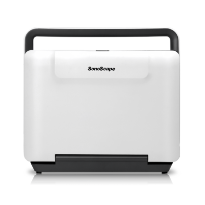
| In Stock A Value Choice beyond Your Expectation. SonoScape’s trolley color Doppler system S11 redefines price and performance with practical design. The S11 will go beyond your expectations but not your budget. Delivery & Availability: Typically 2 working days – excluding furniture and heavy/bulky equipment. Please contact us for further information. | In Stock A feature-rich system inheriting the Wi-Sono high-end platform, the P15 uses an array of advanced tools to help enhance the image quality. It's a cost-effective, simplified console with an intuitive user interface and multiple intelligent functions. Delivery & Availability: Typically 2 working days – excluding furniture and heavy/bulky equipment. Please contact us for further information. | Shipped from Abroad Sonoscape E2 portable ultrasound machine is a color Doppler ultrasound system that reaches beyond your expectations due to its compact and fashionable appearance. It fulfills GI, OB/GYN, Cardiac and POC applications to fit your routine scanning needs while its color mode will help you for more accurate and efficient diagnosis of lesions. E2 provides a wide range of applications to assist users with routine scanning. E2 provides automatic calculations to enhance your diagnostic confidence and save you time for patient communication. Delivery & Availability: Typically 14 working days – excluding furniture and heavy/bulky equipment. Please contact us for further information. | Shipped from Abroad Incorporating innovative technologies, P20’s user-friendly design with a simple operation panel, intuitive user interface and a variety of intelligent auxiliary scanning tools, will significantly improve your daily examination experience. Besides general imaging applications, P20 has entitled with diagnostic 4D technology which has an extraordinary performance in obstetrics and gynecology applications. Delivery & Availability: Typically 5-7 working days – excluding furniture and heavy/bulky equipment. Please contact us for further information. | Shipped from Abroad The P10 color Doppler ultrasound system is a new generation product from SonoScape. It is designed to give high quality images, rich probe configurations, various clinical tools and automatic analysis software to provide you with comprehensive solutions for your growing demand for clinical applications. Delivery & Availability: Typically 5-7 working days – excluding furniture and heavy/bulky equipment. Please contact us for further information. |
| Content | KT88-3200 Digital Brain Electric Activity Mapping collects EEG signal with electrodes, via integrated amplification, A/D transformation, PC auto-analysis, FFT, to form electroencephalogram that displays with color depth. The product is applicable for checking such diseases as epilepsy, intracranial inflammation, cerebrovascular diseases and brain tumors.
Features:
Technical Specifications:
| DETAILS
SonoScape’s trolley colour Doppler system S11 redefines price and performance with practical design. The S11 will go beyond your expectations but not your budget. As an easy-to-use ultrasound system, the S11 is integrated with a new software platform, especially optimized for a smooth workflow and convenient operation. The system speeds up the exam process and makes file management easier.
SPECIFICATION
- 15-inch high definition LCD monitor with articulating arm
- Compact and agile trolley design
- 3 active transducer sockets available for a wide range of applications
- Duplex, Color Doppler, DPI, PW Doppler, tissue harmonic imaging, μ-scan speckle reduction imaging, compound imaging, trapezoidal imaging
- Customized settings based on your own working style
- Full patient database and image management solutions
Click Here To Download Catalogue | DETAILS
Super Wide-bandwidth Platform
Inheriting Wi-sono's ultra-wide system platform and with the advanced probe technology, high-resolution and deep penetration images are provided for precision medicine.
Spatial Compound Imaging
Spatial Compound Imaging utilizes several lines of sight for optimal contrast resolution, speckle reduction and border detection, with which P15 is ideal for superficial and abdominal imaging with better clarity and improved continuity of structures.
μ-Scan+
The new generation μ-Scan imaging technology gives you better image quality by reducing noise, improving signal strength and improving visualization.
Dynamic Color
Dynamic color improves upon already existing color Doppler technologies for a clearer capture of color flow and detailed visualization of even tiny veins with lower velocities.
Real-time Panoramic
With real-time panoramic, you can acquire an extended field of view for large organs or long vessels for easy measurement and diagnostic efficiency. Accomplished in real-time for the convenience of the sonographers, any mistake can also be easily back tracked and corrected without interrupting the scan.
3D/4D
Outstanding volume performance with speed and convenience makes P15 outshine others on volume imaging.
Tissue Doppler Imaging
Tissue Doppler Imaging allows clinical doctors to quantitatively evaluate local myocardial movements and functions, facilitating them with the ability to analyze and compare the motions of the different parts of the patient's heart.
Auto IMT
Quick measurement of intra-media vessel thickness ensures good reproducibility and high diagnostic efficiency.
Click Here To Download Catalogue | SONOSCAPE E2 DETAILS
Auto Image Optimization
A portable ultrasound machine with the press of a button, the image is automatically adjusted and optimized, saving you time with parameter adjustments. Additionally, with Auto Focus on, the focus area follows the depth of the ROI box as it is moved in the scanning field, providing users with excellent image quality in the desired area of interest.
Automated Calculation
Auto IMT is used when determining the level of vascular sclerosis present in the patient by automatically tracing the thickness of the carotid vessels.
Auto trace provides users sensitive and accurate wave tracing, avoiding the error of manual trace and giving out calculation result in no time
In-Build Battery pack
This portable ultrasound machine was equipped with an in-build battery pack which enable the user to perform image scanning when AC power is not available.
Click Here To Download Catalogue | DETAILS
Upgraded Images with More Clarity
SonoScape never stops making progress in improving the image quality of its ultrasound products to enhance the confidence of diagnosis for doctors. With extraordinary images provided by P20, the anatomy structures are clearer than ever.
C-Xlasto Imaging
With C-xlasto Imaging, P20 enables comprehensive quantitative elastic analysis. Meanwhile, C-xlasto on P20 is supported by linear, convex and transvaginal probes, to ensure good reproducibility and highly consistent quantitative elastic results.
S-Live
S-Live allows for detailed visualization of subtle anatomical features, thereby enabling intuitive diagnosis with real-time 3D images and enriching patient communication.
Pelvic Floor 4D
Transperineal 4D pelvic floor ultrasound can provide useful clinical values in assessing the vaginal delivery impact on the female anterior compartment, judging whether the pelvic organs are prolapsed or not and the extent, determining if the pelvic muscles were torn accurately.
Anatomic M Mode
Anatomic M Mode helps you observe the myocardial motion at different phases by freely placing sample lines. It accurately measures the myocardial thickness and the heart size of even difficult patients and supports the myocardial function and LV wall-motion assessment.
Tissue Doppler Imaging
P20 is endowed with Tissue Doppler Imaging which provides velocities and other clinical information on myocardial functions, facilitating clinical doctors with the ability to analyze and compare the motions of different parts of the patient's heart.
Click Here To Download Catalogue | DETAILS
B + Compound
B + Compound utilizes several lines of sight for optimal contrast resolution, speckle reduction and border detection, with which P10 is ideal for superficial and abdominal imaging with better clarity and improved continuity of structures.
μ-Scan
The new generation μ-Scan imaging technology gives you better image quality by reducing noise, improving signal strength and improving visualization.
P10 offers a comprehensive selection of electronic probes to maximize its capabilities to meet a wide range of applications including abdomen, pediatric, OB/GYN, cardiovascular, musculoskeletal, etc. The advanced probe technologies also effectively enhance the image quality and confidence in reaching clinical diagnoses, even in difficult patients.
Convex Probe 3C-A
Ideal for an abundant of application such as abdomen, gynecology, obstetrics, urology and even abdomen biopsy.
Linear Probe L741
This linear probe is designed to satisfy vascular, breast, thyroid, and other small parts diagnosis, and its adjustable parameters could also present users a clear view of MSK and deep vessels.
Phase Array Probe 3P-A
For the purpose of adult and pediatric cardiology and emergency, the phase array probe provides elaborate presets for different exam modes, even for difficult patients.
Intracavitary Probe 6V1
Intracavitary probe could face application of gynecology, urology, prostate, and its temperature detection technology not only protects the patient but also extends the service life.
Click Here To Download Catalogue |
| Weight | N/A | N/A | N/A | N/A | N/A | N/A |
| Dimensions | N/A | N/A | N/A | N/A | N/A | N/A |
| Additional information |


Mbah Prince
Cost of KT88-3200