CV45/CV40 Ultrasound System
$0.00
Shipped From Abroad
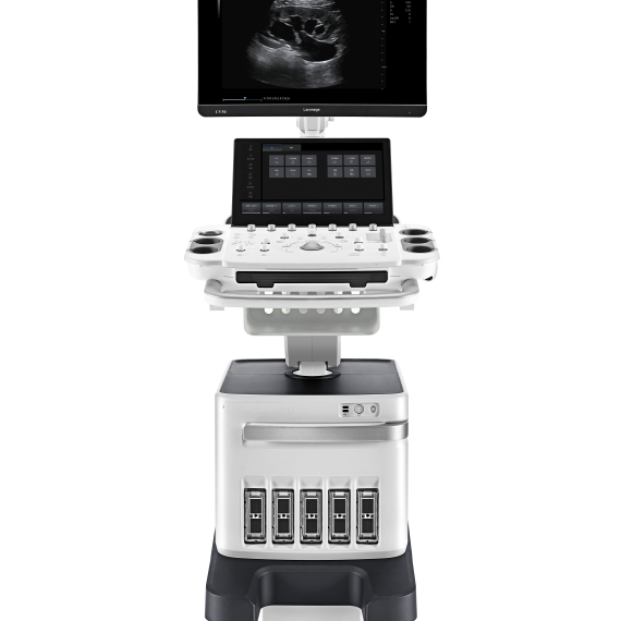
The CV45/CV40 (also known as CU40) is a compact, fully digital B‑mode and spectral Doppler ultrasound system offering high-resolution imaging, intuitive workflow, and versatile clinical presets ideal for general diagnostics in human and veterinary practice.
Typically 10-21 working days – excluding furniture and heavy/bulky equipment. Please contact us for further information.
Description
Key Features
-
High‑Resolution LCD (10″ – 12″ adjustable angle display) – clear visuals with anti‑glare and enhanced brightness
-
Digital Beamforming + Tissue Harmonic Imaging (THI) – for sharper contrast and deeper penetration
-
Speckle Reduction & Compound Imaging – improves image clarity with better edge definition
-
One‑Button Optimization & Biopsy‑Guide Overlay – simplifies setup and assists interventional procedures
-
Dual Probe Ports – supports multiple transducers simultaneously
-
Back‑lit Control Panel & User‑Defined Keys – ergonomic and adaptable for varied environments
-
Portable Power & Connectivity – built‑in battery, DVI/USB/DICOM support, image/video/patient data storage & export
-
Extensive Clinical Presets – packages for general, OB/GYN, cardiac, MSK, urology, small parts, orthopedics
Specifications
| Specification | CV40 / CU40 |
|---|---|
| Display | 10″–12″ non‑interlaced, adjustable angle |
| Imaging Technology | Digital beamforming, THI, compounding |
| Modes | B‑mode, M‑mode, Doppler (spectral, color optional in C6) |
| Probe Connectors | 2 |
| TGC Control | 8 segments |
| Storage | Internal HDD + image/video export formats |
| Connectivity | USB, DVI video, wireless DICOM optional |
| Power | Integrated long-life battery |
| Control Panel | Back-lit keys, user-defined soft keys |
| Ultrasound Presets | General, OB/GYN, Cardiac, Urology, MSK, Small parts, Orthopedics |
| Interventional Aids | Biopsy and lithotripsy guide overlays |
| Video Capture | Cine loop with thumbnail clip viewer |
Quick Comparison
| CV45/CV40 Ultrasound System remove | Sonoscape S8 Exp Portable Ultrasound remove | DrGem Ceiling Mounted Digital X-ray remove | Single X-Ray Viewing Box remove | Sonoscape P15 Ultrasound Machine With Four Probes remove | Sonoscape E2 Ultrasound Machine remove | |||||||||||||||||||||||||||
|---|---|---|---|---|---|---|---|---|---|---|---|---|---|---|---|---|---|---|---|---|---|---|---|---|---|---|---|---|---|---|---|---|
| Name | CV45/CV40 Ultrasound System remove | Sonoscape S8 Exp Portable Ultrasound remove | DrGem Ceiling Mounted Digital X-ray remove | Single X-Ray Viewing Box remove | Sonoscape P15 Ultrasound Machine With Four Probes remove | Sonoscape E2 Ultrasound Machine remove | ||||||||||||||||||||||||||
| Image |  |  |  |  |  |  | ||||||||||||||||||||||||||
| SKU | SF1033560012-15 | SF1033560074-4 | SF1033560084-203 | SF1033560012-8 | SF1033560012-17 | |||||||||||||||||||||||||||
| Rating | ||||||||||||||||||||||||||||||||
| Price |
| $9,350.00 |
| $95.20 | $13,900.00 | $5,500.00 | ||||||||||||||||||||||||||
| Stock | ||||||||||||||||||||||||||||||||
| Availability | ||||||||||||||||||||||||||||||||
| Add to cart | ||||||||||||||||||||||||||||||||
| Description | Shipped From Abroad
The CV45/CV40 (also known as CU40) is a compact, fully digital B‑mode and spectral Doppler ultrasound system offering high-resolution imaging, intuitive workflow, and versatile clinical presets ideal for general diagnostics in human and veterinary practice.
Delivery & Availability:
Typically 10-21 working days – excluding furniture and heavy/bulky equipment. Please contact us for further information.
| Shipped from Abroad With ultra-modern innovative design and the clinically-proven technologies, S8 Exp is portable ultrasound scanner well equipped as a low-physical-effort and enhanced-image-quality ultrasound scanner, which not only provides optimized solutions for versatile applications, but does help to improve the user-experience for both routine and non-traditional challenges. Delivery & Availability: Typically 5-7 working days – excluding furniture and heavy/bulky equipment. Please contact us for further information. | In Stock The GXR-SD is a diagnostic digital radiography system that provides reliable high quality digital radiographic images with a reduced dose. The GXR-SD DR systems offer comprehensive digital solutions to all radiography needs, featuring ACQUIDR digital imaging system with stationary or portable digital flat-panel detectors as well as reliable high-frequency x-ray generators that are known worldwide for their excellent performance, lifetime and stability. Patient tables and wall stands are also offered. Delivery & Availability: Typically 21 working days – excluding furniture and heavy/bulky equipment. Please contact us for further information. | In stock
| In Stock A feature-rich system inheriting the Wi-Sono high-end platform, the P15 uses an array of advanced tools to help enhance the image quality. It's a cost-effective, simplified console with an intuitive user interface and multiple intelligent functions. Delivery & Availability: Typically 2 working days – excluding furniture and heavy/bulky equipment. Please contact us for further information. | Shipped from Abroad Sonoscape E2 portable ultrasound machine is a color Doppler ultrasound system that reaches beyond your expectations due to its compact and fashionable appearance. It fulfills GI, OB/GYN, Cardiac and POC applications to fit your routine scanning needs while its color mode will help you for more accurate and efficient diagnosis of lesions. E2 provides a wide range of applications to assist users with routine scanning. E2 provides automatic calculations to enhance your diagnostic confidence and save you time for patient communication. Delivery & Availability: Typically 14 working days – excluding furniture and heavy/bulky equipment. Please contact us for further information. | ||||||||||||||||||||||||||
| Content | Key Features
Specifications
| Sonoscape S8 Exp Portable Ultrasound scannerDETAILS Agile and Versatile With ultra-modern innovative design and the clinically-proven technologies, S8 Exp Portable Ultrasound scanner is well equipped as a low-physical-effort and enhanced-image-quality ultrasound scanner, which not only provides optimized solutions for versatile applications but does help to improve the user experience for both routine and non-traditional challenges. Working with S8 Exp, it will trigger your unlimited reverie and endow you with endless charm. Carrying forward the classical design of SonoScape's portable ultrasound products, S8 Exp successfully combines the best ergonomics, attractive design and ease of use. This charismatic identity is also enhanced by a sophisticated color palette—with sedate grey as its interior paint color and pearl white as exterior cover, S8 Exp reveals a style of aristocrat and strong character among SonoScape's ultrasound systems. Workflow The S8 Exp is a portable ultrasound scanner that adapts to your workflow, whether you are in the consulting room, at the bedside, or at a remote location. With easy-to-use new platform designed for sonographers' needs and full connection interfaces for easy connectivity and data sharing, S8 Exp leads to improved user comfort and clinical outcome as well as patient throughput and working efficiency. Powerful Platform Embedded with SonoScape's core imaging technologies such as μ-scan, PHI and Spatial Compound, S8 Exp boasts exceptional 2D image, sensitive spectral, Color and Power Doppler, displaying well-defined anatomy and pathology and facilitating a highly optimized diagnostic user environment for conclusive diagnoses. Besides, S8 Exp offers a comprehensive selection of electronic probes to maximally extend its capabilities to meet a wide range of applications including the abdomen, pediatric, OB/GYN, cardiovascular, musculoskeletal, etc. The advanced probe technologies also effectively enhance the image quality and confidence in reaching clinical diagnoses even in difficult patients.Click Here To Download Catalogue | DrGem Ceiling Mounted Digital X-ray is a diagnostic digital radiography system that provides reliable high quality digital radiographic images with a reduced dose. The GXR-SD DR systems offer comprehensive digital solutions to all radiography needs, featuring ACQUIDR digital imaging system with stationary or portable digital flat-panel detectors as well as reliable high-frequency x-ray generators that are known worldwide for their excellent performance, lifetime and stability. Patient tables and wall stands are also offered.
Features:
Click Here To Download Catalogue |
| DETAILS
Super Wide-bandwidth Platform
Inheriting Wi-sono's ultra-wide system platform and with the advanced probe technology, high-resolution and deep penetration images are provided for precision medicine.
Spatial Compound Imaging
Spatial Compound Imaging utilizes several lines of sight for optimal contrast resolution, speckle reduction and border detection, with which P15 is ideal for superficial and abdominal imaging with better clarity and improved continuity of structures.
μ-Scan+
The new generation μ-Scan imaging technology gives you better image quality by reducing noise, improving signal strength and improving visualization.
Dynamic Color
Dynamic color improves upon already existing color Doppler technologies for a clearer capture of color flow and detailed visualization of even tiny veins with lower velocities.
Real-time Panoramic
With real-time panoramic, you can acquire an extended field of view for large organs or long vessels for easy measurement and diagnostic efficiency. Accomplished in real-time for the convenience of the sonographers, any mistake can also be easily back tracked and corrected without interrupting the scan.
3D/4D
Outstanding volume performance with speed and convenience makes P15 outshine others on volume imaging.
Tissue Doppler Imaging
Tissue Doppler Imaging allows clinical doctors to quantitatively evaluate local myocardial movements and functions, facilitating them with the ability to analyze and compare the motions of the different parts of the patient's heart.
Auto IMT
Quick measurement of intra-media vessel thickness ensures good reproducibility and high diagnostic efficiency.
Click Here To Download Catalogue | SONOSCAPE E2 DETAILS
Auto Image Optimization
A portable ultrasound machine with the press of a button, the image is automatically adjusted and optimized, saving you time with parameter adjustments. Additionally, with Auto Focus on, the focus area follows the depth of the ROI box as it is moved in the scanning field, providing users with excellent image quality in the desired area of interest.
Automated Calculation
Auto IMT is used when determining the level of vascular sclerosis present in the patient by automatically tracing the thickness of the carotid vessels.
Auto trace provides users sensitive and accurate wave tracing, avoiding the error of manual trace and giving out calculation result in no time
In-Build Battery pack
This portable ultrasound machine was equipped with an in-build battery pack which enable the user to perform image scanning when AC power is not available.
Click Here To Download Catalogue | ||||||||||||||||||||||||||
| Weight | N/A | N/A | N/A | N/A | N/A | N/A | ||||||||||||||||||||||||||
| Dimensions | N/A | N/A | N/A | N/A | N/A | N/A | ||||||||||||||||||||||||||
| Additional information |


Reviews
There are no reviews yet.