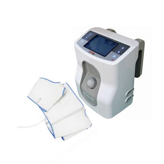
Deep Vein Thrombosis DVT-7700
$0.00
In Stock
DVT-7700 MAIN with HOSE and Accessories (Disposable sleeve thigh(M), Disposable sleeve calf(M), Disposable sleeve foot, and Tube (pair)) – Deep vein thrombosis (DVT) happens when a blood clot forms in a vein deep inside the body. Veins in the thigh, foot, and calf create a natural pumping system that helps flow blood back to the heart. But when an individual is immobile, blood circulation is decreased, and the risk of developing a blood clot goes up.
Delivery & Availability:
Typically 14-21 working days – excluding furniture and heavy/bulky equipment. Please contact us for further information.
Description
Deep vein thrombosis DVT-7700 MAIN is a DVT machine with HOSE and Accessories (Disposable sleeve thigh(M), Disposable sleeve calf(M), Disposable sleeve foot, and Tube (pair)) – Deep vein thrombosis (DVT) happens when a blood clot forms in a vein deep inside the body. Veins in the thigh, foot, and calf create a natural pumping system that helps flow blood back to the heart. But when an individual is immobile, blood circulation is decreased, and the risk of developing a blood clot goes up.
DVT machine Specifications:
Power requiremnet: 100V~240VAC, 50/60Hz
Power Consumption: Maximum 18w
Power Cord: Hospital Grade Plug (Detachable)
Operation Mode: A1,A2,A3,B1,B2,B3,B4,C1,D1,D2
Adjustable Timer: 0-99 Hours
Applied Pressure Leg sleeve: 30~60 mmHg (Caif, Thigh)
Food Cuffs: 130mmHg
Operation Mood: Continuous
Compression Type: Leg sleeve: Gradient, Sequential
Food Cuffs: Uniform
Dimension: 200mm (L) * 190mm (W) * 260 mm (H)
Weight: 2.3 kg (5.07 ib)
Battery: Capacity 10.8 Vdc 4,400 mAh Lithiumion (optional)
Run Time 6-8 Hours
Click Here To Download Catalogue
Reviews(2)
Quick Comparison
| Deep Vein Thrombosis DVT-7700 remove | Sonoscape P15 Ultrasound Machine With Four Probes remove | ASPEL AsCARD Grey ECG Machine remove | DrGem Ceiling Mounted Digital X-ray remove | DrGem Ceiling Analogue X-ray Machine remove | Sonoscape S8 Exp Portable Ultrasound remove | |
|---|---|---|---|---|---|---|
| Name | Deep Vein Thrombosis DVT-7700 remove | Sonoscape P15 Ultrasound Machine With Four Probes remove | ASPEL AsCARD Grey ECG Machine remove | DrGem Ceiling Mounted Digital X-ray remove | DrGem Ceiling Analogue X-ray Machine remove | Sonoscape S8 Exp Portable Ultrasound remove |
| Image |  |  |  |  |  |  |
| SKU | SF1033560084-290 | SF1033560012-8 | SF1033560075-5 | SF1033560074-4 | SF1033560074-7 | SF1033560012-15 |
| Rating | ||||||
| Price |
| $13,900.00 | $1,166.00 |
|
| $9,350.00 |
| Stock | ||||||
| Availability | ||||||
| Add to cart | ||||||
| Description | In Stock DVT-7700 MAIN with HOSE and Accessories (Disposable sleeve thigh(M), Disposable sleeve calf(M), Disposable sleeve foot, and Tube (pair)) - Deep vein thrombosis (DVT) happens when a blood clot forms in a vein deep inside the body. Veins in the thigh, foot, and calf create a natural pumping system that helps flow blood back to the heart. But when an individual is immobile, blood circulation is decreased, and the risk of developing a blood clot goes up. Delivery & Availability: Typically 14-21 working days – excluding furniture and heavy/bulky equipment. Please contact us for further information. | In Stock A feature-rich system inheriting the Wi-Sono high-end platform, the P15 uses an array of advanced tools to help enhance the image quality. It's a cost-effective, simplified console with an intuitive user interface and multiple intelligent functions. Delivery & Availability: Typically 2 working days – excluding furniture and heavy/bulky equipment. Please contact us for further information. | Shipped from Abroad Electrocardiograph AsCARD Grey v.07.225 - is a 1, 3, 6, 12 channel ECG unit which enables to make electrocardiogram in full 12 leads. It is intended to conduct ECG examinations of adults and paediatric patients in all types of health care centres. ECG examination may be recorded in manual or automatic mode, with the possibility of analysis and interpretation. The device can be powered from 100 V ÷ 240 V mains supply or by an internal battery. Delivery & Availability: Typically 10 working days – excluding furniture and heavy/bulky equipment. Please contact us for further information. | In Stock The GXR-SD is a diagnostic digital radiography system that provides reliable high quality digital radiographic images with a reduced dose. The GXR-SD DR systems offer comprehensive digital solutions to all radiography needs, featuring ACQUIDR digital imaging system with stationary or portable digital flat-panel detectors as well as reliable high-frequency x-ray generators that are known worldwide for their excellent performance, lifetime and stability. Patient tables and wall stands are also offered. Delivery & Availability: Typically 21 working days – excluding furniture and heavy/bulky equipment. Please contact us for further information. | Shipped from abroad The DrGem Ceiling Analogue X-ray Machine is a diagnostic radiography system that provides reliable high quality radiographic images with a reduced dose. The reliable high-frequency x-ray generators that are known worldwide for their excellent performance, lifetime and stability. Patient tables and wall stands are also offered. Delivery & Availability: Typically 21 working days – excluding furniture and heavy/bulky equipment. Please contact us for further information. | Shipped from Abroad With ultra-modern innovative design and the clinically-proven technologies, S8 Exp is portable ultrasound scanner well equipped as a low-physical-effort and enhanced-image-quality ultrasound scanner, which not only provides optimized solutions for versatile applications, but does help to improve the user-experience for both routine and non-traditional challenges. Delivery & Availability: Typically 5-7 working days – excluding furniture and heavy/bulky equipment. Please contact us for further information. |
| Content | Deep vein thrombosis DVT-7700 MAIN is a DVT machine with HOSE and Accessories (Disposable sleeve thigh(M), Disposable sleeve calf(M), Disposable sleeve foot, and Tube (pair)) - Deep vein thrombosis (DVT) happens when a blood clot forms in a vein deep inside the body. Veins in the thigh, foot, and calf create a natural pumping system that helps flow blood back to the heart. But when an individual is immobile, blood circulation is decreased, and the risk of developing a blood clot goes up.
DVT machine Specifications:
Power requiremnet: 100V~240VAC, 50/60Hz
Power Consumption: Maximum 18w
Power Cord: Hospital Grade Plug (Detachable)
Operation Mode: A1,A2,A3,B1,B2,B3,B4,C1,D1,D2
Adjustable Timer: 0-99 Hours
Applied Pressure Leg sleeve: 30~60 mmHg (Caif, Thigh)
Food Cuffs: 130mmHg
Operation Mood: Continuous
Compression Type: Leg sleeve: Gradient, Sequential
Food Cuffs: Uniform
Dimension: 200mm (L) * 190mm (W) * 260 mm (H)
Weight: 2.3 kg (5.07 ib)
Battery: Capacity 10.8 Vdc 4,400 mAh Lithiumion (optional)
Run Time 6-8 Hours
Click Here To Download Catalogue | DETAILS
Super Wide-bandwidth Platform
Inheriting Wi-sono's ultra-wide system platform and with the advanced probe technology, high-resolution and deep penetration images are provided for precision medicine.
Spatial Compound Imaging
Spatial Compound Imaging utilizes several lines of sight for optimal contrast resolution, speckle reduction and border detection, with which P15 is ideal for superficial and abdominal imaging with better clarity and improved continuity of structures.
μ-Scan+
The new generation μ-Scan imaging technology gives you better image quality by reducing noise, improving signal strength and improving visualization.
Dynamic Color
Dynamic color improves upon already existing color Doppler technologies for a clearer capture of color flow and detailed visualization of even tiny veins with lower velocities.
Real-time Panoramic
With real-time panoramic, you can acquire an extended field of view for large organs or long vessels for easy measurement and diagnostic efficiency. Accomplished in real-time for the convenience of the sonographers, any mistake can also be easily back tracked and corrected without interrupting the scan.
3D/4D
Outstanding volume performance with speed and convenience makes P15 outshine others on volume imaging.
Tissue Doppler Imaging
Tissue Doppler Imaging allows clinical doctors to quantitatively evaluate local myocardial movements and functions, facilitating them with the ability to analyze and compare the motions of the different parts of the patient's heart.
Auto IMT
Quick measurement of intra-media vessel thickness ensures good reproducibility and high diagnostic efficiency.
Click Here To Download Catalogue |
Electrocardiograph AsCARD Grey v.07.225 - is a 1, 3, 6, 12 channel ECG unit which enables to make electrocardiogram in full 12 leads. It is intended to conduct ECG examinations of adults and paediatric patients in all types of health care centres. ECG examination may be recorded in manual or automatic mode, with the possibility of analysis and interpretation. The device can be powered from 100 V ÷ 240 V mains supply or by an internal battery.
Technical Specification:1. Visualisation of 1, 3, 6 or 12 ECG waveforms, analysis results and interpretations, examinations stored in memory.
2. Recording of 12 standard leads.
3. Print out in 1, 3, 6 or 12 ECG waveforms mode. Printing of a selected group:
Click Here To Download Catalogue | DrGem Ceiling Mounted Digital X-ray is a diagnostic digital radiography system that provides reliable high quality digital radiographic images with a reduced dose. The GXR-SD DR systems offer comprehensive digital solutions to all radiography needs, featuring ACQUIDR digital imaging system with stationary or portable digital flat-panel detectors as well as reliable high-frequency x-ray generators that are known worldwide for their excellent performance, lifetime and stability. Patient tables and wall stands are also offered.
Features:
Click Here To Download Catalogue | DrGem Ceiling Analogue X-ray Machine is a diagnostic radiography system X-ray Machine that provides reliable high quality radiographic images with a reduced dose. The reliable high-frequency x-ray generators that are known worldwide for their excellent performance, lifetime and stability. Patient tables and wall stands are also offered.
Features of DrGem Ceiling Analogue X-ray Machine
Click Here To Download Catalogue | Sonoscape S8 Exp Portable Ultrasound scannerDETAILS Agile and Versatile With ultra-modern innovative design and the clinically-proven technologies, S8 Exp Portable Ultrasound scanner is well equipped as a low-physical-effort and enhanced-image-quality ultrasound scanner, which not only provides optimized solutions for versatile applications but does help to improve the user experience for both routine and non-traditional challenges. Working with S8 Exp, it will trigger your unlimited reverie and endow you with endless charm. Carrying forward the classical design of SonoScape's portable ultrasound products, S8 Exp successfully combines the best ergonomics, attractive design and ease of use. This charismatic identity is also enhanced by a sophisticated color palette—with sedate grey as its interior paint color and pearl white as exterior cover, S8 Exp reveals a style of aristocrat and strong character among SonoScape's ultrasound systems. Workflow The S8 Exp is a portable ultrasound scanner that adapts to your workflow, whether you are in the consulting room, at the bedside, or at a remote location. With easy-to-use new platform designed for sonographers' needs and full connection interfaces for easy connectivity and data sharing, S8 Exp leads to improved user comfort and clinical outcome as well as patient throughput and working efficiency. Powerful Platform Embedded with SonoScape's core imaging technologies such as μ-scan, PHI and Spatial Compound, S8 Exp boasts exceptional 2D image, sensitive spectral, Color and Power Doppler, displaying well-defined anatomy and pathology and facilitating a highly optimized diagnostic user environment for conclusive diagnoses. Besides, S8 Exp offers a comprehensive selection of electronic probes to maximally extend its capabilities to meet a wide range of applications including the abdomen, pediatric, OB/GYN, cardiovascular, musculoskeletal, etc. The advanced probe technologies also effectively enhance the image quality and confidence in reaching clinical diagnoses even in difficult patients.Click Here To Download Catalogue |
| Weight | N/A | N/A | N/A | N/A | N/A | N/A |
| Dimensions | N/A | N/A | N/A | N/A | N/A | N/A |
| Additional information |


Learn More
As I web-site possessor I believe the content matter here is rattling fantastic , appreciate it for your hard work. You should keep it up forever! Best of luck.
manhwaland
Really informative article.Much thanks again. Cool.