


DRGEM DR System
$0.00
Ship from abroad
ACQUIDR is the digital imaging system composed of a Flat Panel Detector(FPD) and an imaging workstation with software. The digital FPD and full-feature imaging software with excellent digital image processing, designed for DRGEM X-ray machine.
Delivery & Availability:
Typically 21 working days – excluding furniture and heavy/bulky equipment. Please contact us for further information.

Description
DRGEM ACQUIDR (DRGEM DR System) is the digital imaging system composed of a Flat Panel Detector(FPD) and an imaging workstation with software. The digital FPD and full-feature imaging software with excellent digital image processing will meet all your needs in the diagnostic digital radiographic field.
Features of DRGEM DR System:
- Adaptable with most existing analog x-ray systems
- AED(Auto Exposure Detection) function available
- Simple and convenient installation
- Intuitive graphic user interface
- Over 1300 pre-loaded exam profiles with DRGEM’s generator
- Configurable automatic study
- PMS / EMR /HIS / RIS work list support
- Multiple printing formats
- DICOM 3.0 compliant
- Brightness and contrast adjustment
- Rotation
- Horizontal and vertical flip
- Zoom and pan
- Left / right makers
- Optimized work flow
- Automatic shutters
- Support DAP interface
- Exposure index (EI)
- DR upgrade solution by retrofit.
- Portable and Wireless FPD DR solution for maximum flexibility in virtually all general radiographic specialties.
- Portable and wireless detector fits into most existing analogue x-ray system.
- Faster workflow after DR upgrade.
- With AED(Auto Exposure Detection) function, there is no DR trigger cable between detector and generator providing very easy DR upgrade.
- Easy to interface with any kind of x-ray generator.
- Radmax acquisition workstation
- Turns any analogue X-Ray system into a fully DR system
- Optional image stitching program
- Vet software available
- Detector format: 17×17” / 17×14″, wired/wireless
Quick Comparison
| DRGEM DR System remove | DrGem Ceiling Analogue X-ray Machine remove | SIGNERS SUPiA Dry Thermal X-ray Film Printer remove | Jade Mobile X-ray machine (Analogue) remove | Sonoscape E2 Ultrasound Machine remove | Sonoscape S11 Ultrasound Machine remove | |
|---|---|---|---|---|---|---|
| Name | DRGEM DR System remove | DrGem Ceiling Analogue X-ray Machine remove | SIGNERS SUPiA Dry Thermal X-ray Film Printer remove | Jade Mobile X-ray machine (Analogue) remove | Sonoscape E2 Ultrasound Machine remove | Sonoscape S11 Ultrasound Machine remove |
| Image | 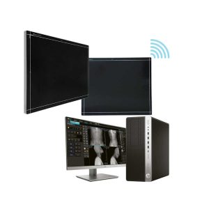 |  | 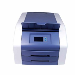 |  |  |  |
| SKU | SF1033560074-8 | SF1033560074-7 | SF1033560050-02 | SF1033560074-2 | SF1033560012-17 | SF1033560012-1 |
| Rating | ||||||
| Price |
|
| $3,520.00 |
| $5,500.00 | $6,380.00 |
| Stock | ||||||
| Availability | ||||||
| Add to cart | ||||||
| Description | Ship from abroad ACQUIDR is the digital imaging system composed of a Flat Panel Detector(FPD) and an imaging workstation with software. The digital FPD and full-feature imaging software with excellent digital image processing, designed for DRGEM X-ray machine. Delivery & Availability: Typically 21 working days – excluding furniture and heavy/bulky equipment. Please contact us for further information. | Shipped from abroad The DrGem Ceiling Analogue X-ray Machine is a diagnostic radiography system that provides reliable high quality radiographic images with a reduced dose. The reliable high-frequency x-ray generators that are known worldwide for their excellent performance, lifetime and stability. Patient tables and wall stands are also offered. Delivery & Availability: Typically 21 working days – excluding furniture and heavy/bulky equipment. Please contact us for further information. | Shipped from Abroad
Signers Digital Dry Thermal X-ray Film Printer - Is a dry imager designed to output information processed through DICOM network protocol, supports 4 films sizes. 8”x10”, 10”x12”, 11”x14” and 14”x17” Achieve the convenience of multiple sizes.
Delivery & Availability: Typically 14 working days – excluding furniture and heavy/bulky equipment. Please contact us for further information. | In Stock JADE is one of the lightest portable X-ray systems on the market, allowing it to be used in any imaginable way including bedside, operating rooms, intensive care units and in veterinary fields. With a simple, easy-to-use operator console, three-way control, two-step foldable stand and auto lock system, JADE is a user-friendly portable X-ray system. Delivery & Availability: Typically 21 working days – excluding furniture and heavy/bulky equipment. Please contact us for further information. | Shipped from Abroad Sonoscape E2 portable ultrasound machine is a color Doppler ultrasound system that reaches beyond your expectations due to its compact and fashionable appearance. It fulfills GI, OB/GYN, Cardiac and POC applications to fit your routine scanning needs while its color mode will help you for more accurate and efficient diagnosis of lesions. E2 provides a wide range of applications to assist users with routine scanning. E2 provides automatic calculations to enhance your diagnostic confidence and save you time for patient communication. Delivery & Availability: Typically 14 working days – excluding furniture and heavy/bulky equipment. Please contact us for further information. | In Stock A Value Choice beyond Your Expectation. SonoScape’s trolley color Doppler system S11 redefines price and performance with practical design. The S11 will go beyond your expectations but not your budget. Delivery & Availability: Typically 2 working days – excluding furniture and heavy/bulky equipment. Please contact us for further information. |
| Content | DRGEM ACQUIDR (DRGEM DR System) is the digital imaging system composed of a Flat Panel Detector(FPD) and an imaging workstation with software. The digital FPD and full-feature imaging software with excellent digital image processing will meet all your needs in the diagnostic digital radiographic field.
Features of DRGEM DR System:
| DrGem Ceiling Analogue X-ray Machine is a diagnostic radiography system X-ray Machine that provides reliable high quality radiographic images with a reduced dose. The reliable high-frequency x-ray generators that are known worldwide for their excellent performance, lifetime and stability. Patient tables and wall stands are also offered.
Features of DrGem Ceiling Analogue X-ray Machine
Click Here To Download Catalogue | Signers Digital Dry Thermal X-ray Film Printer - Is a dry imager designed to output information processed through DICOM network protocol, supports 4 films sizes. 8”x10”, 10”x12”, 11”x14” and 14”x17” Achieve the convenience of multiple sizes.
Technical Specifications:
Click Here To Download Catalogue | JADE Mobile X-ray machine is one of the lightest portable X-ray systems on the market, allowing it to be used in any imaginable way including bedside, operating rooms, intensive care units and veterinary fields. With a simple, easy-to-use operator console, three-way control, two-step foldable stand and auto-lock system, the JADE Mobile X-ray machine is a user-friendly portable X-ray system.
Convenient & Intuitive Operation:
JADE is one of the lightest portable X-ray systems on the market, allowing it to be used in any imaginable way including bedside, operating rooms, intensive care units and in veterinary fields. With a simple, easy-to-use operator console, three-way control, two-step foldable stand and auto-lock system, JADE is a user-friendly portable X-ray system.
Compact & Powerful Design:
JADE Mobile X-ray machine is an innovative, highly versatile portable X-ray system suitable for a variety of clinical uses. Utilizing the unique technology used in DRGEM’s universally recognized X-ray generators, JADE is a compact but powerful unit with a 4kW output and thoughtfully designed components to increase efficiency and maximize workflow. The core part of X-ray source adopts high-quality tube assembly, X-ray collimator and high frequency X-ray generator with excellent performance, lifetime and stability.
Features:
Click Here To Download Catalogue | SONOSCAPE E2 DETAILS
Auto Image Optimization
A portable ultrasound machine with the press of a button, the image is automatically adjusted and optimized, saving you time with parameter adjustments. Additionally, with Auto Focus on, the focus area follows the depth of the ROI box as it is moved in the scanning field, providing users with excellent image quality in the desired area of interest.
Automated Calculation
Auto IMT is used when determining the level of vascular sclerosis present in the patient by automatically tracing the thickness of the carotid vessels.
Auto trace provides users sensitive and accurate wave tracing, avoiding the error of manual trace and giving out calculation result in no time
In-Build Battery pack
This portable ultrasound machine was equipped with an in-build battery pack which enable the user to perform image scanning when AC power is not available.
Click Here To Download Catalogue | DETAILS
SonoScape’s trolley colour Doppler system S11 redefines price and performance with practical design. The S11 will go beyond your expectations but not your budget. As an easy-to-use ultrasound system, the S11 is integrated with a new software platform, especially optimized for a smooth workflow and convenient operation. The system speeds up the exam process and makes file management easier.
SPECIFICATION
- 15-inch high definition LCD monitor with articulating arm
- Compact and agile trolley design
- 3 active transducer sockets available for a wide range of applications
- Duplex, Color Doppler, DPI, PW Doppler, tissue harmonic imaging, μ-scan speckle reduction imaging, compound imaging, trapezoidal imaging
- Customized settings based on your own working style
- Full patient database and image management solutions
Click Here To Download Catalogue |
| Weight | N/A | N/A | N/A | N/A | N/A | N/A |
| Dimensions | N/A | N/A | N/A | N/A | N/A | N/A |
| Additional information |


Reviews
There are no reviews yet.