


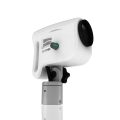
Ecleris HD Digital Video Colposcope
$3,564.00
Shipped from Abroad
Content Includes:
Floor Mounted with Base with 4 Castors,
Video Head,
Positioning Arm,
LED Light Included on Video Head,
110/220V Power Cable and User´s Guide,
Stand For LCD Monitor and Printer,
Digital Capturing System for Images, Videos and Sounds,
USB 2.0. Includes Main Unit Processor with three Camera Inputs,
USB Cable,
Software,
Hands Free Microphone and Footswitch.
Delivery & Availability:
Typically 10 working days – excluding furniture and heavy/bulky equipment. Please contact us for further information.

Description
The Ecleris ColpoHD Digital Video Colposcope offers clinicians an illuminated, high definition magnified view of the cervix and tissues of the vagina and vulva. As a screening tool for sexual assault victims or for the early detection of precancerous lesions, the ColpoHD provides insight into relevant pathology changes of tissue shape and color with magnification of up to 50x and a digital zoom up to 128x. The integrated electronic green filter delivers exceptional pathology visualization, with clear, true color imaging and superb depth of field. The swing arm mounted high-definition (HD) camera can be easily maneuvered into place. Optional integrated image capture is available for documentation along with archiving and printing of images.
A powerful tool for cervical cancer screening
The ColpoHD is precise and easy to handle in all examination situations. High quality optics along bright LED illumination and high-tech CMOS image sensors guarantee fantastic quality HD images are displayed for every patient.
The ColpoHD is also ideally suited for the examination of sexual assault victims who may be uncomfortable with a traditional Colposcope examination with the physician working through the standard microscope binoculars. The ColpoHD offers distance and a level of privacy for these patients who may be in a vulnerable frame of mind. The ColpoHD also offers optional image archiving and documentation of the examination in high definition image quality which is vital for sexual assault cases.
Features at a Glance
- Outstanding image quality.
- Perfect HD 1280X720p color definition.
- Optical zoom.
- LED high power illumination.
- Great maneuverability.
- OSD (on screen display) with indication of real magnification.
- Zoom and focus operated by 4 commands joystick.
- Still function.
- WB (white balance) push bottom control.
- Perfect color discrimination due to CMOS HD technology.
- Floor stand system with 4 antistatic wheels with break.
- Ratched up down pole with double arm for easier and steady positioning.
- Magnification progressive. Optical zoom up to 37x and digital zoom up to 70x.
- Focus automatic and manual.
- Distance 240-300mm.
- Filter optical green.
- Led life 50.000 hs.
- Optional: monitor stand pole / Endodigi HD recording device.
Technical Specification
- Outputs – 2x HDMI
- Magnification – Progressive magnification. Optical zoom up to 50x and digital zoom up to 128x.
- Focus – Automatic and manual
- Resolution – 1280 x 720 P
- Depth of Field – 250 – 300
- Field of View – 8 – 140 mm
- Distance – 200 – 320 mm
- Filter – Optical green
- Light Source – LED High Intensity, 6500 Kelvin
- LED Life – 50.000 hrs
- Illuminated Control – Electronic adjustment. Constant light color.
- Power Supply – 100 – 120 VAC, 50/60 Hz
Content Includes:
Floor Mounted with Base with 4 Castors,
Video Head,
Positioning Arm,
LED Light Included on Video Head,
110/220V Power Cable and User´s Guide,
Stand For LCD Monitor and Printer,
Digital Capturing System for Images, Videos and Sounds,
USB 2.0. Includes Main Unit Processor with three Camera Inputs,
USB Cable,
Software,
Hands Free Microphone and Footswitch.
Click Here To Download Catalogue
Quick Comparison
| Ecleris HD Digital Video Colposcope remove | DrGem Ceiling Mounted Digital X-ray remove | DrGem Diamond All-In-One Digital X-ray Machine remove | Sonoscape S22 Ultrasound Machine remove | ASPEL AsPEKT 712 Holter Monitor and Software remove | Sonoscape P15 Ultrasound Machine With Four Probes remove | |
|---|---|---|---|---|---|---|
| Name | Ecleris HD Digital Video Colposcope remove | DrGem Ceiling Mounted Digital X-ray remove | DrGem Diamond All-In-One Digital X-ray Machine remove | Sonoscape S22 Ultrasound Machine remove | ASPEL AsPEKT 712 Holter Monitor and Software remove | Sonoscape P15 Ultrasound Machine With Four Probes remove |
| Image | 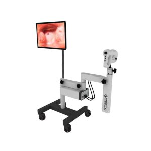 |  |  |  |  |  |
| SKU | SF1033560087-2 | SF1033560074-4 | SF1033560074-3 | SF1033560012-3 | SF1033560075-4 | SF1033560012-8 |
| Rating | ||||||
| Price | $3,564.00 |
|
| $9,350.00 | $1,991.00 | $13,900.00 |
| Stock | ||||||
| Availability | ||||||
| Add to cart | ||||||
| Description | Shipped from Abroad Content Includes: Floor Mounted with Base with 4 Castors, Video Head, Positioning Arm, LED Light Included on Video Head, 110/220V Power Cable and User´s Guide, Stand For LCD Monitor and Printer, Digital Capturing System for Images, Videos and Sounds, USB 2.0. Includes Main Unit Processor with three Camera Inputs, USB Cable, Software, Hands Free Microphone and Footswitch. Delivery & Availability: Typically 10 working days – excluding furniture and heavy/bulky equipment. Please contact us for further information. | In Stock The GXR-SD is a diagnostic digital radiography system that provides reliable high quality digital radiographic images with a reduced dose. The GXR-SD DR systems offer comprehensive digital solutions to all radiography needs, featuring ACQUIDR digital imaging system with stationary or portable digital flat-panel detectors as well as reliable high-frequency x-ray generators that are known worldwide for their excellent performance, lifetime and stability. Patient tables and wall stands are also offered. Delivery & Availability: Typically 21 working days – excluding furniture and heavy/bulky equipment. Please contact us for further information. | Shipped from Abroad DrGem Diamond All-In-One Digital X-ray Machine is a fully automatic digital radiography system providing state-of-the-art image quality, image processing and user interface. With a wide selection of anatomical studies on the imaging software, DIAMOND automatically sets up the x-ray generator’s preprogrammed exposure technique settings, motorized radiographic stand positioning, x-ray collimation and post-image processing for the selected study. Specifically designed to increase workflow, this fully digital system offers convenient auto-positioning and advanced image processing to achieve big performance with little effort. Delivery & Availability: Typically 21 working days – excluding furniture and heavy/bulky equipment. Please contact us for further information. | Shipped from Abroad As SonoScape steps forward to add value and efficiency to ultrasound, the latest S22 was designed in a user-friendly platform to address current and future demanding needs. It represents an excellent mix in performance and price. Delivery & Availability: Typically 5-7 working days – excluding furniture and heavy/bulky equipment. Please contact us for further information. | Shipped from Abroad The Holta Monitor allows quick analysis of ECG examination and detection, reviewing and editing capability in the qualitative assessment of VE, VT, Single SVE, PSVT, Pauses, Irregular Rhythm, VT, IVR, Brady - and Tachycardia, Couplets, ST-segment elevation and depression, Maximum, Minimum and averaged Heart Rates, artifacts Delivery & Availability: Typically 10 working days – excluding furniture and heavy/bulky equipment. Please contact us for further information. | In Stock A feature-rich system inheriting the Wi-Sono high-end platform, the P15 uses an array of advanced tools to help enhance the image quality. It's a cost-effective, simplified console with an intuitive user interface and multiple intelligent functions. Delivery & Availability: Typically 2 working days – excluding furniture and heavy/bulky equipment. Please contact us for further information. |
| Content | The Ecleris ColpoHD Digital Video Colposcope offers clinicians an illuminated, high definition magnified view of the cervix and tissues of the vagina and vulva. As a screening tool for sexual assault victims or for the early detection of precancerous lesions, the ColpoHD provides insight into relevant pathology changes of tissue shape and color with magnification of up to 50x and a digital zoom up to 128x. The integrated electronic green filter delivers exceptional pathology visualization, with clear, true color imaging and superb depth of field. The swing arm mounted high-definition (HD) camera can be easily maneuvered into place. Optional integrated image capture is available for documentation along with archiving and printing of images.
A powerful tool for cervical cancer screening
The ColpoHD is precise and easy to handle in all examination situations. High quality optics along bright LED illumination and high-tech CMOS image sensors guarantee fantastic quality HD images are displayed for every patient.
The ColpoHD is also ideally suited for the examination of sexual assault victims who may be uncomfortable with a traditional Colposcope examination with the physician working through the standard microscope binoculars. The ColpoHD offers distance and a level of privacy for these patients who may be in a vulnerable frame of mind. The ColpoHD also offers optional image archiving and documentation of the examination in high definition image quality which is vital for sexual assault cases.
Features at a Glance
Click Here To Download Catalogue | DrGem Ceiling Mounted Digital X-ray is a diagnostic digital radiography system that provides reliable high quality digital radiographic images with a reduced dose. The GXR-SD DR systems offer comprehensive digital solutions to all radiography needs, featuring ACQUIDR digital imaging system with stationary or portable digital flat-panel detectors as well as reliable high-frequency x-ray generators that are known worldwide for their excellent performance, lifetime and stability. Patient tables and wall stands are also offered.
Features:
Click Here To Download Catalogue | DrGem Diamond All-In-One Digital X-ray Machine is a fully automatic digital radiography system providing state-of-the-art image quality, image processing and user interface. With a wide selection of anatomical studies on the imaging software, DIAMOND automatically sets up the x-ray generator’s pre-programmed exposure technique settings, motorized radiographic stand positioning, x-ray collimation and post-image processing for the selected study. Specifically designed to increase workflow, this fully digital system offers convenient auto-positioning and advanced image processing to achieve big performance with little effort.
Features of DrGem Diamond All-In-One Digital X-ray Machine:
Outstanding Image Quality -
Digital radiography via at panel detector improves your workflow, exam speed and comfort with efficiency. Digital at panel detector with Csl screen provides excellent spatial resolution, MTF, DQE and stability based on ne pixel pitch. A 3-field ion-chamber is provided for AEC function.
Automatic Collimation –
Automatic x-ray eld size control of the motorized collimator corresponds to dierent SIDs. Includes user adjustable lamp timer with on/oswitch.
Automatic Positioning –
Click Here To Download Catalogue | DETAILS
As SonoScape steps forward to add value and efficiency to ultrasound, the latest S22 was designed in a user-friendly platform to address current and future demanding needs. It represents an excellent mix in performance and price.
S22, is a shared service ultrasound system with a slim and elegant package that has combined mobility with utility to fit in specific clinical situations including emergency department, ICU, operating room and so on. Furthermore, its ergonomic design, easy operating and flexible data management will give you a memorable experience.
SPECIFICATION
• Large high-resolution widescreen LED
• Sensitive touch screen
• Four transducer sockets plus one socket for pencil probe
• A comprehensive selection of probes: linear, Convex, Micro-convex, Volumetric, Endocavity, Bi-plane, Phased Array, TEE, Intraoperative, Pencil
• Premium application technology: 4D, μ-scan speckle reduction, compound imaging, Pulse Inversion Harmonic Imaging, Color M-Mode, Steer M-Mode, PDI, TDI, Real-time Panoramic Imaging, Trapezoid Imaging, Auto-IMT…
• Full patient database and image management solutions: DICOM 3.0, AVI/JPG, USB 2.0, HDD, DVD, PDF report
• Multi-Language Input Keyboard
• Built-in battery
Click Here To Download Catalogue | The Holter Monitor allows quick analysis of ECG examination (arrhythmias and ST segment).
Technical specifications:
HolCARD 24W Software:
Click Here To Download Catalogue | DETAILS
Super Wide-bandwidth Platform
Inheriting Wi-sono's ultra-wide system platform and with the advanced probe technology, high-resolution and deep penetration images are provided for precision medicine.
Spatial Compound Imaging
Spatial Compound Imaging utilizes several lines of sight for optimal contrast resolution, speckle reduction and border detection, with which P15 is ideal for superficial and abdominal imaging with better clarity and improved continuity of structures.
μ-Scan+
The new generation μ-Scan imaging technology gives you better image quality by reducing noise, improving signal strength and improving visualization.
Dynamic Color
Dynamic color improves upon already existing color Doppler technologies for a clearer capture of color flow and detailed visualization of even tiny veins with lower velocities.
Real-time Panoramic
With real-time panoramic, you can acquire an extended field of view for large organs or long vessels for easy measurement and diagnostic efficiency. Accomplished in real-time for the convenience of the sonographers, any mistake can also be easily back tracked and corrected without interrupting the scan.
3D/4D
Outstanding volume performance with speed and convenience makes P15 outshine others on volume imaging.
Tissue Doppler Imaging
Tissue Doppler Imaging allows clinical doctors to quantitatively evaluate local myocardial movements and functions, facilitating them with the ability to analyze and compare the motions of the different parts of the patient's heart.
Auto IMT
Quick measurement of intra-media vessel thickness ensures good reproducibility and high diagnostic efficiency.
Click Here To Download Catalogue |
| Weight | N/A | N/A | N/A | N/A | N/A | N/A |
| Dimensions | N/A | N/A | N/A | N/A | N/A | N/A |
| Additional information |


Reviews
There are no reviews yet.