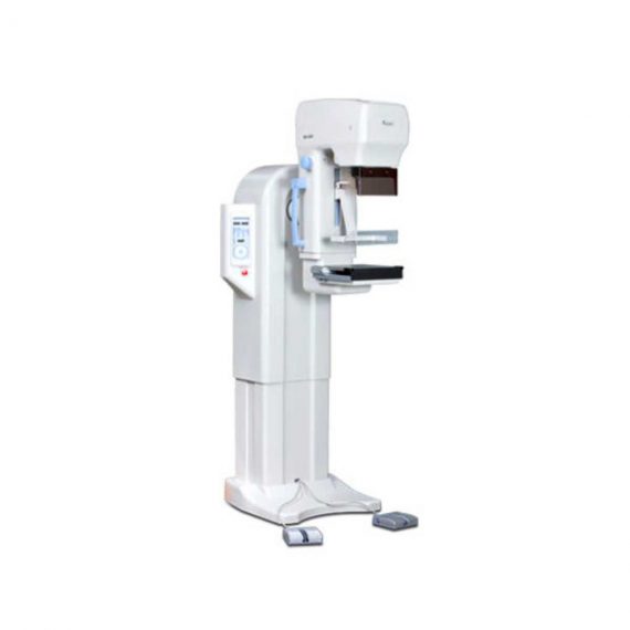
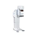
Genoray Analogue Mammography MX-600
$0.00
Shipped from Abroad
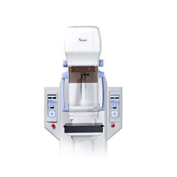
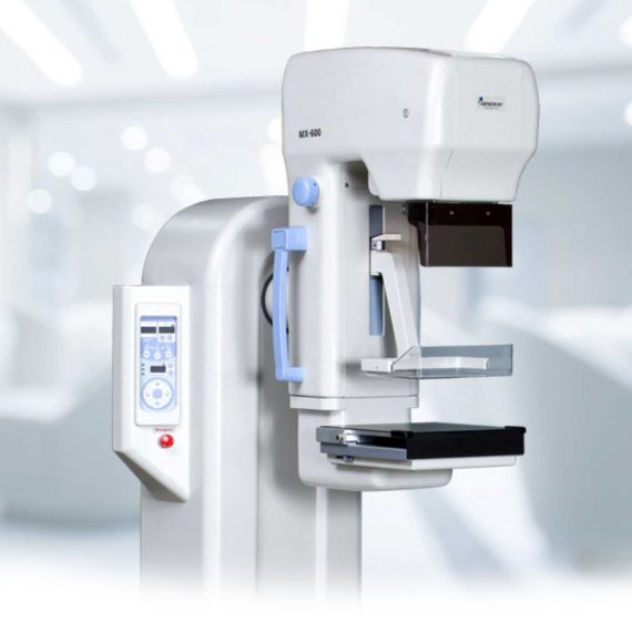
MX-600 is designed compactly for easy install and operation.
Delivery & Availability:
Typically 21 working days – excluding furniture and heavy/bulky equipment. Please contact us for further information.
Description
MX-600 is designed compactly for easy install and operation.
Features:
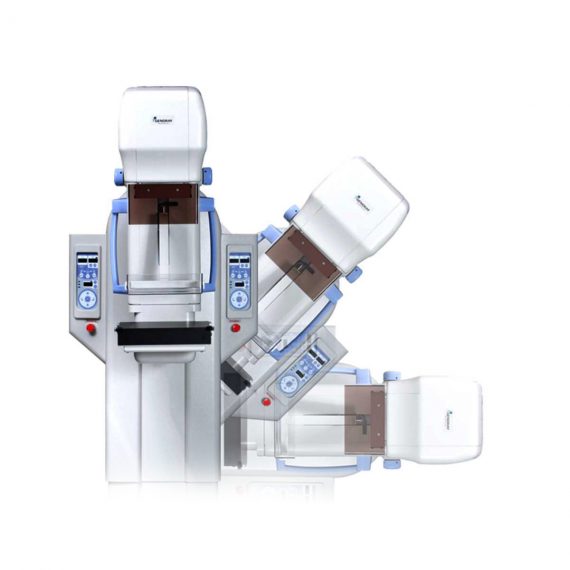
- Auto Standard Positioning system (ASP)
With Auto Standard exposure Positioning systems designed to maximize the convenience of radiography, you can easily adjust positioning by using standard exposure (RCC, LCC, RMLO, LMLO). ASP function makes operators easy to execute 4 axis exposures by software programming. One-touch button controls 4 standard positions (RCC, LCC, RMLO, LMLO) using C-Arm pre-set adjustment (MedioLaterral, Oblique view and CranioCaudal view). The ISO level can adjust the level of MX-600 standing position when it operates from vertical exposure to oblique and vice versa. - Intelligent Automatic Exposure Control (AEC)
Automatic Exposure Control system enables production of images with reliable intensity for any film, screen, or digital radiography. Furthermore, it greatly enhances the convenience of radiography by embedding the Full-AEC function which utilizes Auto kV control. It sets the best diagnosis environment by reducing manual operation and increasing patient throughput. - Compression with comfort in mind
When pressure is required for Mammography, it allows you to apply appropriate amount of pressure (up to maximum of 20kg) and is equipped with MICOM control’s Soft-touch system which minimize the discomfort. - Automatic Conversion (Filter)
Motorized and manual breast compressions are available for MX-600. MX-600 displays compression level and thickness on main body’s panel. - Stable Output
A molybdenum (0.03mm Mo) and Aluminum (0.5mm Al) filter are installed to absorb unnecessary x-ray. Mo filter covers low level kV range (22-35kV) and Al filter covers high kV range (35-39kV). Mo filter is useful for increasing image contrast in large breast with large amounts of glandular tissue. The rhodium filter (0.025mm Rh) can be installed as an option (as replacement of Al filter). - Display of Exposure Condition
When some degree of pressure is required for radiography, it allows you to apply the appropriate pressure (up to a maximum of 20kg) and is equipped with MICOM control’s Soft-touch system which is designed to minimize the discomfort of the examine with in the pressure range.
Technical Specifications:
Generator
- Type: High frequency inverter (40 kHz).
- Radiographic qualification:
- Great focus 22-39kV / 1-600mAs.
- Small bulb 22-35kV / 1-100mAs.
Tube
- Focal point size: 0.1 / 0.3mm.
- Anode heat capacity: 300KHU (molybdenum).
- Filtration: Mo.
Radiographic support
- Movement up / down: 720-1420mm (motorized).
- Rotating movement: ± 180˚ (Motorized) .
- Automatic standard positioning (ASP).
- SID: 650 mm.
Bucky device
- Cassette size: 18x24cm, Grid: 4: 1, 91 lines / inches.
Automatic exposure control (AEC)
- Type: solid state detector.
- Mode: 3 modes (Full / Semi / Manual).
- Density adjustment: 19 steps.
Collimator: Automatic.
Accessories
- Face protection.
- Film marker, hand switch, point compression paddle.
- 18x24cm Bucky device with compression paddle.
- Collimating device (18×24 and dotted plate).
Options
- Set of Bucky devices of 24×30 cm.
- Set of 1.5X or 1.8X magnification devices.
Click Here To Download Catalogue
Quick Comparison
| Genoray Analogue Mammography MX-600 remove | Sonoscape P10 Ultrasound Machine remove | ASPEL AsCARD Green B/W ECG Machine remove | DrGem Diamond All-In-One Digital X-ray Machine remove | SIGNERS SUPiA X-ray Digitizer ( CR Scanner) remove | DrGem Ceiling Mounted Digital X-ray remove | |||||||||||||||||||||||||
|---|---|---|---|---|---|---|---|---|---|---|---|---|---|---|---|---|---|---|---|---|---|---|---|---|---|---|---|---|---|---|
| Name | Genoray Analogue Mammography MX-600 remove | Sonoscape P10 Ultrasound Machine remove | ASPEL AsCARD Green B/W ECG Machine remove | DrGem Diamond All-In-One Digital X-ray Machine remove | SIGNERS SUPiA X-ray Digitizer ( CR Scanner) remove | DrGem Ceiling Mounted Digital X-ray remove | ||||||||||||||||||||||||
| Image |  |  |  |  |  |  | ||||||||||||||||||||||||
| SKU | SF1033560097-6 | SF1033560012-7 | SF1033560075-8 | SF1033560074-3 | SF1033560050-01 | SF1033560074-4 | ||||||||||||||||||||||||
| Rating | ||||||||||||||||||||||||||||||
| Price |
| $9,350.00 |
|
| $6,930.00 |
| ||||||||||||||||||||||||
| Stock | ||||||||||||||||||||||||||||||
| Availability | ||||||||||||||||||||||||||||||
| Add to cart | ||||||||||||||||||||||||||||||
| Description | Shipped from Abroad
MX-600 is designed compactly for easy install and operation.
| Shipped from Abroad The P10 color Doppler ultrasound system is a new generation product from SonoScape. It is designed to give high quality images, rich probe configurations, various clinical tools and automatic analysis software to provide you with comprehensive solutions for your growing demand for clinical applications. Delivery & Availability: Typically 5-7 working days – excluding furniture and heavy/bulky equipment. Please contact us for further information. | Shipped from Abroad AsCARD Green electrocardiograph is a 1- and 3-channel ECG unit which enables to make electrocardiogram in full 12 leads. Intended for ECG examinations of adult and paediatric patients aimed at identification of cardiological abnormalities, myocardial ischaemia or infarction. The device is intended for use in healthcare facilities by duly trained personnel. ECG examination may be recorded in manual or automatic mode with the ability to perform the analysis and interpretation. Delivery & Availability: Typically 10 working days – excluding furniture and heavy/bulky equipment. Please contact us for further information. | Shipped from Abroad DrGem Diamond All-In-One Digital X-ray Machine is a fully automatic digital radiography system providing state-of-the-art image quality, image processing and user interface. With a wide selection of anatomical studies on the imaging software, DIAMOND automatically sets up the x-ray generator’s preprogrammed exposure technique settings, motorized radiographic stand positioning, x-ray collimation and post-image processing for the selected study. Specifically designed to increase workflow, this fully digital system offers convenient auto-positioning and advanced image processing to achieve big performance with little effort. Delivery & Availability: Typically 21 working days – excluding furniture and heavy/bulky equipment. Please contact us for further information. | Shipped from Abroad SUPiA made by Signers offers such a better clinic environment with no chemicals, ideal space, high-resolution image quality, and affordability. Delivery & Availability: Typically 14 working days – excluding furniture and heavy/bulky equipment. Please contact us for further information. | In Stock The GXR-SD is a diagnostic digital radiography system that provides reliable high quality digital radiographic images with a reduced dose. The GXR-SD DR systems offer comprehensive digital solutions to all radiography needs, featuring ACQUIDR digital imaging system with stationary or portable digital flat-panel detectors as well as reliable high-frequency x-ray generators that are known worldwide for their excellent performance, lifetime and stability. Patient tables and wall stands are also offered. Delivery & Availability: Typically 21 working days – excluding furniture and heavy/bulky equipment. Please contact us for further information. | ||||||||||||||||||||||||
| Content | MX-600 is designed compactly for easy install and operation.
Features:
Generator
Tube
Radiographic support
Bucky device
Automatic exposure control (AEC)
Collimator: Automatic. Accessories
Options
Click Here To Download Catalogue | DETAILS
B + Compound
B + Compound utilizes several lines of sight for optimal contrast resolution, speckle reduction and border detection, with which P10 is ideal for superficial and abdominal imaging with better clarity and improved continuity of structures.
μ-Scan
The new generation μ-Scan imaging technology gives you better image quality by reducing noise, improving signal strength and improving visualization.
P10 offers a comprehensive selection of electronic probes to maximize its capabilities to meet a wide range of applications including abdomen, pediatric, OB/GYN, cardiovascular, musculoskeletal, etc. The advanced probe technologies also effectively enhance the image quality and confidence in reaching clinical diagnoses, even in difficult patients.
Convex Probe 3C-A
Ideal for an abundant of application such as abdomen, gynecology, obstetrics, urology and even abdomen biopsy.
Linear Probe L741
This linear probe is designed to satisfy vascular, breast, thyroid, and other small parts diagnosis, and its adjustable parameters could also present users a clear view of MSK and deep vessels.
Phase Array Probe 3P-A
For the purpose of adult and pediatric cardiology and emergency, the phase array probe provides elaborate presets for different exam modes, even for difficult patients.
Intracavitary Probe 6V1
Intracavitary probe could face application of gynecology, urology, prostate, and its temperature detection technology not only protects the patient but also extends the service life.
Click Here To Download Catalogue | AsCARD Green electrocardiograph is a 1- and 3-channel ECG unit which enables to make electrocardiogram in full 12 leads. Intended for ECG examinations of adult and paediatric patients aimed at identification of cardiological abnormalities, myocardial ischaemia or infarction. The device is intended for use in healthcare facilities by duly trained personnel. ECG examination may be recorded in manual or automatic mode with the ability to perform the analysis and interpretation.
Electrocardiograph is based on advanced microprocessor technology. It is equipped with a thermal printer with high-resolution head and graphical LCD display. A hightech membrane keyboard makes the AsCARD Green device operation intuitive, and its menu navigation exceptionally easy. This light-weight, small-footprint and battery powered cause that device can be easily transported to any location. With plastic casing and foil covered keyboard, the device is neat and easy to clean.
Technical Specifications:
Click Here To Download Catalogue | DrGem Diamond All-In-One Digital X-ray Machine is a fully automatic digital radiography system providing state-of-the-art image quality, image processing and user interface. With a wide selection of anatomical studies on the imaging software, DIAMOND automatically sets up the x-ray generator’s pre-programmed exposure technique settings, motorized radiographic stand positioning, x-ray collimation and post-image processing for the selected study. Specifically designed to increase workflow, this fully digital system offers convenient auto-positioning and advanced image processing to achieve big performance with little effort.
Features of DrGem Diamond All-In-One Digital X-ray Machine:
Outstanding Image Quality -
Digital radiography via at panel detector improves your workflow, exam speed and comfort with efficiency. Digital at panel detector with Csl screen provides excellent spatial resolution, MTF, DQE and stability based on ne pixel pitch. A 3-field ion-chamber is provided for AEC function.
Automatic Collimation –
Automatic x-ray eld size control of the motorized collimator corresponds to dierent SIDs. Includes user adjustable lamp timer with on/oswitch.
Automatic Positioning –
Click Here To Download Catalogue | SUPiA X-ray Digitizer made by Signers offers such a better clinic environment with no chemicals, ideal space, high-resolution image quality, and affordability
FEATURE
Rigid Type
• No damage or scratch on image plates during scanning & erasing
• Scanning & Erasing without a roller
• No cut-off image during winter and cold period
Durability
• Extremely simple structure design
• Strong aluminum base plate
• Flip covers preventing dust from inside scanner
Barcode System
• Automatically recognising cassette sizes(14x17", 10x12", 18x24cm) by barcode reader
Compact & lightweight design
Click Here To Download Catalogue | DrGem Ceiling Mounted Digital X-ray is a diagnostic digital radiography system that provides reliable high quality digital radiographic images with a reduced dose. The GXR-SD DR systems offer comprehensive digital solutions to all radiography needs, featuring ACQUIDR digital imaging system with stationary or portable digital flat-panel detectors as well as reliable high-frequency x-ray generators that are known worldwide for their excellent performance, lifetime and stability. Patient tables and wall stands are also offered.
Features:
Click Here To Download Catalogue | ||||||||||||||||||||||||
| Weight | N/A | N/A | N/A | N/A | N/A | N/A | ||||||||||||||||||||||||
| Dimensions | N/A | N/A | N/A | N/A | N/A | N/A | ||||||||||||||||||||||||
| Additional information |


Reviews
There are no reviews yet.