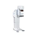
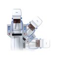
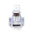
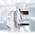
Genoray Analogue Mammography MX-600
Ask for Price$0.00
Shipped from Abroad
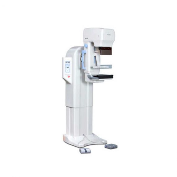
MX-600 is designed compactly for easy install and operation.
Delivery & Availability:
Typically 21 working days – excluding furniture and heavy/bulky equipment. Please contact us for further information.
Description
MX-600 is designed compactly for easy install and operation.
Features:
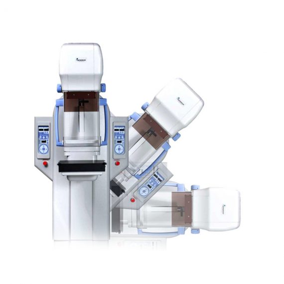
- Auto Standard Positioning system (ASP)
With Auto Standard exposure Positioning systems designed to maximize the convenience of radiography, you can easily adjust positioning by using standard exposure (RCC, LCC, RMLO, LMLO). ASP function makes operators easy to execute 4 axis exposures by software programming. One-touch button controls 4 standard positions (RCC, LCC, RMLO, LMLO) using C-Arm pre-set adjustment (MedioLaterral, Oblique view and CranioCaudal view). The ISO level can adjust the level of MX-600 standing position when it operates from vertical exposure to oblique and vice versa. - Intelligent Automatic Exposure Control (AEC)
Automatic Exposure Control system enables production of images with reliable intensity for any film, screen, or digital radiography. Furthermore, it greatly enhances the convenience of radiography by embedding the Full-AEC function which utilizes Auto kV control. It sets the best diagnosis environment by reducing manual operation and increasing patient throughput. - Compression with comfort in mind
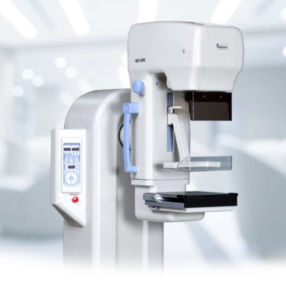
When pressure is required for Mammography, it allows you to apply appropriate amount of pressure (up to maximum of 20kg) and is equipped with MICOM control’s Soft-touch system which minimize the discomfort. - Automatic Conversion (Filter)
Motorized and manual breast compressions are available for MX-600. MX-600 displays compression level and thickness on main body’s panel. - Stable Output
A molybdenum (0.03mm Mo) and Aluminum (0.5mm Al) filter are installed to absorb unnecessary x-ray. Mo filter covers low level kV range (22-35kV) and Al filter covers high kV range (35-39kV). Mo filter is useful for increasing image contrast in large breast with large amounts of glandular tissue. The rhodium filter (0.025mm Rh) can be installed as an option (as replacement of Al filter). - Display of Exposure Condition
When some degree of pressure is required for radiography, it allows you to apply the appropriate pressure (up to a maximum of 20kg) and is equipped with MICOM control’s Soft-touch system which is designed to minimize the discomfort of the examine with in the pressure range.
Technical Specifications:
Generator
- Type: High frequency inverter (40 kHz).
- Radiographic qualification:
- Great focus 22-39kV / 1-600mAs.
- Small bulb 22-35kV / 1-100mAs.
Tube
- Focal point size: 0.1 / 0.3mm.
- Anode heat capacity: 300KHU (molybdenum).
- Filtration: Mo.
Radiographic support
- Movement up / down: 720-1420mm (motorized).
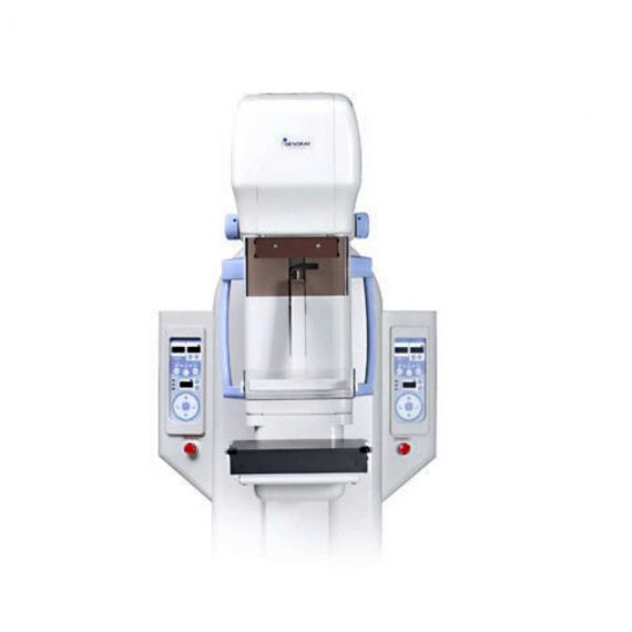
- Rotating movement: ± 180˚ (Motorized) .
- Automatic standard positioning (ASP).
- SID: 650 mm.
Bucky device
- Cassette size: 18x24cm, Grid: 4: 1, 91 lines / inches.
Automatic exposure control (AEC)
- Type: solid state detector.
- Mode: 3 modes (Full / Semi / Manual).
- Density adjustment: 19 steps.
Collimator: Automatic.
Accessories
- Face protection.
- Film marker, hand switch, point compression paddle.
- 18x24cm Bucky device with compression paddle.
- Collimating device (18×24 and dotted plate).
Options
- Set of Bucky devices of 24×30 cm.
- Set of 1.5X or 1.8X magnification devices.
Click Here To Download Catalogue
Quick Comparison
| Settings | Genoray Analogue Mammography MX-600 remove | Sonoscape S8 Exp Portable Ultrasound remove | DrGem Ceiling Analogue X-ray Machine remove | Jade Mobile X-ray machine (Analogue) remove | Sonoscape P20 Ultrasound Machine remove | Sonoscape S11 Ultrasound Machine remove |
|---|---|---|---|---|---|---|
| Name | Genoray Analogue Mammography MX-600 remove | Sonoscape S8 Exp Portable Ultrasound remove | DrGem Ceiling Analogue X-ray Machine remove | Jade Mobile X-ray machine (Analogue) remove | Sonoscape P20 Ultrasound Machine remove | Sonoscape S11 Ultrasound Machine remove |
| Image |  |  |  |  |  |  |
| SKU | SF1033560097-6 | SF1033560012-15 | SF1033560074-7 | SF1033560074-2 | SF1033560012-9 | SF1033560012-1 |
| Rating | ||||||
| Price | Ask for Price | $7,700.00 | Ask for Price | $6,930.00 | Ask for Price | $6,950.00 |
| Stock | ||||||
| Availability | ||||||
| Add to cart | ||||||
| Description | Shipped from Abroad
MX-600 is designed compactly for easy install and operation.
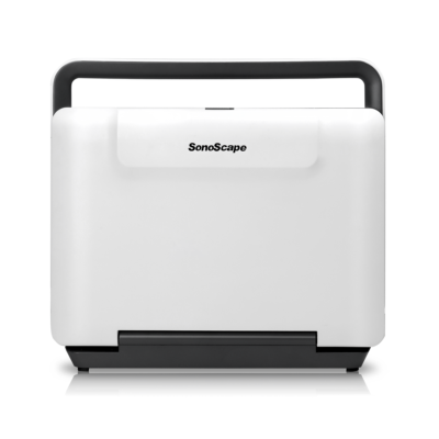
| Shipped from Abroad With ultra-modern innovative design and the clinically-proven technologies, S8 Exp is portable ultrasound scanner well equipped as a low-physical-effort and enhanced-image-quality ultrasound scanner, which not only provides optimized solutions for versatile applications, but does help to improve the user-experience for both routine and non-traditional challenges. Delivery & Availability: Typically 5-7 working days – excluding furniture and heavy/bulky equipment. Please contact us for further information. | Shipped from abroad The DrGem Ceiling Analogue X-ray Machine is a diagnostic radiography system that provides reliable high quality radiographic images with a reduced dose. The reliable high-frequency x-ray generators that are known worldwide for their excellent performance, lifetime and stability. Patient tables and wall stands are also offered. Delivery & Availability: Typically 21 working days – excluding furniture and heavy/bulky equipment. Please contact us for further information. | In Stock JADE is one of the lightest portable X-ray systems on the market, allowing it to be used in any imaginable way including bedside, operating rooms, intensive care units and in veterinary fields. With a simple, easy-to-use operator console, three-way control, two-step foldable stand and auto lock system, JADE is a user-friendly portable X-ray system. Delivery & Availability: Typically 21 working days – excluding furniture and heavy/bulky equipment. Please contact us for further information. | Shipped from Abroad Incorporating innovative technologies, P20’s user-friendly design with a simple operation panel, intuitive user interface and a variety of intelligent auxiliary scanning tools, will significantly improve your daily examination experience. Besides general imaging applications, P20 has entitled with diagnostic 4D technology which has an extraordinary performance in obstetrics and gynecology applications. Delivery & Availability: Typically 5-7 working days – excluding furniture and heavy/bulky equipment. Please contact us for further information. | In Stock A Value Choice beyond Your Expectation. SonoScape’s trolley color Doppler system S11 redefines price and performance with practical design. The S11 will go beyond your expectations but not your budget. Delivery & Availability: Typically 2 working days – excluding furniture and heavy/bulky equipment. Please contact us for further information. |
| Content | MX-600 is designed compactly for easy install and operation.
Features:
Generator
Tube
Radiographic support
Bucky device
Automatic exposure control (AEC)
Collimator: Automatic. Accessories
Options
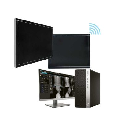
Click Here To Download Catalogue | Sonoscape S8 Exp Portable Ultrasound scannerDETAILS Agile and Versatile With ultra-modern innovative design and the clinically-proven technologies, S8 Exp Portable Ultrasound scanner is well equipped as a low-physical-effort and enhanced-image-quality ultrasound scanner, which not only provides optimized solutions for versatile applications but does help to improve the user experience for both routine and non-traditional challenges. Working with S8 Exp, it will trigger your unlimited reverie and endow you with endless charm. Carrying forward the classical design of SonoScape's portable ultrasound products, S8 Exp successfully combines the best ergonomics, attractive design and ease of use. This charismatic identity is also enhanced by a sophisticated color palette—with sedate grey as its interior paint color and pearl white as exterior cover, S8 Exp reveals a style of aristocrat and strong character among SonoScape's ultrasound systems. Workflow The S8 Exp is a portable ultrasound scanner that adapts to your workflow, whether you are in the consulting room, at the bedside, or at a remote location. With easy-to-use new platform designed for sonographers' needs and full connection interfaces for easy connectivity and data sharing, S8 Exp leads to improved user comfort and clinical outcome as well as patient throughput and working efficiency. Powerful Platform Embedded with SonoScape's core imaging technologies such as μ-scan, PHI and Spatial Compound, S8 Exp boasts exceptional 2D image, sensitive spectral, Color and Power Doppler, displaying well-defined anatomy and pathology and facilitating a highly optimized diagnostic user environment for conclusive diagnoses. Besides, S8 Exp offers a comprehensive selection of electronic probes to maximally extend its capabilities to meet a wide range of applications including the abdomen, pediatric, OB/GYN, cardiovascular, musculoskeletal, etc. The advanced probe technologies also effectively enhance the image quality and confidence in reaching clinical diagnoses even in difficult patients.Click Here To Download Catalogue | DrGem Ceiling Analogue X-ray Machine is a diagnostic radiography system X-ray Machine that provides reliable high quality radiographic images with a reduced dose. The reliable high-frequency x-ray generators that are known worldwide for their excellent performance, lifetime and stability. Patient tables and wall stands are also offered.
Features of DrGem Ceiling Analogue X-ray Machine
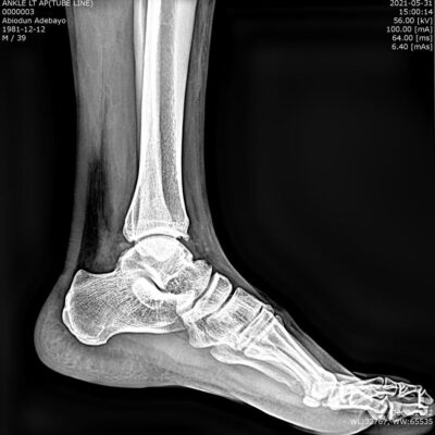
Click Here To Download Catalogue | JADE Mobile X-ray machine is one of the lightest portable X-ray systems on the market, allowing it to be used in any imaginable way including bedside, operating rooms, intensive care units and veterinary fields. With a simple, easy-to-use operator console, three-way control, two-step foldable stand and auto-lock system, the JADE Mobile X-ray machine is a user-friendly portable X-ray system.
Convenient & Intuitive Operation:
JADE is one of the lightest portable X-ray systems on the market, allowing it to be used in any imaginable way including bedside, operating rooms, intensive care units and in veterinary fields. With a simple, easy-to-use operator console, three-way control, two-step foldable stand and auto-lock system, JADE is a user-friendly portable X-ray system.
Compact & Powerful Design:
JADE Mobile X-ray machine is an innovative, highly versatile portable X-ray system suitable for a variety of clinical uses. Utilizing the unique technology used in DRGEM’s universally recognized X-ray generators, JADE is a compact but powerful unit with a 4kW output and thoughtfully designed components to increase efficiency and maximize workflow. The core part of X-ray source adopts high-quality tube assembly, X-ray collimator and high frequency X-ray generator with excellent performance, lifetime and stability.
Features:
Click Here To Download Catalogue | DETAILS
Upgraded Images with More Clarity
SonoScape never stops making progress in improving the image quality of its ultrasound products to enhance the confidence of diagnosis for doctors. With extraordinary images provided by P20, the anatomy structures are clearer than ever.
C-Xlasto Imaging
With C-xlasto Imaging, P20 enables comprehensive quantitative elastic analysis. Meanwhile, C-xlasto on P20 is supported by linear, convex and transvaginal probes, to ensure good reproducibility and highly consistent quantitative elastic results.
S-Live
S-Live allows for detailed visualization of subtle anatomical features, thereby enabling intuitive diagnosis with real-time 3D images and enriching patient communication.
Pelvic Floor 4D
Transperineal 4D pelvic floor ultrasound can provide useful clinical values in assessing the vaginal delivery impact on the female anterior compartment, judging whether the pelvic organs are prolapsed or not and the extent, determining if the pelvic muscles were torn accurately.
Anatomic M Mode
Anatomic M Mode helps you observe the myocardial motion at different phases by freely placing sample lines. It accurately measures the myocardial thickness and the heart size of even difficult patients and supports the myocardial function and LV wall-motion assessment.
Tissue Doppler Imaging
P20 is endowed with Tissue Doppler Imaging which provides velocities and other clinical information on myocardial functions, facilitating clinical doctors with the ability to analyze and compare the motions of different parts of the patient's heart.
Click Here To Download Catalogue | DETAILS
SonoScape’s trolley colour Doppler system S11 redefines price and performance with practical design. The S11 will go beyond your expectations but not your budget. As an easy-to-use ultrasound system, the S11 is integrated with a new software platform, especially optimized for a smooth workflow and convenient operation. The system speeds up the exam process and makes file management easier.
SPECIFICATION
- 15-inch high definition LCD monitor with articulating arm
- Compact and agile trolley design
- 3 active transducer sockets available for a wide range of applications
- Duplex, Color Doppler, DPI, PW Doppler, tissue harmonic imaging, μ-scan speckle reduction imaging, compound imaging, trapezoidal imaging
- Customized settings based on your own working style
- Full patient database and image management solutions
Click Here To Download Catalogue |
| Weight | N/A | N/A | N/A | N/A | N/A | N/A |
| Dimensions | N/A | N/A | N/A | N/A | N/A | N/A |
| Additional information |


Reviews
There are no reviews yet.