
Genoray OSCAR 15 Surgical C-Arm Machine
$0.00
Shipped from Abroad
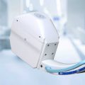
The OSCAR 15 is a culmination of several years worth of developmental experience from Genoray. With CMOS imaging excellence & 15kW HFG you diagnosis need will be met while improving your productivity especially DSA (Digital Subtraction Angiography).
Delivery & Availability:
Typically 21 working days – excluding furniture and heavy/bulky equipment. Please contact us for further information.
Description
The OSCAR 15 is a culmination of several years worth of developmental experience from Genoray. With CMOS imaging excellence & 15kW HFG you diagnosis need will be met while improving your productivity especially DSA (Digital Subtraction Angiography).
APPLICATION
- General Surgery
- Office based Vascular Center
- Pain Management
- Orthopdics
- Urology
- Cardiac Procedures
- Hybrid OR
- Neuro & Spine Surgery
- Pain Management
- Orthopedic Surgery
- Trauma Procedure -Urology Procedure
- Cardiac Surgery
- Peripheral Artery Diseases -Vascular Surgery
SPECIFICATION
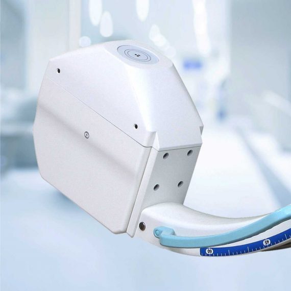
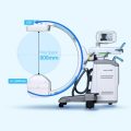
- 260 x 260 mm CMOS Type Flat-Panel detector for distortion-free imaging (High resolution images, Wide FOV, Low Noise)
- 15kW HFG
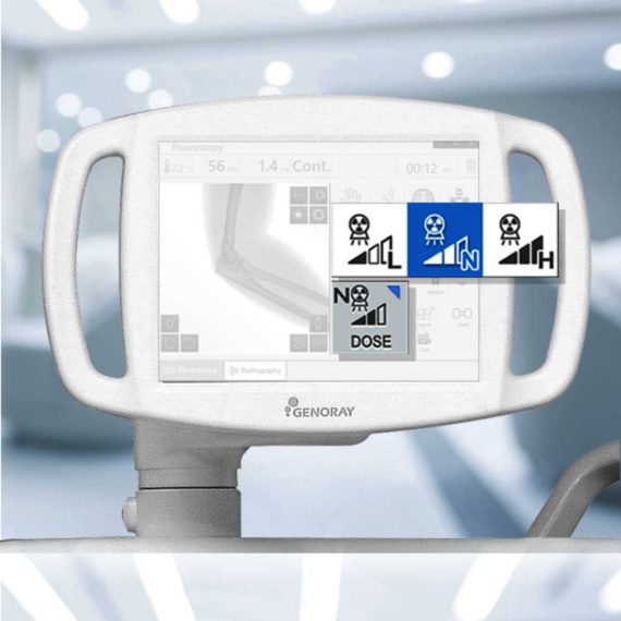
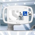
- 4″Touch LCD monitor
- 43″ LCD Monitor
- Dual Foot Switch
- DICOM 3.0 -CD/DVD Burner – USB Port
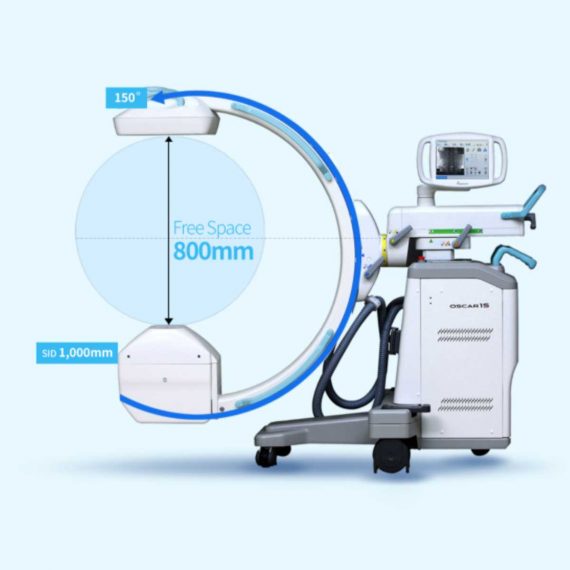
- 800mm free space and 150° (+90°,-60°) orbital rotation, SID 1,000mm
- 155º Dynamic Orbital Rotation
- 2 kW Stationary Anode X-Ray Tube
- 5 Million Images Storage Capacity
FEATURE
Exceptional Image Quality
- With an optimal flat panel detector size of 26 x 26cm you won’t miss a thing with its high quality resolution. It makes accurate diagnosis in a variety of departments especially DSA
Low Dose Mode
- Low dose mode is desgined to acquire reasonable image to diagnose the patient with minimum dosage.
Edge Enhancenment
- For user to get more accurate diagnosis result enhancing edge of image.
Motion Correction
- This function detects the movement and reduce the after image while exposing X-ray.
Metal Correction
- To prevent over-dose radiation or low quality image casued by metal inturrption on field of view.
Virtual Collimator
- The virtual collimator allows for the selection of your desired field of view, while reducing the amount of radiation exposure by limiting the X-Ray beam.
Auto Collimation
- Prevention of unncessary X-Ray exposure by focusing on the area of interest while autmoatically collimating the remaining areas.
POWERFULL SOFTWARE ZENIS
A total solution from acquisition, storage, management, communication to print out. Provide convenient environment from user-centric interface. Diagnosis and confirm from recognizable simple icons. Convenience of database management.
- Convenient diagnostic functions for easy patient / image management
- Accurate diagnostic tools
- Improve the efficiency of your hospital management
- Perfect compatibility with all PACS
- A must have for a digitally equipped hospital
- Convenient communication and management for your customers
- Dicom Support
DIGITAL SUBTRACTION ANGIOGRAPHY
Native DSA
- Pairing fluoroscopy with constrast media to display the basic angiography views
Motion Matching
- Selects the proper mask to apply and remove artifacts made by a patient’s movement or breathing
Post-Processing
- Processing: Improvement of the processed image after the DSA procedure
Landmarking / Brightness / Contrast
- After setting the position for a vessel, the subject can be placed back to their original position by using the shift function to compensate for any movement. Allows for various functions that assist with accurately inserting a catheter.
Peak Opacification
- Ability to diagnose a blood vessel with only a small amount of contrast media
Road Mapping, Land Mark
- After setting a position for a vessel, the subject can be moved back to their original place by using the shift function to compensate for any movement. Provides various functions that helps accurately to insert a guide wire, catheter is compatible with the hybrid operating room.
Auto Roadmap Mask
- Obtain blood vessel type information while only using a small amount of contrast media
Manual Roadmap Mask
- Roadmap your vessels using a prevoiusly taken DSA image
Roadmap Pixel Shift
- Re-position the roadmap mask by shifting the pixels to the proper position
Click Here To Download Catalogue
Review(1)
Quick Comparison
| Genoray OSCAR 15 Surgical C-Arm Machine remove | Sonoscape E2 Ultrasound Machine remove | Sonoscape P10 Ultrasound Machine remove | Sonoscape S22 Ultrasound Machine remove | Topaz Digital X-ray Machine remove | DrGem Ceiling Analogue X-ray Machine remove | |
|---|---|---|---|---|---|---|
| Name | Genoray OSCAR 15 Surgical C-Arm Machine remove | Sonoscape E2 Ultrasound Machine remove | Sonoscape P10 Ultrasound Machine remove | Sonoscape S22 Ultrasound Machine remove | Topaz Digital X-ray Machine remove | DrGem Ceiling Analogue X-ray Machine remove |
| Image | 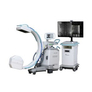 |  |  |  |  |  |
| SKU | SF1033560422-1 | SF1033560012-17 | SF1033560012-7 | SF1033560012-3 | SF1033560074-1 | SF1033560074-7 |
| Rating | ||||||
| Price |
| $5,500.00 | $9,350.00 | $9,350.00 |
|
|
| Stock | ||||||
| Availability | ||||||
| Add to cart | ||||||
| Description | Shipped from Abroad The OSCAR 15 is a culmination of several years worth of developmental experience from Genoray. With CMOS imaging excellence & 15kW HFG you diagnosis need will be met while improving your productivity especially DSA (Digital Subtraction Angiography). Delivery & Availability: Typically 21 working days – excluding furniture and heavy/bulky equipment. Please contact us for further information. | Shipped from Abroad Sonoscape E2 portable ultrasound machine is a color Doppler ultrasound system that reaches beyond your expectations due to its compact and fashionable appearance. It fulfills GI, OB/GYN, Cardiac and POC applications to fit your routine scanning needs while its color mode will help you for more accurate and efficient diagnosis of lesions. E2 provides a wide range of applications to assist users with routine scanning. E2 provides automatic calculations to enhance your diagnostic confidence and save you time for patient communication. Delivery & Availability: Typically 14 working days – excluding furniture and heavy/bulky equipment. Please contact us for further information. | Shipped from Abroad The P10 color Doppler ultrasound system is a new generation product from SonoScape. It is designed to give high quality images, rich probe configurations, various clinical tools and automatic analysis software to provide you with comprehensive solutions for your growing demand for clinical applications. Delivery & Availability: Typically 5-7 working days – excluding furniture and heavy/bulky equipment. Please contact us for further information. | Shipped from Abroad As SonoScape steps forward to add value and efficiency to ultrasound, the latest S22 was designed in a user-friendly platform to address current and future demanding needs. It represents an excellent mix in performance and price. Delivery & Availability: Typically 5-7 working days – excluding furniture and heavy/bulky equipment. Please contact us for further information. | In Stock DRGEM’s TOPAZ X-ray machine is a state-of-the-art mobile digital radiography system, designed with maximum comfort for patients and users in mind. From its user-friendly software to smooth movements, TOPAZ is made to improve your workflow and provide you with high-quality images. Delivery & Availability: Typically 21 working days – excluding furniture and heavy/bulky equipment. Please contact us for further information. | Shipped from abroad The DrGem Ceiling Analogue X-ray Machine is a diagnostic radiography system that provides reliable high quality radiographic images with a reduced dose. The reliable high-frequency x-ray generators that are known worldwide for their excellent performance, lifetime and stability. Patient tables and wall stands are also offered. Delivery & Availability: Typically 21 working days – excluding furniture and heavy/bulky equipment. Please contact us for further information. |
| Content | The OSCAR 15 is a culmination of several years worth of developmental experience from Genoray. With CMOS imaging excellence & 15kW HFG you diagnosis need will be met while improving your productivity especially DSA (Digital Subtraction Angiography).
APPLICATION
Click Here To Download Catalogue | SONOSCAPE E2 DETAILS
Auto Image Optimization
A portable ultrasound machine with the press of a button, the image is automatically adjusted and optimized, saving you time with parameter adjustments. Additionally, with Auto Focus on, the focus area follows the depth of the ROI box as it is moved in the scanning field, providing users with excellent image quality in the desired area of interest.
Automated Calculation
Auto IMT is used when determining the level of vascular sclerosis present in the patient by automatically tracing the thickness of the carotid vessels.
Auto trace provides users sensitive and accurate wave tracing, avoiding the error of manual trace and giving out calculation result in no time
In-Build Battery pack
This portable ultrasound machine was equipped with an in-build battery pack which enable the user to perform image scanning when AC power is not available.
Click Here To Download Catalogue | DETAILS
B + Compound
B + Compound utilizes several lines of sight for optimal contrast resolution, speckle reduction and border detection, with which P10 is ideal for superficial and abdominal imaging with better clarity and improved continuity of structures.
μ-Scan
The new generation μ-Scan imaging technology gives you better image quality by reducing noise, improving signal strength and improving visualization.
P10 offers a comprehensive selection of electronic probes to maximize its capabilities to meet a wide range of applications including abdomen, pediatric, OB/GYN, cardiovascular, musculoskeletal, etc. The advanced probe technologies also effectively enhance the image quality and confidence in reaching clinical diagnoses, even in difficult patients.
Convex Probe 3C-A
Ideal for an abundant of application such as abdomen, gynecology, obstetrics, urology and even abdomen biopsy.
Linear Probe L741
This linear probe is designed to satisfy vascular, breast, thyroid, and other small parts diagnosis, and its adjustable parameters could also present users a clear view of MSK and deep vessels.
Phase Array Probe 3P-A
For the purpose of adult and pediatric cardiology and emergency, the phase array probe provides elaborate presets for different exam modes, even for difficult patients.
Intracavitary Probe 6V1
Intracavitary probe could face application of gynecology, urology, prostate, and its temperature detection technology not only protects the patient but also extends the service life.
Click Here To Download Catalogue | DETAILS
As SonoScape steps forward to add value and efficiency to ultrasound, the latest S22 was designed in a user-friendly platform to address current and future demanding needs. It represents an excellent mix in performance and price.
S22, is a shared service ultrasound system with a slim and elegant package that has combined mobility with utility to fit in specific clinical situations including emergency department, ICU, operating room and so on. Furthermore, its ergonomic design, easy operating and flexible data management will give you a memorable experience.
SPECIFICATION
• Large high-resolution widescreen LED
• Sensitive touch screen
• Four transducer sockets plus one socket for pencil probe
• A comprehensive selection of probes: linear, Convex, Micro-convex, Volumetric, Endocavity, Bi-plane, Phased Array, TEE, Intraoperative, Pencil
• Premium application technology: 4D, μ-scan speckle reduction, compound imaging, Pulse Inversion Harmonic Imaging, Color M-Mode, Steer M-Mode, PDI, TDI, Real-time Panoramic Imaging, Trapezoid Imaging, Auto-IMT…
• Full patient database and image management solutions: DICOM 3.0, AVI/JPG, USB 2.0, HDD, DVD, PDF report
• Multi-Language Input Keyboard
• Built-in battery
Click Here To Download Catalogue | TOPAZ X-ray machine is among the high end X-ray machine manufactured by DRGEM, a digital X-ray system that provides quality images with little or no effort.
It begins with Advanced Technology
Integrating high technology and over a decade of experience in conventional and digital radiography systems, DRGEM’s TOPAZ X-ray machine is a state-of-the-art mobile digital radiography system, designed with maximum comfort for patients and users. From its user-friendly software to smooth movements, TOPAZ X-ray machine is made to improve your workflow and provide you with high-quality images.
Full Featured Imaging Software & Excellent Digital Image Processing
With a high-performance, built-in touchscreen, TOPAZ X-ray machine offers a user-friendly interface and powerful software for easy operation and increased workflow. The anatomical view-based digital image processing, automatically optimizes and enhances the quality of the image. it also comes with automatic image storage and print with DICOM 3.0 networking capability. additionally, the system offers increasing exam throughput while decreasing examination time.
Click Here To Download Catalogue | DrGem Ceiling Analogue X-ray Machine is a diagnostic radiography system X-ray Machine that provides reliable high quality radiographic images with a reduced dose. The reliable high-frequency x-ray generators that are known worldwide for their excellent performance, lifetime and stability. Patient tables and wall stands are also offered.
Features of DrGem Ceiling Analogue X-ray Machine
Click Here To Download Catalogue |
| Weight | N/A | N/A | N/A | N/A | N/A | N/A |
| Dimensions | N/A | N/A | N/A | N/A | N/A | N/A |
| Additional information |


Aiden
Excellent web site you’ve got here.. It’s difficult to
find high quality writing like yours nowadays. I truly appreciate individuals like you!
Take care!!
samson faluro
Thanks for your comment