
Genoray OSCAR 15 Surgical C-Arm Machine
$0.00
Shipped from Abroad
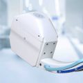
The OSCAR 15 is a culmination of several years worth of developmental experience from Genoray. With CMOS imaging excellence & 15kW HFG you diagnosis need will be met while improving your productivity especially DSA (Digital Subtraction Angiography).
Delivery & Availability:
Typically 21 working days – excluding furniture and heavy/bulky equipment. Please contact us for further information.
Description
The OSCAR 15 is a culmination of several years worth of developmental experience from Genoray. With CMOS imaging excellence & 15kW HFG you diagnosis need will be met while improving your productivity especially DSA (Digital Subtraction Angiography).
APPLICATION
- General Surgery
- Office based Vascular Center
- Pain Management
- Orthopdics
- Urology
- Cardiac Procedures
- Hybrid OR
- Neuro & Spine Surgery
- Pain Management
- Orthopedic Surgery
- Trauma Procedure -Urology Procedure
- Cardiac Surgery
- Peripheral Artery Diseases -Vascular Surgery
SPECIFICATION
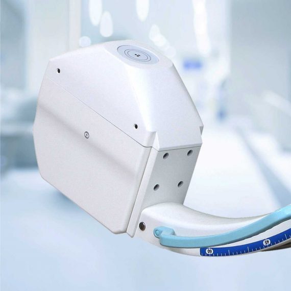
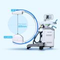
- 260 x 260 mm CMOS Type Flat-Panel detector for distortion-free imaging (High resolution images, Wide FOV, Low Noise)
- 15kW HFG
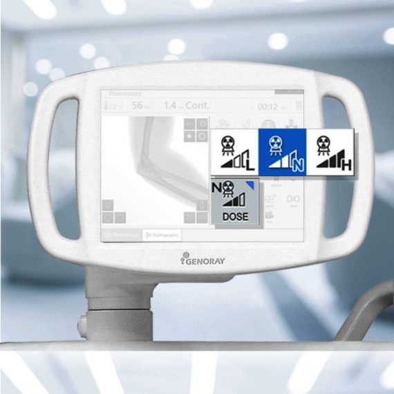
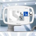
- 4″Touch LCD monitor
- 43″ LCD Monitor
- Dual Foot Switch
- DICOM 3.0 -CD/DVD Burner – USB Port
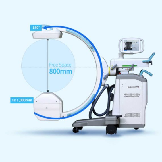
- 800mm free space and 150° (+90°,-60°) orbital rotation, SID 1,000mm
- 155º Dynamic Orbital Rotation
- 2 kW Stationary Anode X-Ray Tube
- 5 Million Images Storage Capacity
FEATURE
Exceptional Image Quality
- With an optimal flat panel detector size of 26 x 26cm you won’t miss a thing with its high quality resolution. It makes accurate diagnosis in a variety of departments especially DSA
Low Dose Mode
- Low dose mode is desgined to acquire reasonable image to diagnose the patient with minimum dosage.
Edge Enhancenment
- For user to get more accurate diagnosis result enhancing edge of image.
Motion Correction
- This function detects the movement and reduce the after image while exposing X-ray.
Metal Correction
- To prevent over-dose radiation or low quality image casued by metal inturrption on field of view.
Virtual Collimator
- The virtual collimator allows for the selection of your desired field of view, while reducing the amount of radiation exposure by limiting the X-Ray beam.
Auto Collimation
- Prevention of unncessary X-Ray exposure by focusing on the area of interest while autmoatically collimating the remaining areas.
POWERFULL SOFTWARE ZENIS
A total solution from acquisition, storage, management, communication to print out. Provide convenient environment from user-centric interface. Diagnosis and confirm from recognizable simple icons. Convenience of database management.
- Convenient diagnostic functions for easy patient / image management
- Accurate diagnostic tools
- Improve the efficiency of your hospital management
- Perfect compatibility with all PACS
- A must have for a digitally equipped hospital
- Convenient communication and management for your customers
- Dicom Support
DIGITAL SUBTRACTION ANGIOGRAPHY
Native DSA
- Pairing fluoroscopy with constrast media to display the basic angiography views
Motion Matching
- Selects the proper mask to apply and remove artifacts made by a patient’s movement or breathing
Post-Processing
- Processing: Improvement of the processed image after the DSA procedure
Landmarking / Brightness / Contrast
- After setting the position for a vessel, the subject can be placed back to their original position by using the shift function to compensate for any movement. Allows for various functions that assist with accurately inserting a catheter.
Peak Opacification
- Ability to diagnose a blood vessel with only a small amount of contrast media
Road Mapping, Land Mark
- After setting a position for a vessel, the subject can be moved back to their original place by using the shift function to compensate for any movement. Provides various functions that helps accurately to insert a guide wire, catheter is compatible with the hybrid operating room.
Auto Roadmap Mask
- Obtain blood vessel type information while only using a small amount of contrast media
Manual Roadmap Mask
- Roadmap your vessels using a prevoiusly taken DSA image
Roadmap Pixel Shift
- Re-position the roadmap mask by shifting the pixels to the proper position
Click Here To Download Catalogue
Review(1)
Quick Comparison
| Genoray OSCAR 15 Surgical C-Arm Machine remove | DrGem Ceiling Analogue X-ray Machine remove | ASPEL AsPEKT 712 Holter Monitor and Software remove | Sonoscape P15 Ultrasound Machine With Four Probes remove | ASPEL AsCARD Coral PC Based ECG Machine remove | Sonoscape S8 Exp Portable Ultrasound remove | |
|---|---|---|---|---|---|---|
| Name | Genoray OSCAR 15 Surgical C-Arm Machine remove | DrGem Ceiling Analogue X-ray Machine remove | ASPEL AsPEKT 712 Holter Monitor and Software remove | Sonoscape P15 Ultrasound Machine With Four Probes remove | ASPEL AsCARD Coral PC Based ECG Machine remove | Sonoscape S8 Exp Portable Ultrasound remove |
| Image | 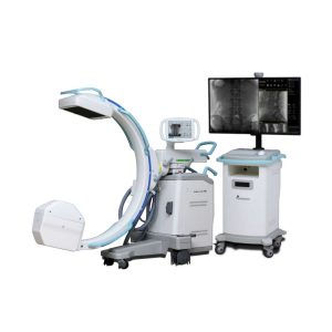 |  |  |  | 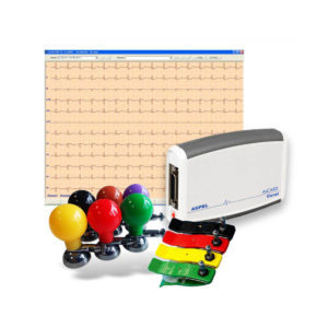 |  |
| SKU | SF1033560422-1 | SF1033560074-7 | SF1033560075-4 | SF1033560012-8 | SF1033560075-11 | SF1033560012-15 |
| Rating | ||||||
| Price |
|
| $1,991.00 | $13,900.00 | $486.00 | $9,350.00 |
| Stock | ||||||
| Availability | ||||||
| Add to cart | ||||||
| Description | Shipped from Abroad The OSCAR 15 is a culmination of several years worth of developmental experience from Genoray. With CMOS imaging excellence & 15kW HFG you diagnosis need will be met while improving your productivity especially DSA (Digital Subtraction Angiography). Delivery & Availability: Typically 21 working days – excluding furniture and heavy/bulky equipment. Please contact us for further information. | Shipped from abroad The DrGem Ceiling Analogue X-ray Machine is a diagnostic radiography system that provides reliable high quality radiographic images with a reduced dose. The reliable high-frequency x-ray generators that are known worldwide for their excellent performance, lifetime and stability. Patient tables and wall stands are also offered. Delivery & Availability: Typically 21 working days – excluding furniture and heavy/bulky equipment. Please contact us for further information. | Shipped from Abroad The Holta Monitor allows quick analysis of ECG examination and detection, reviewing and editing capability in the qualitative assessment of VE, VT, Single SVE, PSVT, Pauses, Irregular Rhythm, VT, IVR, Brady - and Tachycardia, Couplets, ST-segment elevation and depression, Maximum, Minimum and averaged Heart Rates, artifacts Delivery & Availability: Typically 10 working days – excluding furniture and heavy/bulky equipment. Please contact us for further information. | In Stock A feature-rich system inheriting the Wi-Sono high-end platform, the P15 uses an array of advanced tools to help enhance the image quality. It's a cost-effective, simplified console with an intuitive user interface and multiple intelligent functions. Delivery & Availability: Typically 2 working days – excluding furniture and heavy/bulky equipment. Please contact us for further information. | Shipped from Abroad AsCARD Coral electrocardiograph is a 3-, 6-, 12-channel ECG equipped with CardioTEKA software allows transmission of full 12 ECG leads to the user PC through USB interface. It is intended for carrying out ECG examinations in adults and pediatric patients in all types of health care centres. ECG procedures can be performed by qualified personnel only. AsCARD Coral can cooperate also with CardioTEST system as 12-channel ECG device allows transmission of full 12 ECG leads to the user PC through USB interface. Delivery & Availability: Typically 10 working days – excluding furniture and heavy/bulky equipment. Please contact us for further information. | Shipped from Abroad With ultra-modern innovative design and the clinically-proven technologies, S8 Exp is portable ultrasound scanner well equipped as a low-physical-effort and enhanced-image-quality ultrasound scanner, which not only provides optimized solutions for versatile applications, but does help to improve the user-experience for both routine and non-traditional challenges. Delivery & Availability: Typically 5-7 working days – excluding furniture and heavy/bulky equipment. Please contact us for further information. |
| Content | The OSCAR 15 is a culmination of several years worth of developmental experience from Genoray. With CMOS imaging excellence & 15kW HFG you diagnosis need will be met while improving your productivity especially DSA (Digital Subtraction Angiography).
APPLICATION
Click Here To Download Catalogue | DrGem Ceiling Analogue X-ray Machine is a diagnostic radiography system X-ray Machine that provides reliable high quality radiographic images with a reduced dose. The reliable high-frequency x-ray generators that are known worldwide for their excellent performance, lifetime and stability. Patient tables and wall stands are also offered.
Features of DrGem Ceiling Analogue X-ray Machine
Click Here To Download Catalogue | The Holter Monitor allows quick analysis of ECG examination (arrhythmias and ST segment).
Technical specifications:
HolCARD 24W Software:
Click Here To Download Catalogue | DETAILS
Super Wide-bandwidth Platform
Inheriting Wi-sono's ultra-wide system platform and with the advanced probe technology, high-resolution and deep penetration images are provided for precision medicine.
Spatial Compound Imaging
Spatial Compound Imaging utilizes several lines of sight for optimal contrast resolution, speckle reduction and border detection, with which P15 is ideal for superficial and abdominal imaging with better clarity and improved continuity of structures.
μ-Scan+
The new generation μ-Scan imaging technology gives you better image quality by reducing noise, improving signal strength and improving visualization.
Dynamic Color
Dynamic color improves upon already existing color Doppler technologies for a clearer capture of color flow and detailed visualization of even tiny veins with lower velocities.
Real-time Panoramic
With real-time panoramic, you can acquire an extended field of view for large organs or long vessels for easy measurement and diagnostic efficiency. Accomplished in real-time for the convenience of the sonographers, any mistake can also be easily back tracked and corrected without interrupting the scan.
3D/4D
Outstanding volume performance with speed and convenience makes P15 outshine others on volume imaging.
Tissue Doppler Imaging
Tissue Doppler Imaging allows clinical doctors to quantitatively evaluate local myocardial movements and functions, facilitating them with the ability to analyze and compare the motions of the different parts of the patient's heart.
Auto IMT
Quick measurement of intra-media vessel thickness ensures good reproducibility and high diagnostic efficiency.
Click Here To Download Catalogue |
AsCARD Coral electrocardiograph is a 3-, 6-, 12-channel ECG equipped with CardioTEKA software allows transmission of full 12 ECG leads to the user PC through USB interface. It is intended for carrying out ECG examinations in adults and pediatric patients in all types of health care centres. ECG procedures can be performed by qualified personnel only. AsCARD Coral can cooperate also with CardioTEST system as 12-channel ECG device allows transmission of full 12 ECG leads to the user PC through USB interface.
Technical Specification:
Click Here To Download Catalogue | Sonoscape S8 Exp Portable Ultrasound scannerDETAILS Agile and Versatile With ultra-modern innovative design and the clinically-proven technologies, S8 Exp Portable Ultrasound scanner is well equipped as a low-physical-effort and enhanced-image-quality ultrasound scanner, which not only provides optimized solutions for versatile applications but does help to improve the user experience for both routine and non-traditional challenges. Working with S8 Exp, it will trigger your unlimited reverie and endow you with endless charm. Carrying forward the classical design of SonoScape's portable ultrasound products, S8 Exp successfully combines the best ergonomics, attractive design and ease of use. This charismatic identity is also enhanced by a sophisticated color palette—with sedate grey as its interior paint color and pearl white as exterior cover, S8 Exp reveals a style of aristocrat and strong character among SonoScape's ultrasound systems. Workflow The S8 Exp is a portable ultrasound scanner that adapts to your workflow, whether you are in the consulting room, at the bedside, or at a remote location. With easy-to-use new platform designed for sonographers' needs and full connection interfaces for easy connectivity and data sharing, S8 Exp leads to improved user comfort and clinical outcome as well as patient throughput and working efficiency. Powerful Platform Embedded with SonoScape's core imaging technologies such as μ-scan, PHI and Spatial Compound, S8 Exp boasts exceptional 2D image, sensitive spectral, Color and Power Doppler, displaying well-defined anatomy and pathology and facilitating a highly optimized diagnostic user environment for conclusive diagnoses. Besides, S8 Exp offers a comprehensive selection of electronic probes to maximally extend its capabilities to meet a wide range of applications including the abdomen, pediatric, OB/GYN, cardiovascular, musculoskeletal, etc. The advanced probe technologies also effectively enhance the image quality and confidence in reaching clinical diagnoses even in difficult patients.Click Here To Download Catalogue |
| Weight | N/A | N/A | N/A | N/A | N/A | N/A |
| Dimensions | N/A | N/A | N/A | N/A | N/A | N/A |
| Additional information |


Aiden
Excellent web site you’ve got here.. It’s difficult to
find high quality writing like yours nowadays. I truly appreciate individuals like you!
Take care!!
samson faluro
Thanks for your comment