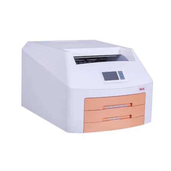
HU.Q hq-460dy Dry Image X-Ray Film Printer
$3,850.00
Shipped From Abroad
- It is the one and only domestically engineered medical dry thermal imager. The HQ-DY series Dry Imager uses the latest direct dry thermal imaging technology which accommodates for a full range of applications, including CT, MR, DSA and US, as well as CR/DR applications for GenRad, Orthopaedics, Dental Imaging and more
Delivery & Availability:
Typically 14-21 working days – excluding furniture and heavy/bulky equipment. Please contact us for further information.
Description
It is the one and only domestically engineered medical dry thermal imager. The HQ-DY series Dry Imager uses the latest direct dry thermal imaging technology which accommodates for a full range of applications, including CT, MR, DSA and US, as well as CR/DR applications for GenRad, Orthopaedics, Dental Imaging and more. The HQ-Series Dry Imager dedicates to precision in diagnosis with its outstanding image quality, and offers affordable imaging catering to your needs.
– Dry thermal technology
– Daylight load film cartridges
– Double tray, supports 4 film sizes
– Speed printing, higher efficiency
– Economical, stable, reliable
– Compact design, easy installation
– Straight forward operation, user-friendly
Usage
The HQ-DY series dry imager is a medical imaging output device. It is engineered to achieve its optimal performance when used with HQ-brand medical dry films. Different from the old method of film processors, our dry imager can be operated under daylight condition. With the elimination of chemical liquid, this thermal dry printing technology is significantly more environmentally friendly. However, to ensure the quality of output image, please keep away from heat source, direct sunlight, and acid and alkaline gas such as hydrogen sulfide, ammonia, sulfur dioxide, and formaldehyde, etc.
Technical Specification:
|
Print Technology |
Direct thermal (dry, daylight-load film) |
|
Spatial Resolution |
320dpi (12.6 pixels/mm) |
|
Grayscale Contrast Resolution |
14 bits |
|
Film Tray |
Two supply trays, 200-sheet capacity total |
|
Film Sizes |
8’’×10’’, 10’’×12’’, 11’’×14’’, 14’’×17’’ |
|
Applicable film |
Medical Dry Thermal Film (blue or clear base) |
|
Interface |
10/100/1000 Base-T Ethernet (RJ-45) |
|
Network Protocol |
Standard DICOM 3.0 connection |
|
Image Quality |
Automatic calibration using built-in densitometer |
|
Control Panel |
Touch Screen, Online Display, Alert, Fault and Active |
|
Power Supply |
100-240VAC 50/60Hz 600W |
|
Weight |
50Kg |
|
Operating Temperature |
5℃-35℃ |
|
Storage Humidity |
30%-95% |
|
Storage Temperature |
-22℃-50℃ |
Click Here To Download Catalogue
Review(1)
Quick Comparison
| HU.Q hq-460dy Dry Image X-Ray Film Printer remove | SIGNERS SUPiA X-ray Digitizer ( CR Scanner) remove | Sonoscape E1 Ultrasound Machine With Two Probes remove | Sonoscape P15 Ultrasound Machine With Four Probes remove | Sonoscape P20 Ultrasound Machine remove | Single X-Ray Viewing Box remove | |||||||||||||||||||||||||||||||||||||||||||||||||||||||
|---|---|---|---|---|---|---|---|---|---|---|---|---|---|---|---|---|---|---|---|---|---|---|---|---|---|---|---|---|---|---|---|---|---|---|---|---|---|---|---|---|---|---|---|---|---|---|---|---|---|---|---|---|---|---|---|---|---|---|---|---|
| Name | HU.Q hq-460dy Dry Image X-Ray Film Printer remove | SIGNERS SUPiA X-ray Digitizer ( CR Scanner) remove | Sonoscape E1 Ultrasound Machine With Two Probes remove | Sonoscape P15 Ultrasound Machine With Four Probes remove | Sonoscape P20 Ultrasound Machine remove | Single X-Ray Viewing Box remove | ||||||||||||||||||||||||||||||||||||||||||||||||||||||
| Image |  | 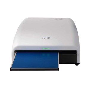 |  |  |  |  | ||||||||||||||||||||||||||||||||||||||||||||||||||||||
| SKU | SF10335601102 | SF1033560050-01 | SF1033560012-20 | SF1033560012-8 | SF1033560012-9 | SF1033560084-203 | ||||||||||||||||||||||||||||||||||||||||||||||||||||||
| Rating | ||||||||||||||||||||||||||||||||||||||||||||||||||||||||||||
| Price | $3,850.00 | $6,930.00 | $4,620.00 | $13,900.00 |
| $95.20 | ||||||||||||||||||||||||||||||||||||||||||||||||||||||
| Stock | ||||||||||||||||||||||||||||||||||||||||||||||||||||||||||||
| Availability | ||||||||||||||||||||||||||||||||||||||||||||||||||||||||||||
| Add to cart | ||||||||||||||||||||||||||||||||||||||||||||||||||||||||||||
| Description | Shipped From Abroad
| Shipped from Abroad SUPiA made by Signers offers such a better clinic environment with no chemicals, ideal space, high-resolution image quality, and affordability. Delivery & Availability: Typically 14 working days – excluding furniture and heavy/bulky equipment. Please contact us for further information. | Shipped from Abroad SonoScape has developed a new probe and function for the E1 Exp. With these additions the E1 Exp will bring users a more efficient examination experience with satisfying image quality and a smooth workflow. Delivery & Availability: Typically 5-7 working days – excluding furniture and heavy/bulky equipment. Please contact us for further information. | In Stock A feature-rich system inheriting the Wi-Sono high-end platform, the P15 uses an array of advanced tools to help enhance the image quality. It's a cost-effective, simplified console with an intuitive user interface and multiple intelligent functions. Delivery & Availability: Typically 2 working days – excluding furniture and heavy/bulky equipment. Please contact us for further information. | Shipped from Abroad Incorporating innovative technologies, P20’s user-friendly design with a simple operation panel, intuitive user interface and a variety of intelligent auxiliary scanning tools, will significantly improve your daily examination experience. Besides general imaging applications, P20 has entitled with diagnostic 4D technology which has an extraordinary performance in obstetrics and gynecology applications. Delivery & Availability: Typically 5-7 working days – excluding furniture and heavy/bulky equipment. Please contact us for further information. | In stock
| ||||||||||||||||||||||||||||||||||||||||||||||||||||||
| Content | It is the one and only domestically engineered medical dry thermal imager. The HQ-DY series Dry Imager uses the latest direct dry thermal imaging technology which accommodates for a full range of applications, including CT, MR, DSA and US, as well as CR/DR applications for GenRad, Orthopaedics, Dental Imaging and more. The HQ-Series Dry Imager dedicates to precision in diagnosis with its outstanding image quality, and offers affordable imaging catering to your needs.
- Dry thermal technology
- Daylight load film cartridges
- Double tray, supports 4 film sizes
- Speed printing, higher efficiency
- Economical, stable, reliable
- Compact design, easy installation
- Straight forward operation, user-friendly
UsageThe HQ-DY series dry imager is a medical imaging output device. It is engineered to achieve its optimal performance when used with HQ-brand medical dry films. Different from the old method of film processors, our dry imager can be operated under daylight condition. With the elimination of chemical liquid, this thermal dry printing technology is significantly more environmentally friendly. However, to ensure the quality of output image, please keep away from heat source, direct sunlight, and acid and alkaline gas such as hydrogen sulfide, ammonia, sulfur dioxide, and formaldehyde, etc. Technical Specification:
Click Here To Download Catalogue | SUPiA X-ray Digitizer made by Signers offers such a better clinic environment with no chemicals, ideal space, high-resolution image quality, and affordability
FEATURE
Rigid Type
• No damage or scratch on image plates during scanning & erasing
• Scanning & Erasing without a roller
• No cut-off image during winter and cold period
Durability
• Extremely simple structure design
• Strong aluminum base plate
• Flip covers preventing dust from inside scanner
Barcode System
• Automatically recognising cassette sizes(14x17", 10x12", 18x24cm) by barcode reader
Compact & lightweight design
Click Here To Download Catalogue | DETAILS
Efficient Diagnosis
μ-Scan, Speckle Reduction & Edge Enhancement
Spatial Compound Imaging
PIH - Pure Inversion Harmonic
Wide Scan - Enlarged Image Area
Tissue-Specific Imaging
SR Flow
Ergonomic Designs
Up to 2 Transducer Ports
Light Weight and Compact
15.6 inch Anti-flickering HD LED Screen
Tilting Monitor Angle Adjustment
Backlit Keyboard and Intelligent Panel
Long-lasting Battery for 90 mins
Ease of Use
Quick Boot Up
Auto-Brightness Adjustment
Auto Image Optimization
Auto IMT
Auto Trace
Equipped Accessories
Wi-Fi and Bluetooth Available
DICOM
500GB Hard Disk
Height Adjustable Trolley
Durable, Carry-on Site Suitcase
Click Here To Download Catalogue | DETAILS
Super Wide-bandwidth Platform
Inheriting Wi-sono's ultra-wide system platform and with the advanced probe technology, high-resolution and deep penetration images are provided for precision medicine.
Spatial Compound Imaging
Spatial Compound Imaging utilizes several lines of sight for optimal contrast resolution, speckle reduction and border detection, with which P15 is ideal for superficial and abdominal imaging with better clarity and improved continuity of structures.
μ-Scan+
The new generation μ-Scan imaging technology gives you better image quality by reducing noise, improving signal strength and improving visualization.
Dynamic Color
Dynamic color improves upon already existing color Doppler technologies for a clearer capture of color flow and detailed visualization of even tiny veins with lower velocities.
Real-time Panoramic
With real-time panoramic, you can acquire an extended field of view for large organs or long vessels for easy measurement and diagnostic efficiency. Accomplished in real-time for the convenience of the sonographers, any mistake can also be easily back tracked and corrected without interrupting the scan.
3D/4D
Outstanding volume performance with speed and convenience makes P15 outshine others on volume imaging.
Tissue Doppler Imaging
Tissue Doppler Imaging allows clinical doctors to quantitatively evaluate local myocardial movements and functions, facilitating them with the ability to analyze and compare the motions of the different parts of the patient's heart.
Auto IMT
Quick measurement of intra-media vessel thickness ensures good reproducibility and high diagnostic efficiency.
Click Here To Download Catalogue | DETAILS
Upgraded Images with More Clarity
SonoScape never stops making progress in improving the image quality of its ultrasound products to enhance the confidence of diagnosis for doctors. With extraordinary images provided by P20, the anatomy structures are clearer than ever.
C-Xlasto Imaging
With C-xlasto Imaging, P20 enables comprehensive quantitative elastic analysis. Meanwhile, C-xlasto on P20 is supported by linear, convex and transvaginal probes, to ensure good reproducibility and highly consistent quantitative elastic results.
S-Live
S-Live allows for detailed visualization of subtle anatomical features, thereby enabling intuitive diagnosis with real-time 3D images and enriching patient communication.
Pelvic Floor 4D
Transperineal 4D pelvic floor ultrasound can provide useful clinical values in assessing the vaginal delivery impact on the female anterior compartment, judging whether the pelvic organs are prolapsed or not and the extent, determining if the pelvic muscles were torn accurately.
Anatomic M Mode
Anatomic M Mode helps you observe the myocardial motion at different phases by freely placing sample lines. It accurately measures the myocardial thickness and the heart size of even difficult patients and supports the myocardial function and LV wall-motion assessment.
Tissue Doppler Imaging
P20 is endowed with Tissue Doppler Imaging which provides velocities and other clinical information on myocardial functions, facilitating clinical doctors with the ability to analyze and compare the motions of different parts of the patient's heart.
Click Here To Download Catalogue |
| ||||||||||||||||||||||||||||||||||||||||||||||||||||||
| Weight | N/A | N/A | N/A | N/A | N/A | N/A | ||||||||||||||||||||||||||||||||||||||||||||||||||||||
| Dimensions | N/A | N/A | N/A | N/A | N/A | N/A | ||||||||||||||||||||||||||||||||||||||||||||||||||||||
| Additional information |


Umar Halilu
I need to buy the dry imager 460 and it’s film