HU.Q hq-460dy Dry Image X-Ray Film Printer
$3,850.00
Shipped From Abroad
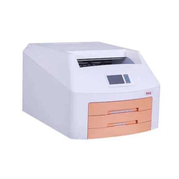
- It is the one and only domestically engineered medical dry thermal imager. The HQ-DY series Dry Imager uses the latest direct dry thermal imaging technology which accommodates for a full range of applications, including CT, MR, DSA and US, as well as CR/DR applications for GenRad, Orthopaedics, Dental Imaging and more
Delivery & Availability:
Typically 14-21 working days – excluding furniture and heavy/bulky equipment. Please contact us for further information.
Description
It is the one and only domestically engineered medical dry thermal imager. The HQ-DY series Dry Imager uses the latest direct dry thermal imaging technology which accommodates for a full range of applications, including CT, MR, DSA and US, as well as CR/DR applications for GenRad, Orthopaedics, Dental Imaging and more. The HQ-Series Dry Imager dedicates to precision in diagnosis with its outstanding image quality, and offers affordable imaging catering to your needs.
– Dry thermal technology
– Daylight load film cartridges
– Double tray, supports 4 film sizes
– Speed printing, higher efficiency
– Economical, stable, reliable
– Compact design, easy installation
– Straight forward operation, user-friendly
Usage
The HQ-DY series dry imager is a medical imaging output device. It is engineered to achieve its optimal performance when used with HQ-brand medical dry films. Different from the old method of film processors, our dry imager can be operated under daylight condition. With the elimination of chemical liquid, this thermal dry printing technology is significantly more environmentally friendly. However, to ensure the quality of output image, please keep away from heat source, direct sunlight, and acid and alkaline gas such as hydrogen sulfide, ammonia, sulfur dioxide, and formaldehyde, etc.
Technical Specification:
|
Print Technology |
Direct thermal (dry, daylight-load film) |
|
Spatial Resolution |
320dpi (12.6 pixels/mm) |
|
Grayscale Contrast Resolution |
14 bits |
|
Film Tray |
Two supply trays, 200-sheet capacity total |
|
Film Sizes |
8’’×10’’, 10’’×12’’, 11’’×14’’, 14’’×17’’ |
|
Applicable film |
Medical Dry Thermal Film (blue or clear base) |
|
Interface |
10/100/1000 Base-T Ethernet (RJ-45) |
|
Network Protocol |
Standard DICOM 3.0 connection |
|
Image Quality |
Automatic calibration using built-in densitometer |
|
Control Panel |
Touch Screen, Online Display, Alert, Fault and Active |
|
Power Supply |
100-240VAC 50/60Hz 600W |
|
Weight |
50Kg |
|
Operating Temperature |
5℃-35℃ |
|
Storage Humidity |
30%-95% |
|
Storage Temperature |
-22℃-50℃ |
Click Here To Download Catalogue
Review(1)
Quick Comparison
| HU.Q hq-460dy Dry Image X-Ray Film Printer remove | DrGem Ceiling Analogue X-ray Machine remove | Single X-Ray Viewing Box remove | IBIS Neeo R9 Digital Surgical C-Arm remove | Sonoscape P10 Ultrasound Machine remove | DrGem Diamond All-In-One Digital X-ray Machine remove | |||||||||||||||||||||||||||||||
|---|---|---|---|---|---|---|---|---|---|---|---|---|---|---|---|---|---|---|---|---|---|---|---|---|---|---|---|---|---|---|---|---|---|---|---|---|
| Name | HU.Q hq-460dy Dry Image X-Ray Film Printer remove | DrGem Ceiling Analogue X-ray Machine remove | Single X-Ray Viewing Box remove | IBIS Neeo R9 Digital Surgical C-Arm remove | Sonoscape P10 Ultrasound Machine remove | DrGem Diamond All-In-One Digital X-ray Machine remove | ||||||||||||||||||||||||||||||
| Image |  |  |  |  |  |  | ||||||||||||||||||||||||||||||
| SKU | SF10335601102 | SF1033560074-7 | SF1033560084-203 | SF1033560011-1 | SF1033560012-7 | SF1033560074-3 | ||||||||||||||||||||||||||||||
| Rating | ||||||||||||||||||||||||||||||||||||
| Price | $3,850.00 |
| $95.20 |
| $9,350.00 |
| ||||||||||||||||||||||||||||||
| Stock | ||||||||||||||||||||||||||||||||||||
| Availability | ||||||||||||||||||||||||||||||||||||
| Add to cart | ||||||||||||||||||||||||||||||||||||
| Description | Shipped From Abroad
| Shipped from abroad The DrGem Ceiling Analogue X-ray Machine is a diagnostic radiography system that provides reliable high quality radiographic images with a reduced dose. The reliable high-frequency x-ray generators that are known worldwide for their excellent performance, lifetime and stability. Patient tables and wall stands are also offered. Delivery & Availability: Typically 21 working days – excluding furniture and heavy/bulky equipment. Please contact us for further information. | In stock
| Shipped from Abroad Our Neeo “C” arms are easy to place, use and are specifically designed to be used in orthopedics, traumatology, abdominal surgery, urology, cardiology and operating rooms. Delivery & Availability: Typically 21 working days – excluding furniture and heavy/bulky equipment. Please contact us for further information. | Shipped from Abroad The P10 color Doppler ultrasound system is a new generation product from SonoScape. It is designed to give high quality images, rich probe configurations, various clinical tools and automatic analysis software to provide you with comprehensive solutions for your growing demand for clinical applications. Delivery & Availability: Typically 5-7 working days – excluding furniture and heavy/bulky equipment. Please contact us for further information. | Shipped from Abroad DrGem Diamond All-In-One Digital X-ray Machine is a fully automatic digital radiography system providing state-of-the-art image quality, image processing and user interface. With a wide selection of anatomical studies on the imaging software, DIAMOND automatically sets up the x-ray generator’s preprogrammed exposure technique settings, motorized radiographic stand positioning, x-ray collimation and post-image processing for the selected study. Specifically designed to increase workflow, this fully digital system offers convenient auto-positioning and advanced image processing to achieve big performance with little effort. Delivery & Availability: Typically 21 working days – excluding furniture and heavy/bulky equipment. Please contact us for further information. | ||||||||||||||||||||||||||||||
| Content | It is the one and only domestically engineered medical dry thermal imager. The HQ-DY series Dry Imager uses the latest direct dry thermal imaging technology which accommodates for a full range of applications, including CT, MR, DSA and US, as well as CR/DR applications for GenRad, Orthopaedics, Dental Imaging and more. The HQ-Series Dry Imager dedicates to precision in diagnosis with its outstanding image quality, and offers affordable imaging catering to your needs.
- Dry thermal technology
- Daylight load film cartridges
- Double tray, supports 4 film sizes
- Speed printing, higher efficiency
- Economical, stable, reliable
- Compact design, easy installation
- Straight forward operation, user-friendly
UsageThe HQ-DY series dry imager is a medical imaging output device. It is engineered to achieve its optimal performance when used with HQ-brand medical dry films. Different from the old method of film processors, our dry imager can be operated under daylight condition. With the elimination of chemical liquid, this thermal dry printing technology is significantly more environmentally friendly. However, to ensure the quality of output image, please keep away from heat source, direct sunlight, and acid and alkaline gas such as hydrogen sulfide, ammonia, sulfur dioxide, and formaldehyde, etc. Technical Specification:
Click Here To Download Catalogue | DrGem Ceiling Analogue X-ray Machine is a diagnostic radiography system X-ray Machine that provides reliable high quality radiographic images with a reduced dose. The reliable high-frequency x-ray generators that are known worldwide for their excellent performance, lifetime and stability. Patient tables and wall stands are also offered.
Features of DrGem Ceiling Analogue X-ray Machine
Click Here To Download Catalogue |
| Our Neeo “C” arms are easy to place, use and are specifically designed to be used in orthopedics, traumatology, abdominal surgery, urology, cardiology and operating rooms.
Using Neeo with the RTP (Real Time Processing) option it is possible to perform vascular, urological and cardiological diagnostics. One of the main functions, digital image subtraction, allows to see, as an example, the passage of contrast liquids in a tissue or in a venous or arterial duct; thanks to the possibility of looping, the acquired video can be reproduced several times to monitor more accurately the passage of the fluid within the area in question. Angiographic measurement is another useful function in the vascular field (QA Quantitative Angiography) that allows the measurement of stenoses. Finally, fluoroscopy allows the correct positioning of stents or expanders.
Neeo is used in various interventional and diagnostic procedures in traumatology and orthopedics wards and operating rooms as well. Thanks to low-dose fluoroscopy, it is possible to use the device for positioning bone or subcutaneous grafts, inserting K-wire (Kirschner wire) for stabilization of bone fragments or for the correct positioning of prostheses. The low dose emitted ensures safe use for both the patient and the surgeon or doctor on the operating field.
On the control panel there is a large touch screen display that allows to adjust the basic functions of the equipment. From this display it is possible to select and adjust the fluoroscopic data for the examination, activate or deactivate the laser pointer, select between pulsed, one shot or standard fluoroscopy, rotate the image and perform all operations on collimator. The four side buttons on the display offer the possibility to move the bow vertically thanks to an extremely silent motor.
Neeo has two 19 “medical grade monitors that can be positioned according to the needs of the medical practitioner. Work monitors and feedback monitors are separated to be managed independently. The possible movements are: rotation, revolution, tilting and possibility of height adjustment.
Features:
Click Here To Download Catalogue | DETAILS
B + Compound
B + Compound utilizes several lines of sight for optimal contrast resolution, speckle reduction and border detection, with which P10 is ideal for superficial and abdominal imaging with better clarity and improved continuity of structures.
μ-Scan
The new generation μ-Scan imaging technology gives you better image quality by reducing noise, improving signal strength and improving visualization.
P10 offers a comprehensive selection of electronic probes to maximize its capabilities to meet a wide range of applications including abdomen, pediatric, OB/GYN, cardiovascular, musculoskeletal, etc. The advanced probe technologies also effectively enhance the image quality and confidence in reaching clinical diagnoses, even in difficult patients.
Convex Probe 3C-A
Ideal for an abundant of application such as abdomen, gynecology, obstetrics, urology and even abdomen biopsy.
Linear Probe L741
This linear probe is designed to satisfy vascular, breast, thyroid, and other small parts diagnosis, and its adjustable parameters could also present users a clear view of MSK and deep vessels.
Phase Array Probe 3P-A
For the purpose of adult and pediatric cardiology and emergency, the phase array probe provides elaborate presets for different exam modes, even for difficult patients.
Intracavitary Probe 6V1
Intracavitary probe could face application of gynecology, urology, prostate, and its temperature detection technology not only protects the patient but also extends the service life.
Click Here To Download Catalogue | DrGem Diamond All-In-One Digital X-ray Machine is a fully automatic digital radiography system providing state-of-the-art image quality, image processing and user interface. With a wide selection of anatomical studies on the imaging software, DIAMOND automatically sets up the x-ray generator’s pre-programmed exposure technique settings, motorized radiographic stand positioning, x-ray collimation and post-image processing for the selected study. Specifically designed to increase workflow, this fully digital system offers convenient auto-positioning and advanced image processing to achieve big performance with little effort.
Features of DrGem Diamond All-In-One Digital X-ray Machine:
Outstanding Image Quality -
Digital radiography via at panel detector improves your workflow, exam speed and comfort with efficiency. Digital at panel detector with Csl screen provides excellent spatial resolution, MTF, DQE and stability based on ne pixel pitch. A 3-field ion-chamber is provided for AEC function.
Automatic Collimation –
Automatic x-ray eld size control of the motorized collimator corresponds to dierent SIDs. Includes user adjustable lamp timer with on/oswitch.
Automatic Positioning –
Click Here To Download Catalogue | ||||||||||||||||||||||||||||||
| Weight | N/A | N/A | N/A | N/A | N/A | N/A | ||||||||||||||||||||||||||||||
| Dimensions | N/A | N/A | N/A | N/A | N/A | N/A | ||||||||||||||||||||||||||||||
| Additional information |


Umar Halilu
I need to buy the dry imager 460 and it’s film