
I-Genius-The Intraoperative MRI System
$0.00
Shipped From Abroad
The intraoperative MRI system for neurosurgery
I-Genius is a game-changing intraoperative MRI system that simplifies workflow by providing real-time MR imaging capabilities and delivering valuable information to assist neurosurgeons in making critical decisions during surgical resections.
Typically 10-21 working days – excluding furniture and heavy/bulky equipment. Please contact us for further information.
Description
Next-generation intraoperative MRI
- 1-ROOM
Everything in one room: no more patient transfers or workflow interruptions.
- 1-SURGICAL TABLE
To meet the neurosurgery operating standards.
- 1.2m SAFE ZONE
Thanks to its smart design, the operator can quickly switch from the surgery to the imaging modality.
- 3-ADVANCED TOOL
Tools to support the surgeon during the surgery: dedicated cranial fixation system, 55” UHD monitor available in the surgical room for real-time imaging display, customized neuronavigation system (BrainNav by MedCom)*.
MR IMAGING DIRECTLY IN THE OPERATING ROOM
A new era of neurosurgery has begun
Every year, more than 500.000* new cases of primary malignant brain tumors are diagnosed worldwide. Gliomas can be difficult to differentiate from surrounding tissue, making it challenging to estimate residual tumor. In gliomas, incomplete resection may lead to recurrence. Intraoperative MRI with I-Genius transforms the practice of brain tumor surgery by allowing MR imaging directly in the operating room and supporting surgical decisions. To guarantee a seamless and safe workflow within the operating room, Esaote has developed a dedicated MRI solution with an integrated intraoperative patient bed.
Confident vision for smarter surgery
In the operating room, every detail counts. Advanced intraoperative imaging provides neurosurgeons with the clarity and confidence required to make important decisions in critical moments. All protocols are designed with optimized sequences, ensuring imaging tailored to surgical needs. I-Genius redefines intraoperative MRI, breaking down barriers and democratizing access to innovation worldwide. By combining efficiency, flexibility, and sustainability, it makes state-of-the-art MRI accessible to more hospitals, more surgeons, and ultimately more patients.
Imaging & Visualization
The I-Genius system is designed to provide imaging directly in the operating room to support real-time surgical decisions.
-
Real-Time MR Imaging: The core feature is providing real-time MR imaging capabilities directly in the operating room (OR), eliminating the need for patient transfer and workflow interruptions.
-
Optimized Protocols: The protocols are designed with optimized sequences specifically tailored to surgical needs, ensuring image clarity and confidence for neurosurgeons in critical moments.
-
Display: It includes a 55-inch UHD monitor available in the surgical room for real-time image display.
-
Workflow Integration: The system facilitates a safe and seamless workflow by integrating a dedicated MR solution with an integrated intraoperative patient bed.
Clinical Applications
While the I-Genius is a specialized neurosurgical tool, the broader Esaote MR product line (listed on the same page) covers a wide range of applications, including:
| System Focus | Primary Applications | I-Genius Specific Role |
| I-Genius | Intraoperative Neurosurgery | Assisting neurosurgeons in critical decisions during surgical resections, particularly for gliomas and brain tumors, by estimating residual tumor and guiding the resection extent. |
| General Esaote MR (e.g., G-scan, S-scan, O-scan) | Musculoskeletal (MSK) | Orthopedics, Sports Medicine, and Rheumatology (some models feature Weight-bearing imaging). |
| General Esaote MR | Whole Body | General radiology and full-body examinations (on applicable open systems like Magnifico Open). |
| General Esaote MR | Neuroimaging | Standard diagnostic neuroimaging and advanced applications. |
Coils (Probe Types)
In Magnetic Resonance Imaging (MRI), coils are used as transducers to send and receive radiofrequency signals, similar to how probes are used in ultrasound. The I-Genius system features dedicated coils for neurosurgical applications:
-
Headrest Coil: The primary coil for stabilizing and imaging the patient’s head.
-
Large Head Coil: A dedicated coil for comprehensive brain imaging.
-
Flex Coil: An optional flexible coil for specific or smaller area imaging needs.
The system also supports advanced tools like a dedicated cranial fixation system and a customized neuronavigation system (BrainNav by MedCom) to support the surgeon during the procedure.
Quick Comparison
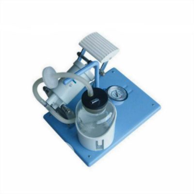
| I-Genius-The Intraoperative MRI System remove | DrGem Ceiling Mounted Digital X-ray remove | Reusable Surgical Gown remove | Keewell FT-1800 Blood & Infusion Warmer remove | Operating Light Four (4) Halogen bulbs remove | Topaz Digital X-ray Machine remove | ||||||||||||||||||||||||||||
|---|---|---|---|---|---|---|---|---|---|---|---|---|---|---|---|---|---|---|---|---|---|---|---|---|---|---|---|---|---|---|---|---|---|
| Name | I-Genius-The Intraoperative MRI System remove | DrGem Ceiling Mounted Digital X-ray remove | Reusable Surgical Gown remove | Keewell FT-1800 Blood & Infusion Warmer remove | Operating Light Four (4) Halogen bulbs remove | Topaz Digital X-ray Machine remove | |||||||||||||||||||||||||||
| Image | 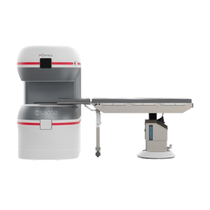 |  |  | 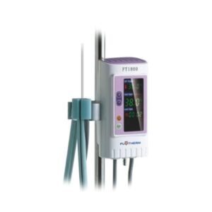 | 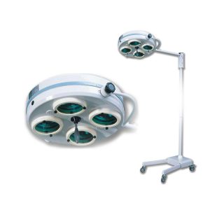 |  | |||||||||||||||||||||||||||
| SKU | SF1033560074-4 | SF1033560084-71 | SF1033560076 | SF1033560084-170 | SF1033560074-1 | ||||||||||||||||||||||||||||
| Rating | |||||||||||||||||||||||||||||||||
| Price |
|
| $12.80 | $781.00 |
|
| |||||||||||||||||||||||||||
| Stock | |||||||||||||||||||||||||||||||||
| Availability | |||||||||||||||||||||||||||||||||
| Add to cart | |||||||||||||||||||||||||||||||||
| Description | Shipped From Abroad
The intraoperative MRI system for neurosurgery
I-Genius is a game-changing intraoperative MRI system that simplifies workflow by providing real-time MR imaging capabilities and delivering valuable information to assist neurosurgeons in making critical decisions during surgical resections. Delivery & Availability:
Typically 10-21 working days – excluding furniture and heavy/bulky equipment. Please contact us for further information.
| In Stock The GXR-SD is a diagnostic digital radiography system that provides reliable high quality digital radiographic images with a reduced dose. The GXR-SD DR systems offer comprehensive digital solutions to all radiography needs, featuring ACQUIDR digital imaging system with stationary or portable digital flat-panel detectors as well as reliable high-frequency x-ray generators that are known worldwide for their excellent performance, lifetime and stability. Patient tables and wall stands are also offered. Delivery & Availability: Typically 21 working days – excluding furniture and heavy/bulky equipment. Please contact us for further information. | In stock Delivery & Availability: Typically 5-7 working days – excluding furniture and heavy/bulky equipment. Please contact us for further information. | Shipped From Abroad
Safety system:Permanent running self-tests, 24 hours continuous operating Double independent over-heating protections and automatic cut off Visual and acoustic alarm for high temperature / low temperature/sensor faultAdvance Dry Heating Technology:Quick Warming up and effective heatingHeating up to the patient:IV tube is completely wrapped in, no heat lossUser friendly interface:Big LED screen showing set temp., actual temp., heating time and fault situation | In stock
| In Stock DRGEM’s TOPAZ X-ray machine is a state-of-the-art mobile digital radiography system, designed with maximum comfort for patients and users in mind. From its user-friendly software to smooth movements, TOPAZ is made to improve your workflow and provide you with high-quality images. Delivery & Availability: Typically 21 working days – excluding furniture and heavy/bulky equipment. Please contact us for further information. | |||||||||||||||||||||||||||
| Content | https://vimeo.com/1124201105?fl=pl&fe=sh
Next-generation intraoperative MRI
Everything in one room: no more patient transfers or workflow interruptions.
To meet the neurosurgery operating standards.
Thanks to its smart design, the operator can quickly switch from the surgery to the imaging modality.
Tools to support the surgeon during the surgery: dedicated cranial fixation system, 55” UHD monitor available in the surgical room for real-time imaging display, customized neuronavigation system (BrainNav by MedCom)*.
MR IMAGING DIRECTLY IN THE OPERATING ROOM A new era of neurosurgery has begunEvery year, more than 500.000* new cases of primary malignant brain tumors are diagnosed worldwide. Gliomas can be difficult to differentiate from surrounding tissue, making it challenging to estimate residual tumor. In gliomas, incomplete resection may lead to recurrence. Intraoperative MRI with I-Genius transforms the practice of brain tumor surgery by allowing MR imaging directly in the operating room and supporting surgical decisions. To guarantee a seamless and safe workflow within the operating room, Esaote has developed a dedicated MRI solution with an integrated intraoperative patient bed. DESIGNED FOR THE OPERATING ROOM
Confident vision for smarter surgeryIn the operating room, every detail counts. Advanced intraoperative imaging provides neurosurgeons with the clarity and confidence required to make important decisions in critical moments. All protocols are designed with optimized sequences, ensuring imaging tailored to surgical needs. I-Genius redefines intraoperative MRI, breaking down barriers and democratizing access to innovation worldwide. By combining efficiency, flexibility, and sustainability, it makes state-of-the-art MRI accessible to more hospitals, more surgeons, and ultimately more patients. Imaging & VisualizationThe I-Genius system is designed to provide imaging directly in the operating room to support real-time surgical decisions.
Clinical ApplicationsWhile the I-Genius is a specialized neurosurgical tool, the broader Esaote MR product line (listed on the same page) covers a wide range of applications, including:
Coils (Probe Types)In Magnetic Resonance Imaging (MRI), coils are used as transducers to send and receive radiofrequency signals, similar to how probes are used in ultrasound. The I-Genius system features dedicated coils for neurosurgical applications:
The system also supports advanced tools like a dedicated cranial fixation system and a customized neuronavigation system (BrainNav by MedCom) to support the surgeon during the procedure. | DrGem Ceiling Mounted Digital X-ray is a diagnostic digital radiography system that provides reliable high quality digital radiographic images with a reduced dose. The GXR-SD DR systems offer comprehensive digital solutions to all radiography needs, featuring ACQUIDR digital imaging system with stationary or portable digital flat-panel detectors as well as reliable high-frequency x-ray generators that are known worldwide for their excellent performance, lifetime and stability. Patient tables and wall stands are also offered.
Features:
Click Here To Download Catalogue |
Products ID: GS-SMS 50A/B/C
User: Unisex
| Features:Safety system:Permanent running self-tests, 24 hours continuous operating Double independent over-heating protections and automatic cut off Visual and acoustic alarm for high temperature / low temperature/sensor faultAdvance Dry Heating Technology:Quick Warming up and effective heatingHeating up to the patient:IV tube is completely wrapped in, no heat lossUser friendly interface:Big LED screen showing set temp., actual temp., heating time and fault situation Easy and quick to set up, ready to use within minutes.Open system:Accepts standard IV tube, no special disposables needed The most economical warming solution without extra consumable costs TECHNICAL SPECIFICATIONS Model: FT1800 Temperature setting: 33˚C - 41˚C Power supply: a.c.100~240V/50~60Hz Power consumption: Max. 120VA Type of protection against electric shock: Class I Degree of protection against electric shock: BF Applied part; Defibrillation-protected Degree of protection against ingress of liquids: IPX2 Temperature accuracy: ±1.0˚C Overheat protection: 42˚C/43˚C Low temperature alarm 32˚C Warming up time: From 20˚C to 36˚C approx. 2 min. Operating mode: Continuous Dimension (W*D*H): 85×65×175mm Net Weight: 1.2kg Warming profile: - length 1400mm - compatible IV tube 3.5-5.0mm O.D.Click Here To Download Catalogue |
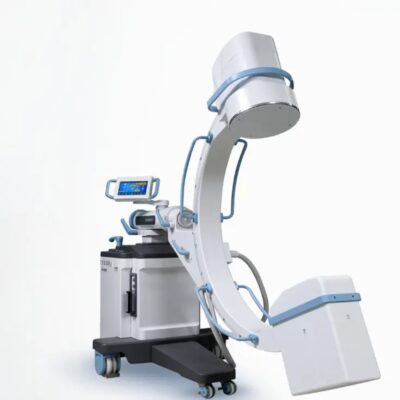
| TOPAZ X-ray machine is among the high end X-ray machine manufactured by DRGEM, a digital X-ray system that provides quality images with little or no effort.
It begins with Advanced Technology
Integrating high technology and over a decade of experience in conventional and digital radiography systems, DRGEM’s TOPAZ X-ray machine is a state-of-the-art mobile digital radiography system, designed with maximum comfort for patients and users. From its user-friendly software to smooth movements, TOPAZ X-ray machine is made to improve your workflow and provide you with high-quality images.
Full Featured Imaging Software & Excellent Digital Image Processing
With a high-performance, built-in touchscreen, TOPAZ X-ray machine offers a user-friendly interface and powerful software for easy operation and increased workflow. The anatomical view-based digital image processing, automatically optimizes and enhances the quality of the image. it also comes with automatic image storage and print with DICOM 3.0 networking capability. additionally, the system offers increasing exam throughput while decreasing examination time.
Click Here To Download Catalogue | |||||||||||||||||||||||||||
| Weight | N/A | N/A | N/A | N/A | N/A | N/A | |||||||||||||||||||||||||||
| Dimensions | N/A | N/A | N/A | N/A | N/A | N/A | |||||||||||||||||||||||||||
| Additional information |


Reviews
There are no reviews yet.