| Description | In Stock
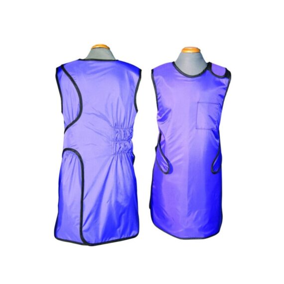
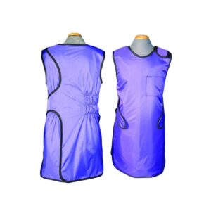
Overlap wrap around x-ray lead apron offers full body protection. Distributes weight between shoulders and hips. • 0.5mm Pb equivalency protection in the front and 0.25mm in the back. Comes with Hook & loop (Velcro) closure and built in 6" Elastic Back-Guard. Front belt assures snug fit and maximum support.
|
Shipped from Abroad
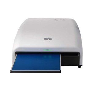
SUPiA made by Signers offers such a better clinic environment with no chemicals, ideal space, high-resolution image quality, and affordability.
Delivery & Availability:
Typically 14 working days – excluding furniture and heavy/bulky equipment. Please contact us for further information. | Shipped from Abroad
The ANATOM 64 CT scanner is the latest innovation for cardiac imaging based on Precision Platform system. The excellent design of Ahart technology which innovatively combined single spiral scan + gated imaging + mA modulation for easy heart imaging at extremely low radiation dose. We provide you ANATOM 64 Clarity/Precision of two models which are low/high configurations for preferences. It also offers you conventional clinical applications of low dose, better image quality and faster exams.
Delivery & Availability:
Typically 90 working days – excluding furniture and heavy/bulky equipment. Please contact us for further information. | Shipped from Abroad
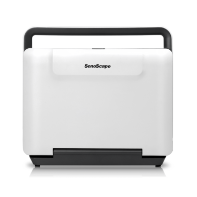
With ultra-modern innovative design and the clinically-proven technologies, S8 Exp is portable ultrasound scanner well equipped as a low-physical-effort and enhanced-image-quality ultrasound scanner, which not only provides optimized solutions for versatile applications, but does help to improve the user-experience for both routine and non-traditional challenges.
Delivery & Availability:
Typically 5-7 working days – excluding furniture and heavy/bulky equipment. Please contact us for further information. | In Stock
GXR Analogue X-ray system matches with a radiographic room which perfectly fits your workow and can be easily upgraded to DR system with the help of DR interface and PC interface in GXR generator as well as Bucky suitable to Flat Panel Detector. GXR X-ray system is equipped with a high frequency X-ray generator which consistently produces high quality radiograph in favor of high quality X-ray output with a very small kV ripple and accurate mA and mAs. GXR X-ray system is designed to provide convenience to operator and comfort to patient.
Delivery & Availability:
Typically 21 working days – excluding furniture and heavy/bulky equipment. Please contact us for further information. | Shipped from Abroad
This Machine gives a possibility to perform computed tomography without any problems and on high quality level. This device is used to conduct exams of internal organs and their functioning. With its help, a physician has a possibility to assess the condition of the human body as a whole.
Delivery & Availability:
Typically 90 working days – excluding furniture and heavy/bulky equipment. Please contact us for further information. |
| Content | Overlap wrap around x-ray lead apron offers full body protection. Distributes weight between shoulders and hips. • 0.5mm Pb equivalency protection in the front and 0.25mm in the back. Comes with Hook & loop (Velcro) closure and built in 6" Elastic Back-Guard. Front belt assures snug fit and maximum support. Our Protective Apparel unique 2-in-1 pocket design allows you to store like a cell phone or markers in the other.
| SUPiA X-ray Digitizer made by Signers offers such a better clinic environment with no chemicals, ideal space, high-resolution image quality, and affordability
FEATURE
Rigid Type
• No damage or scratch on image plates during scanning & erasing
• Scanning & Erasing without a roller
• No cut-off image during winter and cold period
Durability
• Extremely simple structure design
• Strong aluminum base plate
• Flip covers preventing dust from inside scanner
Barcode System
• Automatically recognising cassette sizes(14x17", 10x12", 18x24cm) by barcode reader
Compact & lightweight design
- Very small and compatible CR on desktop (only 63.5cm)
- Only 21.5kg (47.4lbs)
Cassette
Strong structure
• Strong enough against external impact
• Totally metal frame
• Enduring under 150kg on cassette
Featherlight
• 14x17" : 2.05kg 10x12" : 0.99kg 18x24cm : 0.75kg
Dust free & Easy cleaning
• Easy to clean up dust on IPs
• Prevent dust from outside
User friendly design
• Various colors 14x17"(Green),
10x12"(Blue), 18x24cm(Pink))
• Barcode label
TECHNICAL SPECIFICATION
|
SUPiA Specifications |
|
| Cassette |
SUPiA CR Cassette 14x17 (inch)
SUPiA CR Cassette 10x12 (inch)
SUPiA CR Cassette 18x24 (cm) |
| Throughput |
Up to 94 IPs/hour (14x17"/160μm) |
| Slots |
Single Cassette feed |
| Dimensions (W x D x H) |
436 x 636 x 196mm |
| Weight |
21.5kg (47.4lbs) |
| Grayscale Resolution |
Acquisition : 16 bits per pixel
Display : 12 bits per pixel |
| Power Supply Conditions |
Single Phase 50 ~ 60Hz
AC 90 ~ 264V |
| Network |
100 MBit |
| PC Connection |
USB 2.0 |
| Computer Min.
requirements |
OS : Win 7 or 10
CPU : Intel i5
RAM : 4GB
Graphic Card : Intel HD 2500
HDD : 500GB
Monitor : FHD 1920 x 1080
|
| Operating conditions |
Temperature : 10 ~ 30˚C
Humidity : 15 ~ 85% RH |
| The ANATOM 64 CT scanner is the latest innovation for cardiac imaging based on Precision Platform system. The excellent design of Ahart technology which innovatively combined single spiral scan + gated imaging + mA modulation for easy heart imaging at extremely low radiation dose. We provide you ANATOM 64 Clarity/Precision of two models which are low/high configurations for preferences. It also offers you conventional clinical applications of low dose, better image quality and faster exams.
Features:
- Modularized OptiWave HD detector features low-cost & easy maintenance, high spatial resolution and long lifetime
- Admir3D iterative technology delivers optimal dose efficiency and noise reduction without compromising image quality
- High configurations of main components ensure the best results and maximum patient throughput
- Uniquely and creatively uses 140kV and 80kV dual energy scan mode for brain imaging on 16-slice CT to offers you extraordinary image quality both in low and high density resolutions
- AdoseTM mA modulation ensures you low dose imaging without compromising image quality particularly useful in pediatric applications
- Equipped with dedicated Abast and Amast for bone and metal artifacts
- The brilliant Ahart technology enables you to experience so easy and low-dose cardiac imaging applications
Technical Specifications:
| Model |
ANATOM 64 Precision |
ANATOM 64 Fit |
| Rack type |
Low pressure slip ring |
Low pressure slip ring |
| Scan aperture |
70cm |
70cm |
| Rack Physical inclination |
± 30 ° |
N.A |
| Rack digital inclination |
± 50 ° |
± 50 ° |
| cooling method |
Air-cooled |
Air-cooled |
| Focus to the center distance |
56 cm |
53 cm |
| |
|
|
| Maximum power (non-equivalent) |
80kW |
42kW |
| Votage (kV) |
80kV / 100kV / 120kV / 140kV |
70kV / 80kV / 100kV / 120kV /
140kV |
| |
|
|
| Heat capacity |
8MHU |
5.0MHU |
| Heat dissipation rate |
931 kHU / min |
748kHU / min |
| cooling method |
Oil cool |
Oil cool |
| Large focus size |
1.1mm × 1.2mm |
1.2mm × 1.4mm |
| Small focus size |
0.6mm × 1.2mm |
0.7mm × 0.8mm |
| Tube current range |
10-670mA |
10-350mA |
| |
|
|
| Detector type |
Optiwave detectors |
Optiwave detectors |
| Number of Z-axis |
32 |
32 |
| The width of the Z-axis |
20mm |
20mm |
| The number of elements per row |
912 |
848 |
| Total number of detectors |
29184 |
27136 |
| Acquisition mode |
64x0.625, 32x0.625,
16x0.625 |
64x0.625, 32x0.625,
16x0.625 |
| |
|
|
| Scanning range |
1800mm |
1800mm |
| Horizontal positioning accuracy |
± 0.25mm |
± 0.25mm |
| weight capacity |
205kg |
205kg |
| Minimum height |
43cm |
43cm |
| Anti - collision protection device |
Yes |
Yes |
| Foot control switch |
Yes |
Yes |
| IV rack |
Yes |
Yes |
| |
|
|
| CPU |
3.5GHz |
3.5GHz |
| RAM |
16 GB × 4 |
16 GB × 4 |
| Hard drive capacity |
1T × 2 |
1T × 2 |
| Display size |
24 inch LCD monitor |
24 inch LCD monitor |
| Display resolution |
1920 × 1200 |
1920 × 1200 |
| Computer operating system |
Windows 7 |
Windows 7 |
| Image reconstruction speed |
65 frames/ second |
65 frames/ second |
| Number of image store |
1000000 |
1000000 |
| Data external storage mode |
CD / DVD / USB |
CD / DVD / USB |
| |
|
|
| Minimum Scan Time of 360 degree |
0.39sec |
0.75sec |
| Sub-millimeter acquisition layers |
64 |
64 |
| Double sub-millimeter acquisition
layers |
64 |
64 |
| Thinnest acquisition thickness |
0.625mm |
0.625mm |
| The thinnest reconstruction
thickness |
0.3125mm |
0.625mm |
| Conventional reconstruction
thickness (mm) |
0.3125 mm, 0.625 mm, 1.25 mm, 2.5
mm, 5.0 mm, 7.5 mm, 10 mm |
0.625 mm, 1.25 mm, 2.5 mm, 5.0
mm, 7.5 mm, 10 mm |
| The reconstruction matrix |
512 x 512, 1024 x 1024 |
512 x 512, 1024 x 1024 |
| Display matrix |
1024 × 1024 |
1024 × 1024 |
| Max FOV |
52cm |
50cm |
| The maximum display field of view |
70cm |
50cm |
| Maximum scan length |
1800mm |
1800mm |
| Maximum continuous helix scan
time |
120s |
120s |
| Pitch range |
0.5-1.5 |
0.5, 1.0, 1.5 |
| |
|
|
| High contrast resolution |
21 Lp / cm @ 0% MTF |
21 Lp / cm @ 0% MTF |
| Low contrast resolution |
2mm @ 0.3% |
2mm @ 0.3% |
| Image noise |
≤ 0.25 |
≤ 0.29 |
| |
|
|
| MPR |
Yes |
Yes |
| CPR |
Yes |
Yes |
| SSD |
Yes |
Yes |
| VR |
Yes |
Yes |
| MIP |
Yes |
Yes |
| MinIP |
Yes |
Yes |
| Virtual endoscopy |
Yes |
Yes |
| CT angiography |
Yes |
Yes |
| Tissue segmentation |
Yes |
Yes |
| One-key bone removal |
Yes |
Yes |
| Automatically patient table removal |
Yes |
Yes |
| Contrast Agent Automatic Tracking
Technology- bolus tracking |
Yes |
Yes |
| Automatic linkage trigger
technology |
Yes |
Yes |
| Cine mode display |
Yes |
Yes |
| Bone artifact suppression technique |
AbastTM |
AbastTM |
| Metal artifact suppression technique |
AbastTM |
AbastTM |
| Iterative reconstruction technique |
Admir3D global iteration |
Admir3D full-domain iteration |
| Low - dose children 's scanning
technology |
Yes |
Yes |
| Low - dose lung scan |
Yes |
Yes |
| Gray matter enhancement
technology |
AccuHead |
AccuHead |
| High resolution imaging of the lung |
AccuLung |
AccuLung |
| Inner ear high resolution imaging |
AccuOtica |
AccuOtica |
| Body high resolution imaging |
AccuBody |
AccuBody |
| Bone high resolution imaging |
AccuBone |
AccuBone |
| Head dual-energy imaging |
Ahead |
Ahead |
| CT perfusion imaging |
Optional |
Optional |
| Quantitative analysis of blood
vessels |
Optional |
Optional |
| Heart coronary artery imaging |
Aheart |
N.A |
| ECG gated |
Yes |
N.A |
| Low dose cardiac scan |
Yes |
N.A |
| |
|
|
| Green energy saving technology |
AccuSaving |
AccuSaving |
| Dual-energy scan technology |
Optional |
Optional |
| Sonoscape S8 Exp Portable Ultrasound scanner
DETAILS
Agile and Versatile
With ultra-modern innovative design and the clinically-proven technologies, S8 Exp Portable Ultrasound scanner is well equipped as a low-physical-effort and enhanced-image-quality ultrasound scanner, which not only provides optimized solutions for versatile applications but does help to improve the user experience for both routine and non-traditional challenges. Working with S8 Exp, it will trigger your unlimited reverie and endow you with endless charm.
Carrying forward the classical design of SonoScape's portable ultrasound products, S8 Exp successfully combines the best ergonomics, attractive design and ease of use. This charismatic identity is also enhanced by a sophisticated color palette—with sedate grey as its interior paint color and pearl white as exterior cover, S8 Exp reveals a style of aristocrat and strong character among SonoScape's ultrasound systems.
Workflow
The S8 Exp is a portable ultrasound scanner that adapts to your workflow, whether you are in the consulting room, at the bedside, or at a remote location. With easy-to-use new platform designed for sonographers' needs and full connection interfaces for easy connectivity and data sharing, S8 Exp leads to improved user comfort and clinical outcome as well as patient throughput and working efficiency.
Powerful Platform
Embedded with SonoScape's core imaging technologies such as μ-scan, PHI and Spatial Compound, S8 Exp boasts exceptional 2D image, sensitive spectral, Color and Power Doppler, displaying well-defined anatomy and pathology and facilitating a highly optimized diagnostic user environment for conclusive diagnoses. Besides, S8 Exp offers a comprehensive selection of electronic probes to maximally extend its capabilities to meet a wide range of applications including the abdomen, pediatric, OB/GYN, cardiovascular, musculoskeletal, etc. The advanced probe technologies also effectively enhance the image quality and confidence in reaching clinical diagnoses even in difficult patients.
| DrGem GXR Floor Mounted Analogue X-ray system matches with a radiographic room which perfectly fits your workflow and can be easily upgraded to DR system with the help of DR interface and PC interface in GXR generator as well as Bucky suitable to Flat Panel Detector. GXR (Analogue X-ray)system is equipped with a high frequency X-ray generator which consistently produces high quality radiograph in favor of high quality X-ray output with a very small kV ripple and accurate mA and mAs. GXR (Analogue X-ray) system is designed to provide convenience to operator and comfort to patient.
Features of DrGem GXR Floor Mounted Analogue X-ray:
- 4 way Tabletop Patient Table (PBT-4)
A large tabletop with extended travel enables all radiography studies with minimal patient movement, and supporting patient weight up to 300kg. Fully at tabletop without a frame on the edge makes cleanliness and odors free
- Floor Mount Tube Stand (TS-FM6)
Floor Rail type tube stand provides all radiography studies with smooth movement on the rail.
- Wall Bucky Stand (WBS)
Elegant design, durable and easy-to-use Wall Bucky Stand provides full satisfaction.
Technical Specifications of DrGem GXR Floor Mounted Analogue X-ray:
- Power Rating - 32KW
- Generator - GXR-32S
- Rotor - Dual Speed Starter(DSS)
- Input Power - 400/480VAC, Three phase
- Line Frequency - 50/60Hz
- X-ray tube - DXT-12M, (0.6/1.2mm, 300kHU)
- Tube Voltage - 40 to 150kV, 1kV Step
- Tube Current – 10 to 640mA
- Output - 640mA@81kV, 500mA@104kV, 400mA@130kV, 320mA@150kV
- Time Range - 1ms to 10s
- mAs Range - 0.1 to 800mAs
- Reproducibility - Coecient of Variation : kV < 0.005, Time < 0.005,mAs < 0.01
- Accuracy - kV < ±(1%+1kV), mA < ±(3%+1mA), Time <±(1%+0.5ms), mAs < ±(3%+0.1mAs)
- Linearity - Coecient of Linearity < 0.01 : CL = (X1-X2)/(X1+X2), where X is mR/mAs
| This Machine gives a possibility to perform computed tomography without any problems and on high quality level. This device is used to conduct exams of internal organs and their functioning. With its help, a physician has a possibility to assess the condition of the human body as a whole.
Features:
- It is easy to use;
- Convenience;
- Multi functionality;
- Obtained images are of high definition;
- High-definition 3D images of the area under study;
- The procedure is pain-free;
- The data is processed fast;
- The image can be stored in the computer memory;
- The diagnostics does not take a lot of time;
- Acceptable radiation dose.
Technical Specifications:
| No. |
Technical Features |
Descriptions |
| 1 |
Gantry |
|
| 1.01 |
Gantry type |
Low voltage slip-ring |
| 1.02 |
Gantry driven type |
Strap-driven |
| 1.03 |
Patient opening |
70cm |
| 1.04 |
Gantry tilt mode |
Digital gantry tilt |
| 1.05 |
Digital tilt capability |
±50° |
| 1.06 |
Detector type |
OptiWave rare-earth ceramic detector |
| 1.07 |
Numbers of detector rows |
16 |
| 1.08 |
Width of Z-axle detector |
20mm |
| 1.09 |
Detector columns of channels per row |
848 |
| 1.10 |
Numbers of detector columns |
13568 |
| 1.11 |
Data-transfer type |
RF, optical fiber communication |
| 1.12 |
Distance of focus-ISO-center |
53cm |
| 1.13 |
Distance of focus-detector |
94cm |
| 1.14 |
3D laser orientation |
Provided |
| 1.15 |
13" integrated display panel |
Provided |
| 1.16 |
Adose automatic exposure control (mA
Modulation) |
Provided |
| 1.17 |
Auto-voice manager |
Breath Graphical Display
Hold Message (Record/Playback)
Breath Message (Record/Playback) |
| 1.18 |
AccuSaving energy conservation management |
Provided |
| 2 |
HVPS and X-ray tube |
|
| 2.01 |
Maximum continuous output of HVgenerator |
42kW |
| 2.02 |
Tube kV selections |
70kV, 80kV, 100 kV, 120 kV, 140 kV |
| 2.03 |
Tube mA range |
10~350mA |
| 2.04 |
Tube anode heat capacity |
3.5MHU |
| 2.05 |
Max. anode cooling rate |
735kHU/min |
| 2.06 |
Type of cooling |
Oil cooling + Air cooling |
| 2.07 |
Tube focus |
Large: 1.2mm×1.4mm
Small: 0.7mm×0.8mm |
| 2.08 |
Collimator width selection |
4-level election |
| 2.09 |
Focus spot tracking technology |
Provided |
| 3 |
Patient table |
|
| 3.01 |
Maximum horizontal-movable range |
1850mm |
| 3.02 |
Table horizontal-scannablerange |
1800mm |
| 3.03 |
Table horizontal-position repeatability |
±0.25mm |
| 3.04 |
Minimum height above floor |
430mm |
| 3.05 |
Maximum vertical-movable range |
500mm |
| 3.06 |
Maximum speed of vertical movement |
35mm |
| 3.07 |
Maximum speed of horizontal movement |
150mm/s |
| 3.08 |
Maximum patient weight |
205kg |
| 3.09 |
Foot pedal of patient table control |
Provided |
| 4 |
Computer |
|
| 4.01 |
CPU |
3.5GHz |
| 4.02 |
Memory |
32GB |
| 4.03 |
Storage of hard-disk |
1TB×2 |
| 4.04 |
Monitor |
24’’ LCD Monitor |
| 4.05 |
Resolution of monitor |
1920×1200 |
| 4.06 |
Image-data external storage type |
CD/DVD/USB |
| 4.07 |
Time of image reconstruction (512×512) |
33.3ms/image |
| 4.08 |
Speed of image reconstruction (512×12) |
30fps |
| 4.09 |
DICOM 3.0 interface |
Provided |
| 4.10 |
Printer DICOM 3.0 interface |
Provided |
| 4.11 |
Auto filming |
Provided |
| 4.12 |
Worklist function |
Provided |
| 5 |
Scan parameters |
|
| 5.01 |
Shortest 360 degree rotation time |
0.75s |
| 5.02 |
Allowed rotation times |
0.75s, 1.0s, 1.5s, 2.0s, 3.0s, 4.0s |
| 5.03 |
Maximum slice numbers per rotation |
32 |
| 5.04 |
Minimum slice thickness of scan |
1.25mm |
| 5.05 |
Minimum slice thickness of reconstruction |
0.625mm |
| 5.06 |
Maximum slice thickness of scan |
20mm |
| 5.07 |
Nominal reconstruction slice thickness |
0.625mm, 1.25mm, 2.5mm, 5.0mm, 7.5mm,
10mm, 20mm |
| 5.08 |
Speed of image reconstruction (512×512) |
30 frames/s |
| 5.09 |
Scan FOV |
50cm |
| 5.10 |
Image reconstruction matrix |
512×512, 1024×1024 (Optional) |
| 5.11 |
Image reconstruction matrix |
512×512, 1024×1024 (Optional) |
| 5.12 |
Image display matrix |
512×512, 1024×1024 (Optional) |
| 5.13 |
Maximum continuous scan duration |
120s |
| 5.14 |
Maximum continuous scan length |
180cm |
| 5.15 |
Direction of TOPO |
Front-back, Left-right |
| 5.16 |
Max. length of TOPO |
180cm |
| 5.17 |
Range of pitch |
0.5~1.5 |
| 5.18 |
Scan mode |
Scout scan
Axial scan
Helical scan
Cine scan |
| 6 |
Image Quality |
|
| 6.01 |
High contrast resolution |
21lp/cm@0%MTF |
| 6.02 |
Low contrast resolution |
2.0mm@0.30% |
| 6.03 |
Isotropic imaging resolution |
0.24mm |
| 6.04 |
Range of CT numbers |
-32767~32768 |
| 6.05 |
Image noise |
≤0.29@28mGy |
| 7 |
Advanced application |
|
| 7.01 |
Multi-Planar Reconstruction (MPR) |
Provided |
| 7.02 |
Curve Multi-Planar Reconstruction (CPR) |
Provided |
| 7.03 |
Surface Shaded Display (SSD) |
Provided |
| 7.04 |
Volume Rendering (VR) |
Provided |
| 7.05 |
Maximum Intensity Projection (MIP) |
Provided |
| 7.06 |
Minimum Intensity Projection (MinIP) |
Provided |
| 7.07 |
Virtual Endoscopy (VE) |
Provided |
| 7.08 |
CT angiography (CTA) |
Provided |
| 7.09 |
Tissue segmentation |
Provided |
| 7.10 |
One click bone remove |
Provided |
| 7.11 |
One click patient table remove |
Provided |
| 7.12 |
Bolus-tracking Technology |
Provided |
| 7.13 |
Spiral auto start |
Provided |
| 7.14 |
Cine display |
Provided |
| 7.15 |
AbastTM bone artifact suppression technology |
Provided |
| 7.16 |
AmastTM metal artifact suppression technology |
Provided |
| 7.17 |
Admir3D all-domain iterative reconstruction |
Provided |
| 7.18 |
Low-dose pediatric scan technology |
Provided |
| 7.19 |
Low-dose lung scan technology |
Provided |
| 7.20 |
AccuHead grey-white matter enhanced
technology |
Provided |
| 7.21 |
AccuOrgan lung high resolution scan technology |
Provided |
| 7.22 |
AccuOrgan inner-ear high resolution scan
technology |
Provided |
| 7.23 |
AccuOrgan body high resolution scan technology |
Provided |
| 7.24 |
AccuOrgan bone high resolution scan technology |
Provided |
| 7.25 |
AccuMatter dual-energy with Admir3D for new
application |
Provided |
|


Reviews
There are no reviews yet.