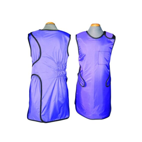
Lead Apron Front and Back
$95.00
In Stock
Overlap wrap around x-ray lead apron offers full body protection. Distributes weight between shoulders and hips. • 0.5mm Pb equivalency protection in the front and 0.25mm in the back. Comes with Hook & loop (Velcro) closure and built in 6″ Elastic Back-Guard. Front belt assures snug fit and maximum support.
Description
Overlap wrap around x-ray lead apron offers full body protection. Distributes weight between shoulders and hips. • 0.5mm Pb equivalency protection in the front and 0.25mm in the back. Comes with Hook & loop (Velcro) closure and built in 6″ Elastic Back-Guard. Front belt assures snug fit and maximum support. Our Protective Apparel unique 2-in-1 pocket design allows you to store like a cell phone or markers in the other.
Quick Comparison
| Settings | Lead Apron Front and Back remove | Sonoscape S11 Ultrasound Machine remove | DrGem Ceiling Analogue X-ray Machine remove | Anke Anatom 32 Fit Multi-Slice Spiral CT Scan remove | IBIS Neeo R9 Digital Surgical C-Arm remove | LED Double X-ray Viewing Box remove | ||||||||||||||||||||||||||||||||||||||||||||||||||||||||||||||||||||||||||||||||||||||||||||||||||||||||||||||||||||||||||||||||||||||||||||||||||||||||||||||||||||||||||||||||||||||||||||||||||||||||||||||||||||||||||||||||||||||||||||||||||||||||||||||||||||||||||||||||||||||||||||||||||||||||||||||||||||||||
|---|---|---|---|---|---|---|---|---|---|---|---|---|---|---|---|---|---|---|---|---|---|---|---|---|---|---|---|---|---|---|---|---|---|---|---|---|---|---|---|---|---|---|---|---|---|---|---|---|---|---|---|---|---|---|---|---|---|---|---|---|---|---|---|---|---|---|---|---|---|---|---|---|---|---|---|---|---|---|---|---|---|---|---|---|---|---|---|---|---|---|---|---|---|---|---|---|---|---|---|---|---|---|---|---|---|---|---|---|---|---|---|---|---|---|---|---|---|---|---|---|---|---|---|---|---|---|---|---|---|---|---|---|---|---|---|---|---|---|---|---|---|---|---|---|---|---|---|---|---|---|---|---|---|---|---|---|---|---|---|---|---|---|---|---|---|---|---|---|---|---|---|---|---|---|---|---|---|---|---|---|---|---|---|---|---|---|---|---|---|---|---|---|---|---|---|---|---|---|---|---|---|---|---|---|---|---|---|---|---|---|---|---|---|---|---|---|---|---|---|---|---|---|---|---|---|---|---|---|---|---|---|---|---|---|---|---|---|---|---|---|---|---|---|---|---|---|---|---|---|---|---|---|---|---|---|---|---|---|---|---|---|---|---|---|---|---|---|---|---|---|---|---|---|---|---|---|---|---|---|---|---|---|---|---|---|---|---|---|---|---|---|---|---|---|---|---|---|---|---|---|---|---|---|---|---|---|---|---|---|---|---|---|---|---|---|---|---|---|
| Name | Lead Apron Front and Back remove | Sonoscape S11 Ultrasound Machine remove | DrGem Ceiling Analogue X-ray Machine remove | Anke Anatom 32 Fit Multi-Slice Spiral CT Scan remove | IBIS Neeo R9 Digital Surgical C-Arm remove | LED Double X-ray Viewing Box remove | ||||||||||||||||||||||||||||||||||||||||||||||||||||||||||||||||||||||||||||||||||||||||||||||||||||||||||||||||||||||||||||||||||||||||||||||||||||||||||||||||||||||||||||||||||||||||||||||||||||||||||||||||||||||||||||||||||||||||||||||||||||||||||||||||||||||||||||||||||||||||||||||||||||||||||||||||||||||||
| Image |  |  |  |  |  |  | ||||||||||||||||||||||||||||||||||||||||||||||||||||||||||||||||||||||||||||||||||||||||||||||||||||||||||||||||||||||||||||||||||||||||||||||||||||||||||||||||||||||||||||||||||||||||||||||||||||||||||||||||||||||||||||||||||||||||||||||||||||||||||||||||||||||||||||||||||||||||||||||||||||||||||||||||||||||||
| SKU | SF1033560072 | SF1033560012-1 | SF1033560074-7 | SF1033560092-1 | SF1033560011-1 | SF1033560084-193 | ||||||||||||||||||||||||||||||||||||||||||||||||||||||||||||||||||||||||||||||||||||||||||||||||||||||||||||||||||||||||||||||||||||||||||||||||||||||||||||||||||||||||||||||||||||||||||||||||||||||||||||||||||||||||||||||||||||||||||||||||||||||||||||||||||||||||||||||||||||||||||||||||||||||||||||||||||||||||
| Rating | ||||||||||||||||||||||||||||||||||||||||||||||||||||||||||||||||||||||||||||||||||||||||||||||||||||||||||||||||||||||||||||||||||||||||||||||||||||||||||||||||||||||||||||||||||||||||||||||||||||||||||||||||||||||||||||||||||||||||||||||||||||||||||||||||||||||||||||||||||||||||||||||||||||||||||||||||||||||||||||||
| Price | $95.00 | $6,380.00 |
|
|
| $151.00 | ||||||||||||||||||||||||||||||||||||||||||||||||||||||||||||||||||||||||||||||||||||||||||||||||||||||||||||||||||||||||||||||||||||||||||||||||||||||||||||||||||||||||||||||||||||||||||||||||||||||||||||||||||||||||||||||||||||||||||||||||||||||||||||||||||||||||||||||||||||||||||||||||||||||||||||||||||||||||
| Stock | ||||||||||||||||||||||||||||||||||||||||||||||||||||||||||||||||||||||||||||||||||||||||||||||||||||||||||||||||||||||||||||||||||||||||||||||||||||||||||||||||||||||||||||||||||||||||||||||||||||||||||||||||||||||||||||||||||||||||||||||||||||||||||||||||||||||||||||||||||||||||||||||||||||||||||||||||||||||||||||||
| Availability | ||||||||||||||||||||||||||||||||||||||||||||||||||||||||||||||||||||||||||||||||||||||||||||||||||||||||||||||||||||||||||||||||||||||||||||||||||||||||||||||||||||||||||||||||||||||||||||||||||||||||||||||||||||||||||||||||||||||||||||||||||||||||||||||||||||||||||||||||||||||||||||||||||||||||||||||||||||||||||||||
| Add to cart | ||||||||||||||||||||||||||||||||||||||||||||||||||||||||||||||||||||||||||||||||||||||||||||||||||||||||||||||||||||||||||||||||||||||||||||||||||||||||||||||||||||||||||||||||||||||||||||||||||||||||||||||||||||||||||||||||||||||||||||||||||||||||||||||||||||||||||||||||||||||||||||||||||||||||||||||||||||||||||||||
| Description | In Stock
Overlap wrap around x-ray lead apron offers full body protection. Distributes weight between shoulders and hips. • 0.5mm Pb equivalency protection in the front and 0.25mm in the back. Comes with Hook & loop (Velcro) closure and built in 6" Elastic Back-Guard. Front belt assures snug fit and maximum support.
| In Stock A Value Choice beyond Your Expectation. SonoScape’s trolley color Doppler system S11 redefines price and performance with practical design. The S11 will go beyond your expectations but not your budget. Delivery & Availability: Typically 2 working days – excluding furniture and heavy/bulky equipment. Please contact us for further information. | Shipped from abroad The DrGem Ceiling Analogue X-ray Machine is a diagnostic radiography system that provides reliable high quality radiographic images with a reduced dose. The reliable high-frequency x-ray generators that are known worldwide for their excellent performance, lifetime and stability. Patient tables and wall stands are also offered. Delivery & Availability: Typically 21 working days – excluding furniture and heavy/bulky equipment. Please contact us for further information. | Shipped from Abroad
This Machine gives a possibility to perform computed tomography without any problems and on high quality level. This device is used to conduct exams of internal organs and their functioning. With its help, a physician has a possibility to assess the condition of the human body as a whole.
Delivery & Availability: Typically 90 working days – excluding furniture and heavy/bulky equipment. Please contact us for further information. | Shipped from Abroad Our Neeo “C” arms are easy to place, use and are specifically designed to be used in orthopedics, traumatology, abdominal surgery, urology, cardiology and operating rooms. Delivery & Availability: Typically 21 working days – excluding furniture and heavy/bulky equipment. Please contact us for further information. | In stock
Double x-ray film viewer, Compact, Solid with Backlight of LED’s based panel, Long Life Approximate LED’s life 50, 000 Hrs., Uniform Light at the total surface area, No Heat Emission, Wall Mounted, Can be used for tracing on X-Ray, Auto-sensor, Screen. Size: 430 mm x 710 mm.
| ||||||||||||||||||||||||||||||||||||||||||||||||||||||||||||||||||||||||||||||||||||||||||||||||||||||||||||||||||||||||||||||||||||||||||||||||||||||||||||||||||||||||||||||||||||||||||||||||||||||||||||||||||||||||||||||||||||||||||||||||||||||||||||||||||||||||||||||||||||||||||||||||||||||||||||||||||||||||
| Content | Overlap wrap around x-ray lead apron offers full body protection. Distributes weight between shoulders and hips. • 0.5mm Pb equivalency protection in the front and 0.25mm in the back. Comes with Hook & loop (Velcro) closure and built in 6" Elastic Back-Guard. Front belt assures snug fit and maximum support. Our Protective Apparel unique 2-in-1 pocket design allows you to store like a cell phone or markers in the other. | DETAILS
SonoScape’s trolley colour Doppler system S11 redefines price and performance with practical design. The S11 will go beyond your expectations but not your budget. As an easy-to-use ultrasound system, the S11 is integrated with a new software platform, especially optimized for a smooth workflow and convenient operation. The system speeds up the exam process and makes file management easier.
SPECIFICATION
- 15-inch high definition LCD monitor with articulating arm
- Compact and agile trolley design
- 3 active transducer sockets available for a wide range of applications
- Duplex, Color Doppler, DPI, PW Doppler, tissue harmonic imaging, μ-scan speckle reduction imaging, compound imaging, trapezoidal imaging
- Customized settings based on your own working style
- Full patient database and image management solutions
Click Here To Download Catalogue | DrGem Ceiling Analogue X-ray Machine is a diagnostic radiography system X-ray Machine that provides reliable high quality radiographic images with a reduced dose. The reliable high-frequency x-ray generators that are known worldwide for their excellent performance, lifetime and stability. Patient tables and wall stands are also offered.
Features of DrGem Ceiling Analogue X-ray Machine
Click Here To Download Catalogue | This Machine gives a possibility to perform computed tomography without any problems and on high quality level. This device is used to conduct exams of internal organs and their functioning. With its help, a physician has a possibility to assess the condition of the human body as a whole.
Features:
Click Here To Download Catalogue | Our Neeo “C” arms are easy to place, use and are specifically designed to be used in orthopedics, traumatology, abdominal surgery, urology, cardiology and operating rooms.
Using Neeo with the RTP (Real Time Processing) option it is possible to perform vascular, urological and cardiological diagnostics. One of the main functions, digital image subtraction, allows to see, as an example, the passage of contrast liquids in a tissue or in a venous or arterial duct; thanks to the possibility of looping, the acquired video can be reproduced several times to monitor more accurately the passage of the fluid within the area in question. Angiographic measurement is another useful function in the vascular field (QA Quantitative Angiography) that allows the measurement of stenoses. Finally, fluoroscopy allows the correct positioning of stents or expanders.
Neeo is used in various interventional and diagnostic procedures in traumatology and orthopedics wards and operating rooms as well. Thanks to low-dose fluoroscopy, it is possible to use the device for positioning bone or subcutaneous grafts, inserting K-wire (Kirschner wire) for stabilization of bone fragments or for the correct positioning of prostheses. The low dose emitted ensures safe use for both the patient and the surgeon or doctor on the operating field.
On the control panel there is a large touch screen display that allows to adjust the basic functions of the equipment. From this display it is possible to select and adjust the fluoroscopic data for the examination, activate or deactivate the laser pointer, select between pulsed, one shot or standard fluoroscopy, rotate the image and perform all operations on collimator. The four side buttons on the display offer the possibility to move the bow vertically thanks to an extremely silent motor.
Neeo has two 19 “medical grade monitors that can be positioned according to the needs of the medical practitioner. Work monitors and feedback monitors are separated to be managed independently. The possible movements are: rotation, revolution, tilting and possibility of height adjustment.
Features:
Click Here To Download Catalogue | Double x-ray film viewer, Compact, Solid with Backlight of LED’s based panel, Long Life Approximate LED’s life 50, 000 Hrs., Uniform Light at the total surface area, No Heat Emission, Wall Mounted, Can be used for tracing on X-Ray, Auto-sensor, Screen. Size: 430 mm x 710 mm. | ||||||||||||||||||||||||||||||||||||||||||||||||||||||||||||||||||||||||||||||||||||||||||||||||||||||||||||||||||||||||||||||||||||||||||||||||||||||||||||||||||||||||||||||||||||||||||||||||||||||||||||||||||||||||||||||||||||||||||||||||||||||||||||||||||||||||||||||||||||||||||||||||||||||||||||||||||||||||
| Weight | N/A | N/A | N/A | N/A | N/A | N/A | ||||||||||||||||||||||||||||||||||||||||||||||||||||||||||||||||||||||||||||||||||||||||||||||||||||||||||||||||||||||||||||||||||||||||||||||||||||||||||||||||||||||||||||||||||||||||||||||||||||||||||||||||||||||||||||||||||||||||||||||||||||||||||||||||||||||||||||||||||||||||||||||||||||||||||||||||||||||||
| Dimensions | N/A | N/A | N/A | N/A | N/A | N/A | ||||||||||||||||||||||||||||||||||||||||||||||||||||||||||||||||||||||||||||||||||||||||||||||||||||||||||||||||||||||||||||||||||||||||||||||||||||||||||||||||||||||||||||||||||||||||||||||||||||||||||||||||||||||||||||||||||||||||||||||||||||||||||||||||||||||||||||||||||||||||||||||||||||||||||||||||||||||||
| Additional information |
|


Reviews
There are no reviews yet.