| Description | In Stock
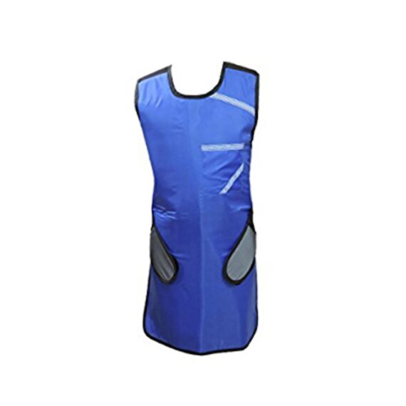
Our x-ray lead aprons provide complete protection to people who are exposed to radiations for prolonged hours. X-ray lead aprons are a must since they reduce the chances of developing cancer which is a common result of radiation exposure. Lead aprons reduce the exposure to these harmful rays and thus the chances of getting cancer.
| Shipped from Abroad
DrGem Diamond All-In-One Digital X-ray Machine is a fully automatic digital radiography system providing state-of-the-art image quality, image processing and user interface. With a wide selection of anatomical studies on the imaging software, DIAMOND automatically sets up the x-ray generator’s preprogrammed exposure technique settings, motorized radiographic stand positioning, x-ray collimation and post-image processing for the selected study. Specifically designed to increase workflow, this fully digital system offers convenient auto-positioning and advanced image processing to achieve big performance with little effort.
Delivery & Availability:
Typically 21 working days – excluding furniture and heavy/bulky equipment. Please contact us for further information. | In stock
- This white coat laboratory coat is a knee-length overcoat/smock is perfect for professionals in the medical field or by those involved in laboratory work.
- This garment is Easy to wash and made from a cotton polyester blend, allowing it to be washed at high temperature and make it easy to see if it is clean.
- Easy to wash.
Delivery & Availability:
Typically 5-7 working days – excluding furniture and heavy/bulky equipment. Please contact us for further information. | Shipped from Abroad
This Machine gives a possibility to perform computed tomography without any problems and on high quality level. This device is used to conduct exams of internal organs and their functioning. With its help, a physician has a possibility to assess the condition of the human body as a whole.
Delivery & Availability:
Typically 90 working days – excluding furniture and heavy/bulky equipment. Please contact us for further information. | In Stock
DRGEM’s TOPAZ X-ray machine is a state-of-the-art mobile digital radiography system, designed with maximum comfort for patients and users in mind. From its user-friendly software to smooth movements, TOPAZ is made to improve your workflow and provide you with high-quality images.
Delivery & Availability:
Typically 21 working days – excluding furniture and heavy/bulky equipment. Please contact us for further information. | Shipped from Abroad
The ANATOM 64 CT scanner is the latest innovation for cardiac imaging based on Precision Platform system. The excellent design of Ahart technology which innovatively combined single spiral scan + gated imaging + mA modulation for easy heart imaging at extremely low radiation dose. We provide you ANATOM 64 Clarity/Precision of two models which are low/high configurations for preferences. It also offers you conventional clinical applications of low dose, better image quality and faster exams.
Delivery & Availability:
Typically 90 working days – excluding furniture and heavy/bulky equipment. Please contact us for further information. |
| Content | Our x-ray lead aprons provide complete protection to people who are exposed to radiations for prolonged hours. X-ray lead aprons are a must since they reduce the chances of developing cancer which is a common result of radiation exposure. Lead aprons reduce the exposure to these harmful rays and thus the chances of getting cancer.
Features:
- 0.5mm Lead (pb) Equivalency Protection at 100 kVp of X-ray beam energy
- With ROBUST HANGER, Protection Material-Lead
- Front Velcro Closure, Includes Single Front pocket
- Padded Shoulders to Prevent Strain
- Stain Resistant and Washable Material for Minimum Maintenance
| DrGem Diamond All-In-One Digital X-ray Machine is a fully automatic digital radiography system providing state-of-the-art image quality, image processing and user interface. With a wide selection of anatomical studies on the imaging software, DIAMOND automatically sets up the x-ray generator’s pre-programmed exposure technique settings, motorized radiographic stand positioning, x-ray collimation and post-image processing for the selected study. Specifically designed to increase workflow, this fully digital system offers convenient auto-positioning and advanced image processing to achieve big performance with little effort.
Features of DrGem Diamond All-In-One Digital X-ray Machine:
Outstanding Image Quality -
Digital radiography via at panel detector improves your workflow, exam speed and comfort with efficiency. Digital at panel detector with Csl screen provides excellent spatial resolution, MTF, DQE and stability based on ne pixel pitch. A 3-field ion-chamber is provided for AEC function.
Automatic Collimation –
Automatic x-ray eld size control of the motorized collimator corresponds to dierent SIDs. Includes user adjustable lamp timer with on/oswitch.
Automatic Positioning –
- DIAMOND is a fully-automatic diagnostic system with motorized movement and pre-programmed data for automatic positioning that can be easily reprogrammed by the operator.
- Seven safety sensors have been integrated into the DIAMOND system to protect patients from accidental collision.
Automatic Stitching –
DIAMOND 5A provides outstanding automatic stitching function via source tilting for creating one long composite image.
DIAMOND Positioning Guide –
The U-arm rotation ranges from +120 ° to +30 ° with SID movements from 100cm to 180cm. The mobile patient table can also be used to take patient images in a variety of positions for a total of over fifteen different positions (Chest PA, Skull Towne’s, Abdomen Supine, Hand AP, C-T Spine Swimmers and Abdomen Decubitus etc.)
- Included – Software, HP Laptop Computer
- CPU≥3.2GHz
- Memory capacity:≥4GB
- Hard drive capacity :≥500 GB
- Resolution: 1280 x 1024
- Display size: 21 inch color LCD screen
- 64 bit Windows 10 operation system
- Core: i5
Technical Specification:
- Power Rating - 52KW
- mA - 10 to 640mA
- mAs - 0.1 to 500mAs
- kV - 40 to 150kV, 1kV Step
- Generator - GXR-52
- Rotor - Dual Speed (High and Low)
- Input Power - 400/480VAC±10%, 3Ø
- Line Frequency - 50/60Hz
- X-ray tube – DXT-12M 0.6/1.2mm, 300kHU
|
- This white coat laboratory coat is a knee-length overcoat/smock is perfect for professionals in the medical field or by those involved in laboratory work.
- This garment is Easy to wash and made from a cotton polyester blend, allowing it to be washed at high temperature and make it easy to see if it is clean.
- Easy to wash.
| This Machine gives a possibility to perform computed tomography without any problems and on high quality level. This device is used to conduct exams of internal organs and their functioning. With its help, a physician has a possibility to assess the condition of the human body as a whole.
Features:
- It is easy to use;
- Convenience;
- Multi functionality;
- Obtained images are of high definition;
- High-definition 3D images of the area under study;
- The procedure is pain-free;
- The data is processed fast;
- The image can be stored in the computer memory;
- The diagnostics does not take a lot of time;
- Acceptable radiation dose.
Technical Specifications:
| No. |
Technical Features |
Descriptions |
| 1 |
Gantry |
|
| 1.01 |
Gantry type |
Low voltage slip-ring |
| 1.02 |
Gantry driven type |
Strap-driven |
| 1.03 |
Patient opening |
70cm |
| 1.04 |
Gantry tilt mode |
Digital gantry tilt |
| 1.05 |
Digital tilt capability |
±50° |
| 1.06 |
Detector type |
OptiWave rare-earth ceramic detector |
| 1.07 |
Numbers of detector rows |
16 |
| 1.08 |
Width of Z-axle detector |
20mm |
| 1.09 |
Detector columns of channels per row |
848 |
| 1.10 |
Numbers of detector columns |
13568 |
| 1.11 |
Data-transfer type |
RF, optical fiber communication |
| 1.12 |
Distance of focus-ISO-center |
53cm |
| 1.13 |
Distance of focus-detector |
94cm |
| 1.14 |
3D laser orientation |
Provided |
| 1.15 |
13" integrated display panel |
Provided |
| 1.16 |
Adose automatic exposure control (mA
Modulation) |
Provided |
| 1.17 |
Auto-voice manager |
Breath Graphical Display
Hold Message (Record/Playback)
Breath Message (Record/Playback) |
| 1.18 |
AccuSaving energy conservation management |
Provided |
| 2 |
HVPS and X-ray tube |
|
| 2.01 |
Maximum continuous output of HVgenerator |
42kW |
| 2.02 |
Tube kV selections |
70kV, 80kV, 100 kV, 120 kV, 140 kV |
| 2.03 |
Tube mA range |
10~350mA |
| 2.04 |
Tube anode heat capacity |
3.5MHU |
| 2.05 |
Max. anode cooling rate |
735kHU/min |
| 2.06 |
Type of cooling |
Oil cooling + Air cooling |
| 2.07 |
Tube focus |
Large: 1.2mm×1.4mm
Small: 0.7mm×0.8mm |
| 2.08 |
Collimator width selection |
4-level election |
| 2.09 |
Focus spot tracking technology |
Provided |
| 3 |
Patient table |
|
| 3.01 |
Maximum horizontal-movable range |
1850mm |
| 3.02 |
Table horizontal-scannablerange |
1800mm |
| 3.03 |
Table horizontal-position repeatability |
±0.25mm |
| 3.04 |
Minimum height above floor |
430mm |
| 3.05 |
Maximum vertical-movable range |
500mm |
| 3.06 |
Maximum speed of vertical movement |
35mm |
| 3.07 |
Maximum speed of horizontal movement |
150mm/s |
| 3.08 |
Maximum patient weight |
205kg |
| 3.09 |
Foot pedal of patient table control |
Provided |
| 4 |
Computer |
|
| 4.01 |
CPU |
3.5GHz |
| 4.02 |
Memory |
32GB |
| 4.03 |
Storage of hard-disk |
1TB×2 |
| 4.04 |
Monitor |
24’’ LCD Monitor |
| 4.05 |
Resolution of monitor |
1920×1200 |
| 4.06 |
Image-data external storage type |
CD/DVD/USB |
| 4.07 |
Time of image reconstruction (512×512) |
33.3ms/image |
| 4.08 |
Speed of image reconstruction (512×12) |
30fps |
| 4.09 |
DICOM 3.0 interface |
Provided |
| 4.10 |
Printer DICOM 3.0 interface |
Provided |
| 4.11 |
Auto filming |
Provided |
| 4.12 |
Worklist function |
Provided |
| 5 |
Scan parameters |
|
| 5.01 |
Shortest 360 degree rotation time |
0.75s |
| 5.02 |
Allowed rotation times |
0.75s, 1.0s, 1.5s, 2.0s, 3.0s, 4.0s |
| 5.03 |
Maximum slice numbers per rotation |
32 |
| 5.04 |
Minimum slice thickness of scan |
1.25mm |
| 5.05 |
Minimum slice thickness of reconstruction |
0.625mm |
| 5.06 |
Maximum slice thickness of scan |
20mm |
| 5.07 |
Nominal reconstruction slice thickness |
0.625mm, 1.25mm, 2.5mm, 5.0mm, 7.5mm,
10mm, 20mm |
| 5.08 |
Speed of image reconstruction (512×512) |
30 frames/s |
| 5.09 |
Scan FOV |
50cm |
| 5.10 |
Image reconstruction matrix |
512×512, 1024×1024 (Optional) |
| 5.11 |
Image reconstruction matrix |
512×512, 1024×1024 (Optional) |
| 5.12 |
Image display matrix |
512×512, 1024×1024 (Optional) |
| 5.13 |
Maximum continuous scan duration |
120s |
| 5.14 |
Maximum continuous scan length |
180cm |
| 5.15 |
Direction of TOPO |
Front-back, Left-right |
| 5.16 |
Max. length of TOPO |
180cm |
| 5.17 |
Range of pitch |
0.5~1.5 |
| 5.18 |
Scan mode |
Scout scan
Axial scan
Helical scan
Cine scan |
| 6 |
Image Quality |
|
| 6.01 |
High contrast resolution |
21lp/cm@0%MTF |
| 6.02 |
Low contrast resolution |
2.0mm@0.30% |
| 6.03 |
Isotropic imaging resolution |
0.24mm |
| 6.04 |
Range of CT numbers |
-32767~32768 |
| 6.05 |
Image noise |
≤0.29@28mGy |
| 7 |
Advanced application |
|
| 7.01 |
Multi-Planar Reconstruction (MPR) |
Provided |
| 7.02 |
Curve Multi-Planar Reconstruction (CPR) |
Provided |
| 7.03 |
Surface Shaded Display (SSD) |
Provided |
| 7.04 |
Volume Rendering (VR) |
Provided |
| 7.05 |
Maximum Intensity Projection (MIP) |
Provided |
| 7.06 |
Minimum Intensity Projection (MinIP) |
Provided |
| 7.07 |
Virtual Endoscopy (VE) |
Provided |
| 7.08 |
CT angiography (CTA) |
Provided |
| 7.09 |
Tissue segmentation |
Provided |
| 7.10 |
One click bone remove |
Provided |
| 7.11 |
One click patient table remove |
Provided |
| 7.12 |
Bolus-tracking Technology |
Provided |
| 7.13 |
Spiral auto start |
Provided |
| 7.14 |
Cine display |
Provided |
| 7.15 |
AbastTM bone artifact suppression technology |
Provided |
| 7.16 |
AmastTM metal artifact suppression technology |
Provided |
| 7.17 |
Admir3D all-domain iterative reconstruction |
Provided |
| 7.18 |
Low-dose pediatric scan technology |
Provided |
| 7.19 |
Low-dose lung scan technology |
Provided |
| 7.20 |
AccuHead grey-white matter enhanced
technology |
Provided |
| 7.21 |
AccuOrgan lung high resolution scan technology |
Provided |
| 7.22 |
AccuOrgan inner-ear high resolution scan
technology |
Provided |
| 7.23 |
AccuOrgan body high resolution scan technology |
Provided |
| 7.24 |
AccuOrgan bone high resolution scan technology |
Provided |
| 7.25 |
AccuMatter dual-energy with Admir3D for new
application |
Provided |
| TOPAZ X-ray machine is among the high end X-ray machine manufactured by DRGEM, a digital X-ray system that provides quality images with little or no effort.
It begins with Advanced Technology
Integrating high technology and over a decade of experience in conventional and digital radiography systems, DRGEM’s TOPAZ X-ray machine is a state-of-the-art mobile digital radiography system, designed with maximum comfort for patients and users. From its user-friendly software to smooth movements, TOPAZ X-ray machine is made to improve your workflow and provide you with high-quality images.
Full Featured Imaging Software & Excellent Digital Image Processing
With a high-performance, built-in touchscreen, TOPAZ X-ray machine offers a user-friendly interface and powerful software for easy operation and increased workflow. The anatomical view-based digital image processing, automatically optimizes and enhances the quality of the image. it also comes with automatic image storage and print with DICOM 3.0 networking capability. additionally, the system offers increasing exam throughput while decreasing examination time.
- Provides convenient user interface and easy operation
- Anatomical view-based digital image processing
- Radiographic stand and automatic collimator control function
- DICOM 3.0 networking interface features include: work-list, print, store, and query for
- integration with any PACS or RIS.
Features of Topaz X-ray Machine:
- Outstanding image quality by optimized digital image processing
- Easy driving and maneuverable with ergonomic and compact design
- Convenient and enough space for detector, battery and other necessary stuff
- Swift mobility with 5km/h speed allows you to save time, cost and satisfy your patient with quick processing
- Accurate positioning and precise movement provided with 4 direction buttons on this control panel
- Longer arm stroke and high column provide wider coverage and patient-friendly operation service
- A safety function with front safety bumper & brake, spring loaded front wheel and status LED indicator
- Provide best satisfaction and convenience for your patient and operator. It will prevent any unexpected and secondary accident
Technical Specification:
Topaz X-ray Machine
- Output Rating - 32kW/40kW
- kVRange - 40 to 125V/150kV
- mA Range - 10 to 400mA/500mA
- mAs Range - 0.1 to 400mA/500mAs
Digital Flat-panel Detector
- Size – 14X17inch
- Scintillator - Csl/GOS
- Type - Wireless
| The ANATOM 64 CT scanner is the latest innovation for cardiac imaging based on Precision Platform system. The excellent design of Ahart technology which innovatively combined single spiral scan + gated imaging + mA modulation for easy heart imaging at extremely low radiation dose. We provide you ANATOM 64 Clarity/Precision of two models which are low/high configurations for preferences. It also offers you conventional clinical applications of low dose, better image quality and faster exams.
Features:
- Modularized OptiWave HD detector features low-cost & easy maintenance, high spatial resolution and long lifetime
- Admir3D iterative technology delivers optimal dose efficiency and noise reduction without compromising image quality
- High configurations of main components ensure the best results and maximum patient throughput
- Uniquely and creatively uses 140kV and 80kV dual energy scan mode for brain imaging on 16-slice CT to offers you extraordinary image quality both in low and high density resolutions
- AdoseTM mA modulation ensures you low dose imaging without compromising image quality particularly useful in pediatric applications
- Equipped with dedicated Abast and Amast for bone and metal artifacts
- The brilliant Ahart technology enables you to experience so easy and low-dose cardiac imaging applications
Technical Specifications:
| Model |
ANATOM 64 Precision |
ANATOM 64 Fit |
| Rack type |
Low pressure slip ring |
Low pressure slip ring |
| Scan aperture |
70cm |
70cm |
| Rack Physical inclination |
± 30 ° |
N.A |
| Rack digital inclination |
± 50 ° |
± 50 ° |
| cooling method |
Air-cooled |
Air-cooled |
| Focus to the center distance |
56 cm |
53 cm |
| |
|
|
| Maximum power (non-equivalent) |
80kW |
42kW |
| Votage (kV) |
80kV / 100kV / 120kV / 140kV |
70kV / 80kV / 100kV / 120kV /
140kV |
| |
|
|
| Heat capacity |
8MHU |
5.0MHU |
| Heat dissipation rate |
931 kHU / min |
748kHU / min |
| cooling method |
Oil cool |
Oil cool |
| Large focus size |
1.1mm × 1.2mm |
1.2mm × 1.4mm |
| Small focus size |
0.6mm × 1.2mm |
0.7mm × 0.8mm |
| Tube current range |
10-670mA |
10-350mA |
| |
|
|
| Detector type |
Optiwave detectors |
Optiwave detectors |
| Number of Z-axis |
32 |
32 |
| The width of the Z-axis |
20mm |
20mm |
| The number of elements per row |
912 |
848 |
| Total number of detectors |
29184 |
27136 |
| Acquisition mode |
64x0.625, 32x0.625,
16x0.625 |
64x0.625, 32x0.625,
16x0.625 |
| |
|
|
| Scanning range |
1800mm |
1800mm |
| Horizontal positioning accuracy |
± 0.25mm |
± 0.25mm |
| weight capacity |
205kg |
205kg |
| Minimum height |
43cm |
43cm |
| Anti - collision protection device |
Yes |
Yes |
| Foot control switch |
Yes |
Yes |
| IV rack |
Yes |
Yes |
| |
|
|
| CPU |
3.5GHz |
3.5GHz |
| RAM |
16 GB × 4 |
16 GB × 4 |
| Hard drive capacity |
1T × 2 |
1T × 2 |
| Display size |
24 inch LCD monitor |
24 inch LCD monitor |
| Display resolution |
1920 × 1200 |
1920 × 1200 |
| Computer operating system |
Windows 7 |
Windows 7 |
| Image reconstruction speed |
65 frames/ second |
65 frames/ second |
| Number of image store |
1000000 |
1000000 |
| Data external storage mode |
CD / DVD / USB |
CD / DVD / USB |
| |
|
|
| Minimum Scan Time of 360 degree |
0.39sec |
0.75sec |
| Sub-millimeter acquisition layers |
64 |
64 |
| Double sub-millimeter acquisition
layers |
64 |
64 |
| Thinnest acquisition thickness |
0.625mm |
0.625mm |
| The thinnest reconstruction
thickness |
0.3125mm |
0.625mm |
| Conventional reconstruction
thickness (mm) |
0.3125 mm, 0.625 mm, 1.25 mm, 2.5
mm, 5.0 mm, 7.5 mm, 10 mm |
0.625 mm, 1.25 mm, 2.5 mm, 5.0
mm, 7.5 mm, 10 mm |
| The reconstruction matrix |
512 x 512, 1024 x 1024 |
512 x 512, 1024 x 1024 |
| Display matrix |
1024 × 1024 |
1024 × 1024 |
| Max FOV |
52cm |
50cm |
| The maximum display field of view |
70cm |
50cm |
| Maximum scan length |
1800mm |
1800mm |
| Maximum continuous helix scan
time |
120s |
120s |
| Pitch range |
0.5-1.5 |
0.5, 1.0, 1.5 |
| |
|
|
| High contrast resolution |
21 Lp / cm @ 0% MTF |
21 Lp / cm @ 0% MTF |
| Low contrast resolution |
2mm @ 0.3% |
2mm @ 0.3% |
| Image noise |
≤ 0.25 |
≤ 0.29 |
| |
|
|
| MPR |
Yes |
Yes |
| CPR |
Yes |
Yes |
| SSD |
Yes |
Yes |
| VR |
Yes |
Yes |
| MIP |
Yes |
Yes |
| MinIP |
Yes |
Yes |
| Virtual endoscopy |
Yes |
Yes |
| CT angiography |
Yes |
Yes |
| Tissue segmentation |
Yes |
Yes |
| One-key bone removal |
Yes |
Yes |
| Automatically patient table removal |
Yes |
Yes |
| Contrast Agent Automatic Tracking
Technology- bolus tracking |
Yes |
Yes |
| Automatic linkage trigger
technology |
Yes |
Yes |
| Cine mode display |
Yes |
Yes |
| Bone artifact suppression technique |
AbastTM |
AbastTM |
| Metal artifact suppression technique |
AbastTM |
AbastTM |
| Iterative reconstruction technique |
Admir3D global iteration |
Admir3D full-domain iteration |
| Low - dose children 's scanning
technology |
Yes |
Yes |
| Low - dose lung scan |
Yes |
Yes |
| Gray matter enhancement
technology |
AccuHead |
AccuHead |
| High resolution imaging of the lung |
AccuLung |
AccuLung |
| Inner ear high resolution imaging |
AccuOtica |
AccuOtica |
| Body high resolution imaging |
AccuBody |
AccuBody |
| Bone high resolution imaging |
AccuBone |
AccuBone |
| Head dual-energy imaging |
Ahead |
Ahead |
| CT perfusion imaging |
Optional |
Optional |
| Quantitative analysis of blood
vessels |
Optional |
Optional |
| Heart coronary artery imaging |
Aheart |
N.A |
| ECG gated |
Yes |
N.A |
| Low dose cardiac scan |
Yes |
N.A |
| |
|
|
| Green energy saving technology |
AccuSaving |
AccuSaving |
| Dual-energy scan technology |
Optional |
Optional |
|


Reviews
There are no reviews yet.