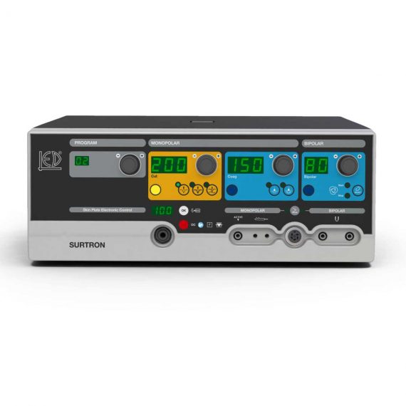

LED Surtron 200 HP Diathermy Machine
$1,750.00
Shipped from Abroad
SURTRON 200 is a high frequency electrosurgical equipment which is suitable for light and medium surgery and also allows to execute surgical minimally invasive procedure of resection, evaporation and coagulation.
Delivery & Availability:
Typically 10 working days – excluding furniture and heavy/bulky equipment. Please contact us for further information.
Description
SURTRON 200 is a high frequency electrosurgical equipment which is suitable to light and medium surgery.
SURTRON 200 allows to execute surgical minimally invasive procedure of resection, evaporation and coagulation.
Through its performances, SURTRON 200, allows pure CUT, cut-coagulation BLEND, incision with reduced prodution of eschar ENHANCED, superficial coagulation FORCED COAG, deep coagulation in absence of necrosis SOFT COAG and BIPOLAR coagulation.
The digital reading of the delivered power and the overseeing through microcontroller of the operational functions, assure the absolute reliability of the conditions of job.
SURTRON 200 allows a highly professional surgery thanks to the user-friendly and safety solutions normally used. The connection of neutral electrode is constantly monitored. Safety control of patient/plate contact using split neutral electrode. The possibility to control by the handle the output functions as well as the delivery of output power, allows to implement the surgical operation without turning away the surgeon attention from the surgical field.
FEATURES
Code 10100.401
Max output power CUT: 200W – 250Ω
Max output power ENHANCED: 120W – 250Ω
Max output power BLEND: 120W – 200Ω
Max output power FORCED COAG: 150W – 150Ω
Max output power SOFT COAG: 90W – 100Ω
Max output power BIPOLAR COAG: 80W – 50Ω
Working frequency: 600 kHz
Patient circuit: F
Mains voltage: 115-230 Vac
Mains frequency: 50-60 Hz
Electrical input power: 350 VA
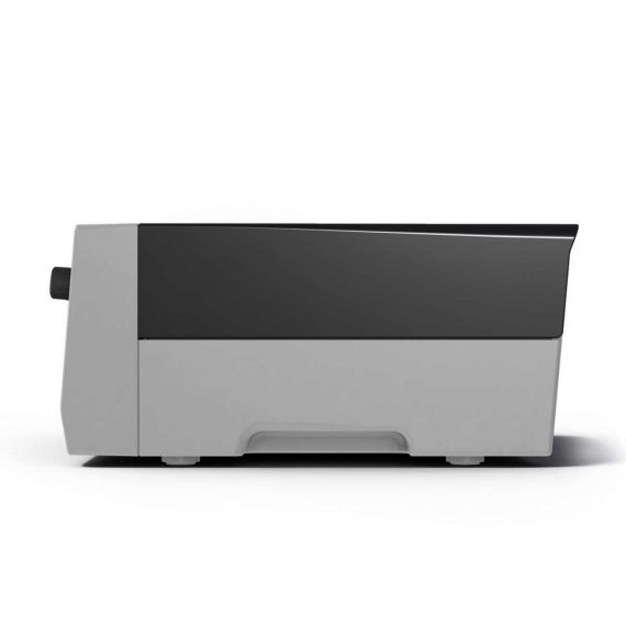
Size WxHxD: 370x144x319 mm
Weight: 6 Kgs
CONTROLS
Patient/Plate circuit monitoring
Output power monitoring
Self check control
SAFETY
EN60601-1
EN60601-1-2
EN60601-2-2
Electrical classification: CF
MDD 93/42 / EC: II b
Click Here To Download Catalogue
Quick Comparison
| LED Surtron 200 HP Diathermy Machine remove | DrGem Diamond All-In-One Digital X-ray Machine remove | Jade Mobile X-ray machine (Analogue) remove | Sonoscape S11 Ultrasound Machine remove | Sonoscape P10 Ultrasound Machine remove | ASPEL AsPEKT 712 Holter Monitor and Software remove | |
|---|---|---|---|---|---|---|
| Name | LED Surtron 200 HP Diathermy Machine remove | DrGem Diamond All-In-One Digital X-ray Machine remove | Jade Mobile X-ray machine (Analogue) remove | Sonoscape S11 Ultrasound Machine remove | Sonoscape P10 Ultrasound Machine remove | ASPEL AsPEKT 712 Holter Monitor and Software remove |
| Image | 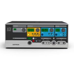 |  |  |  |  |  |
| SKU | SF1033560034-3 | SF1033560074-3 | SF1033560074-2 | SF1033560012-1 | SF1033560012-7 | SF1033560075-4 |
| Rating | ||||||
| Price | $1,750.00 |
|
| $6,380.00 | $9,350.00 | $1,991.00 |
| Stock | ||||||
| Availability | ||||||
| Add to cart | ||||||
| Description | Shipped from Abroad SURTRON 200 is a high frequency electrosurgical equipment which is suitable for light and medium surgery and also allows to execute surgical minimally invasive procedure of resection, evaporation and coagulation. Delivery & Availability: Typically 10 working days – excluding furniture and heavy/bulky equipment. Please contact us for further information. | Shipped from Abroad DrGem Diamond All-In-One Digital X-ray Machine is a fully automatic digital radiography system providing state-of-the-art image quality, image processing and user interface. With a wide selection of anatomical studies on the imaging software, DIAMOND automatically sets up the x-ray generator’s preprogrammed exposure technique settings, motorized radiographic stand positioning, x-ray collimation and post-image processing for the selected study. Specifically designed to increase workflow, this fully digital system offers convenient auto-positioning and advanced image processing to achieve big performance with little effort. Delivery & Availability: Typically 21 working days – excluding furniture and heavy/bulky equipment. Please contact us for further information. | In Stock JADE is one of the lightest portable X-ray systems on the market, allowing it to be used in any imaginable way including bedside, operating rooms, intensive care units and in veterinary fields. With a simple, easy-to-use operator console, three-way control, two-step foldable stand and auto lock system, JADE is a user-friendly portable X-ray system. Delivery & Availability: Typically 21 working days – excluding furniture and heavy/bulky equipment. Please contact us for further information. | In Stock A Value Choice beyond Your Expectation. SonoScape’s trolley color Doppler system S11 redefines price and performance with practical design. The S11 will go beyond your expectations but not your budget. Delivery & Availability: Typically 2 working days – excluding furniture and heavy/bulky equipment. Please contact us for further information. | Shipped from Abroad The P10 color Doppler ultrasound system is a new generation product from SonoScape. It is designed to give high quality images, rich probe configurations, various clinical tools and automatic analysis software to provide you with comprehensive solutions for your growing demand for clinical applications. Delivery & Availability: Typically 5-7 working days – excluding furniture and heavy/bulky equipment. Please contact us for further information. | Shipped from Abroad The Holta Monitor allows quick analysis of ECG examination and detection, reviewing and editing capability in the qualitative assessment of VE, VT, Single SVE, PSVT, Pauses, Irregular Rhythm, VT, IVR, Brady - and Tachycardia, Couplets, ST-segment elevation and depression, Maximum, Minimum and averaged Heart Rates, artifacts Delivery & Availability: Typically 10 working days – excluding furniture and heavy/bulky equipment. Please contact us for further information. |
| Content | SURTRON 200 is a high frequency electrosurgical equipment which is suitable to light and medium surgery.
SURTRON 200 allows to execute surgical minimally invasive procedure of resection, evaporation and coagulation.
Through its performances, SURTRON 200, allows pure CUT, cut-coagulation BLEND, incision with reduced prodution of eschar ENHANCED, superficial coagulation FORCED COAG, deep coagulation in absence of necrosis SOFT COAG and BIPOLAR coagulation.
The digital reading of the delivered power and the overseeing through microcontroller of the operational functions, assure the absolute reliability of the conditions of job.
SURTRON 200 allows a highly professional surgery thanks to the user-friendly and safety solutions normally used. The connection of neutral electrode is constantly monitored. Safety control of patient/plate contact using split neutral electrode. The possibility to control by the handle the output functions as well as the delivery of output power, allows to implement the surgical operation without turning away the surgeon attention from the surgical field.
FEATURES
Code 10100.401
Max output power CUT: 200W – 250Ω
Max output power ENHANCED: 120W – 250Ω
Max output power BLEND: 120W – 200Ω
Max output power FORCED COAG: 150W – 150Ω
Max output power SOFT COAG: 90W – 100Ω
Max output power BIPOLAR COAG: 80W – 50Ω
Working frequency: 600 kHz
Patient circuit: F
Mains voltage: 115-230 Vac
Mains frequency: 50-60 Hz
Electrical input power: 350 VA
Size WxHxD: 370x144x319 mm
Weight: 6 Kgs
CONTROLS
Patient/Plate circuit monitoring
Output power monitoring
Self check control
SAFETY
EN60601-1
EN60601-1-2
EN60601-2-2
Electrical classification: CF
MDD 93/42 / EC: II b
Click Here To Download Catalogue | DrGem Diamond All-In-One Digital X-ray Machine is a fully automatic digital radiography system providing state-of-the-art image quality, image processing and user interface. With a wide selection of anatomical studies on the imaging software, DIAMOND automatically sets up the x-ray generator’s pre-programmed exposure technique settings, motorized radiographic stand positioning, x-ray collimation and post-image processing for the selected study. Specifically designed to increase workflow, this fully digital system offers convenient auto-positioning and advanced image processing to achieve big performance with little effort.
Features of DrGem Diamond All-In-One Digital X-ray Machine:
Outstanding Image Quality -
Digital radiography via at panel detector improves your workflow, exam speed and comfort with efficiency. Digital at panel detector with Csl screen provides excellent spatial resolution, MTF, DQE and stability based on ne pixel pitch. A 3-field ion-chamber is provided for AEC function.
Automatic Collimation –
Automatic x-ray eld size control of the motorized collimator corresponds to dierent SIDs. Includes user adjustable lamp timer with on/oswitch.
Automatic Positioning –
Click Here To Download Catalogue | JADE Mobile X-ray machine is one of the lightest portable X-ray systems on the market, allowing it to be used in any imaginable way including bedside, operating rooms, intensive care units and veterinary fields. With a simple, easy-to-use operator console, three-way control, two-step foldable stand and auto-lock system, the JADE Mobile X-ray machine is a user-friendly portable X-ray system.
Convenient & Intuitive Operation:
JADE is one of the lightest portable X-ray systems on the market, allowing it to be used in any imaginable way including bedside, operating rooms, intensive care units and in veterinary fields. With a simple, easy-to-use operator console, three-way control, two-step foldable stand and auto-lock system, JADE is a user-friendly portable X-ray system.
Compact & Powerful Design:
JADE Mobile X-ray machine is an innovative, highly versatile portable X-ray system suitable for a variety of clinical uses. Utilizing the unique technology used in DRGEM’s universally recognized X-ray generators, JADE is a compact but powerful unit with a 4kW output and thoughtfully designed components to increase efficiency and maximize workflow. The core part of X-ray source adopts high-quality tube assembly, X-ray collimator and high frequency X-ray generator with excellent performance, lifetime and stability.
Features:
Click Here To Download Catalogue | DETAILS
SonoScape’s trolley colour Doppler system S11 redefines price and performance with practical design. The S11 will go beyond your expectations but not your budget. As an easy-to-use ultrasound system, the S11 is integrated with a new software platform, especially optimized for a smooth workflow and convenient operation. The system speeds up the exam process and makes file management easier.
SPECIFICATION
- 15-inch high definition LCD monitor with articulating arm
- Compact and agile trolley design
- 3 active transducer sockets available for a wide range of applications
- Duplex, Color Doppler, DPI, PW Doppler, tissue harmonic imaging, μ-scan speckle reduction imaging, compound imaging, trapezoidal imaging
- Customized settings based on your own working style
- Full patient database and image management solutions
Click Here To Download Catalogue | DETAILS
B + Compound
B + Compound utilizes several lines of sight for optimal contrast resolution, speckle reduction and border detection, with which P10 is ideal for superficial and abdominal imaging with better clarity and improved continuity of structures.
μ-Scan
The new generation μ-Scan imaging technology gives you better image quality by reducing noise, improving signal strength and improving visualization.
P10 offers a comprehensive selection of electronic probes to maximize its capabilities to meet a wide range of applications including abdomen, pediatric, OB/GYN, cardiovascular, musculoskeletal, etc. The advanced probe technologies also effectively enhance the image quality and confidence in reaching clinical diagnoses, even in difficult patients.
Convex Probe 3C-A
Ideal for an abundant of application such as abdomen, gynecology, obstetrics, urology and even abdomen biopsy.
Linear Probe L741
This linear probe is designed to satisfy vascular, breast, thyroid, and other small parts diagnosis, and its adjustable parameters could also present users a clear view of MSK and deep vessels.
Phase Array Probe 3P-A
For the purpose of adult and pediatric cardiology and emergency, the phase array probe provides elaborate presets for different exam modes, even for difficult patients.
Intracavitary Probe 6V1
Intracavitary probe could face application of gynecology, urology, prostate, and its temperature detection technology not only protects the patient but also extends the service life.
Click Here To Download Catalogue | The Holter Monitor allows quick analysis of ECG examination (arrhythmias and ST segment).
Technical specifications:
HolCARD 24W Software:
Click Here To Download Catalogue |
| Weight | N/A | N/A | N/A | N/A | N/A | N/A |
| Dimensions | N/A | N/A | N/A | N/A | N/A | N/A |
| Additional information |


Reviews
There are no reviews yet.