
MCC-E1 Electric-driven 3D Chest Compressor
$6,435.00
Shipped From Abroad
The MCC-E1 provides high-quality 3D chest compressions during CPR. It offers customizable settings, real-time data display, and up to 60 minutes of battery operation.
Delivery & Availability:
Typically 10-21 working days – excluding furniture and heavy/bulky equipment. Please contact us for further information.
Typically 10-21 working days – excluding furniture and heavy/bulky equipment. Please contact us for further information.
Description
Features
- 3D compression technology: This technology combines the cardiac pump and thoracic pump mechanisms to improve resuscitation efficiency.
- Electronic control unit: The MCC-E1 has a 2.8-inch OLED screen that displays compression data in real-time. It also allows the user to adjust the compression rate and depth.
- Low compression risk: The MCC-E1 has a soft start to reduce the risk of fracture. It also has a low gravity center to prevent toppling and hurting patients.
- Suitable for complex environments: The MCC-E1 has IP34 and EN1789 certification.
- Data capacity: The MCC-E1 has 180 hours of data capacity.
- Multi-channel transmission: The MCC-E1 can transmit data using USB, WiFi, and Bluetooth.
- CPR data cloud: The MCC-E1 has a CPR data cloud.
- Lightweight: The MCC-E1 weighs only 3 kg, and the full setup weighs less than 7 kg.
- Adjustable compression: The MCC-E1 allows the user to adjust the compression depth and rate.
- Charge while in use: The MCC-E1 can be used while charging.
Data storage
The MCC-E1 can store resuscitation data automatically and export it for analysis and reporting. It has a data capacity of 180 hours and can transmit data via USB, WiFi, and Bluetooth.UsesThe MCC-E1 is suitable for a variety of uses, including:
- Preparing ECPR
- Hypothermia treatment
- PCI treatment
- Head-up CPR
- Complex environments
- Preparing ECPR
Quick Comparison
| MCC-E1 Electric-driven 3D Chest Compressor remove | Sonoscape E1 Ultrasound Machine With Two Probes remove | DrGem Ceiling Analogue X-ray Machine remove | Sonoscape P20 Ultrasound Machine remove | Sonoscape E2 Ultrasound Machine remove | DrGem GXR-SD 400mA Floor Mounted Digital X-ray remove | |
|---|---|---|---|---|---|---|
| Name | MCC-E1 Electric-driven 3D Chest Compressor remove | Sonoscape E1 Ultrasound Machine With Two Probes remove | DrGem Ceiling Analogue X-ray Machine remove | Sonoscape P20 Ultrasound Machine remove | Sonoscape E2 Ultrasound Machine remove | DrGem GXR-SD 400mA Floor Mounted Digital X-ray remove |
| Image | 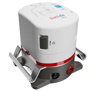 |  |  |  |  |  |
| SKU | SF1033560130160-1 | SF1033560012-20 | SF1033560074-7 | SF1033560012-9 | SF1033560012-17 | SF1033560074-5 |
| Rating | ||||||
| Price | $6,435.00 | $4,620.00 |
|
| $5,500.00 |
|
| Stock | ||||||
| Availability | ||||||
| Add to cart | ||||||
| Description | Shipped From Abroad
The MCC-E1 provides high-quality 3D chest compressions during CPR. It offers customizable settings, real-time data display, and up to 60 minutes of battery operation.
Delivery & Availability:
Typically 10-21 working days – excluding furniture and heavy/bulky equipment. Please contact us for further information.
| Shipped from Abroad SonoScape has developed a new probe and function for the E1 Exp. With these additions the E1 Exp will bring users a more efficient examination experience with satisfying image quality and a smooth workflow. Delivery & Availability: Typically 5-7 working days – excluding furniture and heavy/bulky equipment. Please contact us for further information. | Shipped from abroad The DrGem Ceiling Analogue X-ray Machine is a diagnostic radiography system that provides reliable high quality radiographic images with a reduced dose. The reliable high-frequency x-ray generators that are known worldwide for their excellent performance, lifetime and stability. Patient tables and wall stands are also offered. Delivery & Availability: Typically 21 working days – excluding furniture and heavy/bulky equipment. Please contact us for further information. | Shipped from Abroad Incorporating innovative technologies, P20’s user-friendly design with a simple operation panel, intuitive user interface and a variety of intelligent auxiliary scanning tools, will significantly improve your daily examination experience. Besides general imaging applications, P20 has entitled with diagnostic 4D technology which has an extraordinary performance in obstetrics and gynecology applications. Delivery & Availability: Typically 5-7 working days – excluding furniture and heavy/bulky equipment. Please contact us for further information. | Shipped from Abroad Sonoscape E2 portable ultrasound machine is a color Doppler ultrasound system that reaches beyond your expectations due to its compact and fashionable appearance. It fulfills GI, OB/GYN, Cardiac and POC applications to fit your routine scanning needs while its color mode will help you for more accurate and efficient diagnosis of lesions. E2 provides a wide range of applications to assist users with routine scanning. E2 provides automatic calculations to enhance your diagnostic confidence and save you time for patient communication. Delivery & Availability: Typically 14 working days – excluding furniture and heavy/bulky equipment. Please contact us for further information. | In Stock The GXR-SD Digital X-ray is a diagnostic digital radiography system that provides reliable high quality digital radiographic images with a reduced dose. The GXR-SD DR systems offer comprehensive digital solutions to all radiography needs, featuring ACQUIDR digital imaging system with stationary or portable digital flat-panel detectors as well as reliable high-frequency x-ray generators that are known worldwide for their excellent performance, lifetime and stability. Patient tables and wall stands are also offered. Delivery & Availability: Typically 21 working days – excluding furniture and heavy/bulky equipment. Please contact us for further information. |
| Content | https://youtu.be/8dyQXvobeuA?si=_wnelIZ_c2YmQEGL
https://youtu.be/67kQOIOsE34?si=zRhDbGv5CIwkXOJA
Features
| DETAILS
Efficient Diagnosis
μ-Scan, Speckle Reduction & Edge Enhancement
Spatial Compound Imaging
PIH - Pure Inversion Harmonic
Wide Scan - Enlarged Image Area
Tissue-Specific Imaging
SR Flow
Ergonomic Designs
Up to 2 Transducer Ports
Light Weight and Compact
15.6 inch Anti-flickering HD LED Screen
Tilting Monitor Angle Adjustment
Backlit Keyboard and Intelligent Panel
Long-lasting Battery for 90 mins
Ease of Use
Quick Boot Up
Auto-Brightness Adjustment
Auto Image Optimization
Auto IMT
Auto Trace
Equipped Accessories
Wi-Fi and Bluetooth Available
DICOM
500GB Hard Disk
Height Adjustable Trolley
Durable, Carry-on Site Suitcase
Click Here To Download Catalogue | DrGem Ceiling Analogue X-ray Machine is a diagnostic radiography system X-ray Machine that provides reliable high quality radiographic images with a reduced dose. The reliable high-frequency x-ray generators that are known worldwide for their excellent performance, lifetime and stability. Patient tables and wall stands are also offered.
Features of DrGem Ceiling Analogue X-ray Machine
Click Here To Download Catalogue | DETAILS
Upgraded Images with More Clarity
SonoScape never stops making progress in improving the image quality of its ultrasound products to enhance the confidence of diagnosis for doctors. With extraordinary images provided by P20, the anatomy structures are clearer than ever.
C-Xlasto Imaging
With C-xlasto Imaging, P20 enables comprehensive quantitative elastic analysis. Meanwhile, C-xlasto on P20 is supported by linear, convex and transvaginal probes, to ensure good reproducibility and highly consistent quantitative elastic results.
S-Live
S-Live allows for detailed visualization of subtle anatomical features, thereby enabling intuitive diagnosis with real-time 3D images and enriching patient communication.
Pelvic Floor 4D
Transperineal 4D pelvic floor ultrasound can provide useful clinical values in assessing the vaginal delivery impact on the female anterior compartment, judging whether the pelvic organs are prolapsed or not and the extent, determining if the pelvic muscles were torn accurately.
Anatomic M Mode
Anatomic M Mode helps you observe the myocardial motion at different phases by freely placing sample lines. It accurately measures the myocardial thickness and the heart size of even difficult patients and supports the myocardial function and LV wall-motion assessment.
Tissue Doppler Imaging
P20 is endowed with Tissue Doppler Imaging which provides velocities and other clinical information on myocardial functions, facilitating clinical doctors with the ability to analyze and compare the motions of different parts of the patient's heart.
Click Here To Download Catalogue | SONOSCAPE E2 DETAILS
Auto Image Optimization
A portable ultrasound machine with the press of a button, the image is automatically adjusted and optimized, saving you time with parameter adjustments. Additionally, with Auto Focus on, the focus area follows the depth of the ROI box as it is moved in the scanning field, providing users with excellent image quality in the desired area of interest.
Automated Calculation
Auto IMT is used when determining the level of vascular sclerosis present in the patient by automatically tracing the thickness of the carotid vessels.
Auto trace provides users sensitive and accurate wave tracing, avoiding the error of manual trace and giving out calculation result in no time
In-Build Battery pack
This portable ultrasound machine was equipped with an in-build battery pack which enable the user to perform image scanning when AC power is not available.
Click Here To Download Catalogue | DrGem GXR-SD 400mA Floor Mounted Digital X-ray system matches with a radiographic room which perfectly fits your workow and can be easily upgraded to DR system with the help of DR interface and PC interface in GXR generator as well as Bucky suitable to Flat Panel Detector. GXR X-ray system is equipped with a high frequency X-ray generator which consistently produces high quality radiograph in favor of high quality X-ray output with a very small kV ripple and accurate mA and mAs. GXR X-ray system is designed to provide convenience to operator and comfort to patient
Features of DrGem GXR-SD 400mA Floor Mounted Digital X-ray:
Click Here To Download Catalogue |
| Weight | N/A | N/A | N/A | N/A | N/A | N/A |
| Dimensions | N/A | N/A | N/A | N/A | N/A | N/A |
| Additional information |


Reviews
There are no reviews yet.