Portable Slit Lamp
Ask for Price$0.00
Shipped from abroad
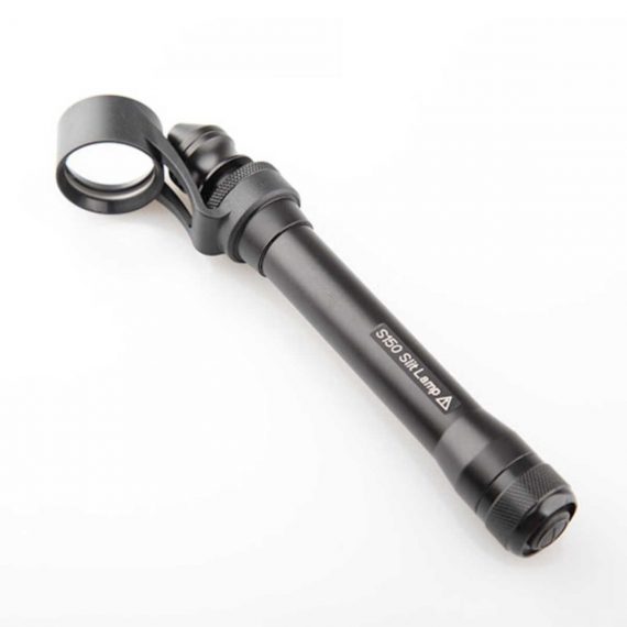
This ultra-portable is an excellent diagnostic instrument for the examination of anterior segment structures and ocular abnormalities.
Delivery & Availability:
Typically 14 working days – excluding furniture and heavy/bulky equipment. Please contact us for further information.
Description
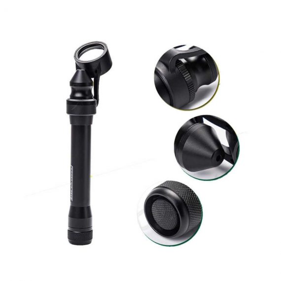
Features:
- Ultra-portable: This ultra-portable is an excellent diagnostic instrument for the examination of anterior segment structures and ocular abnormalities. Its easy-to-operate optical system produces a high-brightness, continuously adjustable slit image ideal for pediatric and geriatric setting, emergency department screenings, ward rounds, beside examination, post-op evaluations, and mission work;
- LED illumination: the first and the only portable slit lamp in the world applying LED illumination system. The most prominent advantage of our LED illumination system gives the examiner the clearest image without glare;
- Comfortable using experience: The low heat radiation from our LED lamp makes the most comfortable examine experience for the patient;
- Sharpest Slit: With the blade imaging system S150 gives equally sharpest slit as the best classic slit lamp in the world;
- No need to replace illumination lamp: The LED lamp used in S150 portable slit lamp is rated 20,000 hours at full power. Its lifetime is almost 10 times longer than a normal halogen lamp;
- Power-saving: S150 can work for long hours without changing batteries.
Technical Specifications:
- Magnification: 5x
- Range of slit length: 5mm~11mm
- Minimum slit width: 0.2mm
- Working distance: 12~32mm
- Power supply: AA batteries x 2
- Illumination: LED blub(3.3V/1W)
- Battery life: More than 3h(AA alkaline batteries x 2)
- Net weight: 150g(without batteries)
How to Operate:
- Press the Power Switch to turn the slit lamp on/off
- Loosen the Locking Screw
- Twist the Lamp Body while holding the Magnifier Bracket still till the slit is in the required angle
- Push or pull the Lamp Body while holding the Magnifier Bracket still to set the Magnifier into a position comfortable for observation
- Fasten the Locking Screw
- Pull the Lamp Head back and forth for adjusting the width and the length of the slit, as well as the intensity of the light
Quick Comparison
| Settings | Portable Slit Lamp remove | Sonoscape S11 Ultrasound Machine remove | Sonoscape E2 Ultrasound Machine remove | DrGem GXR-SD 400mA Floor Mounted Digital X-ray remove | Sonoscape P10 Ultrasound Machine remove | ASPEL AsCARD Green B/W ECG Machine remove |
|---|---|---|---|---|---|---|
| Name | Portable Slit Lamp remove | Sonoscape S11 Ultrasound Machine remove | Sonoscape E2 Ultrasound Machine remove | DrGem GXR-SD 400mA Floor Mounted Digital X-ray remove | Sonoscape P10 Ultrasound Machine remove | ASPEL AsCARD Green B/W ECG Machine remove |
| Image | 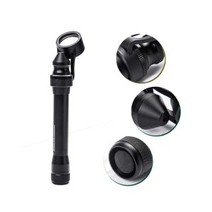 |  |  |  |  |  |
| SKU | SF1033560107-6 | SF1033560012-1 | SF1033560012-17 | SF1033560074-5 | SF1033560012-7 | SF1033560075-8 |
| Rating | ||||||
| Price | Ask for Price | $6,950.00 | $4,300.00 | $42,310.00 | Ask for Price | Ask for Price |
| Stock | ||||||
| Availability | ||||||
| Add to cart | ||||||
| Description | Shipped from abroad
This ultra-portable is an excellent diagnostic instrument for the examination of anterior segment structures and ocular abnormalities.
| In Stock A Value Choice beyond Your Expectation. SonoScape’s trolley color Doppler system S11 redefines price and performance with practical design. The S11 will go beyond your expectations but not your budget. Delivery & Availability: Typically 2 working days – excluding furniture and heavy/bulky equipment. Please contact us for further information. | Shipped from Abroad Sonoscape E2 portable ultrasound machine is a color Doppler ultrasound system that reaches beyond your expectations due to its compact and fashionable appearance. It fulfills GI, OB/GYN, Cardiac and POC applications to fit your routine scanning needs while its color mode will help you for more accurate and efficient diagnosis of lesions. E2 provides a wide range of applications to assist users with routine scanning. E2 provides automatic calculations to enhance your diagnostic confidence and save you time for patient communication. Delivery & Availability: Typically 14 working days – excluding furniture and heavy/bulky equipment. Please contact us for further information. | In Stock The GXR-SD Digital X-ray is a diagnostic digital radiography system that provides reliable high quality digital radiographic images with a reduced dose. The GXR-SD DR systems offer comprehensive digital solutions to all radiography needs, featuring ACQUIDR digital imaging system with stationary or portable digital flat-panel detectors as well as reliable high-frequency x-ray generators that are known worldwide for their excellent performance, lifetime and stability. Patient tables and wall stands are also offered. Delivery & Availability: Typically 21 working days – excluding furniture and heavy/bulky equipment. Please contact us for further information. | Shipped from Abroad The P10 color Doppler ultrasound system is a new generation product from SonoScape. It is designed to give high quality images, rich probe configurations, various clinical tools and automatic analysis software to provide you with comprehensive solutions for your growing demand for clinical applications. Delivery & Availability: Typically 5-7 working days – excluding furniture and heavy/bulky equipment. Please contact us for further information. | Shipped from Abroad AsCARD Green electrocardiograph is a 1- and 3-channel ECG unit which enables to make electrocardiogram in full 12 leads. Intended for ECG examinations of adult and paediatric patients aimed at identification of cardiological abnormalities, myocardial ischaemia or infarction. The device is intended for use in healthcare facilities by duly trained personnel. ECG examination may be recorded in manual or automatic mode with the ability to perform the analysis and interpretation. Delivery & Availability: Typically 10 working days – excluding furniture and heavy/bulky equipment. Please contact us for further information. |
| Content | Features:
| DETAILS
SonoScape’s trolley colour Doppler system S11 redefines price and performance with practical design. The S11 will go beyond your expectations but not your budget. As an easy-to-use ultrasound system, the S11 is integrated with a new software platform, especially optimized for a smooth workflow and convenient operation. The system speeds up the exam process and makes file management easier.
SPECIFICATION
- 15-inch high definition LCD monitor with articulating arm
- Compact and agile trolley design
- 3 active transducer sockets available for a wide range of applications
- Duplex, Color Doppler, DPI, PW Doppler, tissue harmonic imaging, μ-scan speckle reduction imaging, compound imaging, trapezoidal imaging
- Customized settings based on your own working style
- Full patient database and image management solutions
Click Here To Download Catalogue | SONOSCAPE E2 DETAILS
Auto Image Optimization
A portable ultrasound machine with the press of a button, the image is automatically adjusted and optimized, saving you time with parameter adjustments. Additionally, with Auto Focus on, the focus area follows the depth of the ROI box as it is moved in the scanning field, providing users with excellent image quality in the desired area of interest.
Automated Calculation
Auto IMT is used when determining the level of vascular sclerosis present in the patient by automatically tracing the thickness of the carotid vessels.
Auto trace provides users sensitive and accurate wave tracing, avoiding the error of manual trace and giving out calculation result in no time
In-Build Battery pack
This portable ultrasound machine was equipped with an in-build battery pack which enable the user to perform image scanning when AC power is not available.
Click Here To Download Catalogue | DrGem GXR-SD 400mA Floor Mounted Digital X-ray system matches with a radiographic room which perfectly fits your workow and can be easily upgraded to DR system with the help of DR interface and PC interface in GXR generator as well as Bucky suitable to Flat Panel Detector. GXR X-ray system is equipped with a high frequency X-ray generator which consistently produces high quality radiograph in favor of high quality X-ray output with a very small kV ripple and accurate mA and mAs. GXR X-ray system is designed to provide convenience to operator and comfort to patient
Features of DrGem GXR-SD 400mA Floor Mounted Digital X-ray:
Click Here To Download Catalogue | DETAILS
B + Compound
B + Compound utilizes several lines of sight for optimal contrast resolution, speckle reduction and border detection, with which P10 is ideal for superficial and abdominal imaging with better clarity and improved continuity of structures.
μ-Scan
The new generation μ-Scan imaging technology gives you better image quality by reducing noise, improving signal strength and improving visualization.
P10 offers a comprehensive selection of electronic probes to maximize its capabilities to meet a wide range of applications including abdomen, pediatric, OB/GYN, cardiovascular, musculoskeletal, etc. The advanced probe technologies also effectively enhance the image quality and confidence in reaching clinical diagnoses, even in difficult patients.
Convex Probe 3C-A
Ideal for an abundant of application such as abdomen, gynecology, obstetrics, urology and even abdomen biopsy.
Linear Probe L741
This linear probe is designed to satisfy vascular, breast, thyroid, and other small parts diagnosis, and its adjustable parameters could also present users a clear view of MSK and deep vessels.
Phase Array Probe 3P-A
For the purpose of adult and pediatric cardiology and emergency, the phase array probe provides elaborate presets for different exam modes, even for difficult patients.
Intracavitary Probe 6V1
Intracavitary probe could face application of gynecology, urology, prostate, and its temperature detection technology not only protects the patient but also extends the service life.
Click Here To Download Catalogue | AsCARD Green electrocardiograph is a 1- and 3-channel ECG unit which enables to make electrocardiogram in full 12 leads. Intended for ECG examinations of adult and paediatric patients aimed at identification of cardiological abnormalities, myocardial ischaemia or infarction. The device is intended for use in healthcare facilities by duly trained personnel. ECG examination may be recorded in manual or automatic mode with the ability to perform the analysis and interpretation.
Electrocardiograph is based on advanced microprocessor technology. It is equipped with a thermal printer with high-resolution head and graphical LCD display. A hightech membrane keyboard makes the AsCARD Green device operation intuitive, and its menu navigation exceptionally easy. This light-weight, small-footprint and battery powered cause that device can be easily transported to any location. With plastic casing and foil covered keyboard, the device is neat and easy to clean.
Technical Specifications:
Click Here To Download Catalogue |
| Weight | N/A | N/A | N/A | N/A | N/A | N/A |
| Dimensions | N/A | N/A | N/A | N/A | N/A | N/A |
| Additional information |


Reviews
There are no reviews yet.