
Neuro MEP Micro-Portable EMG and NCS System
$0.00
Shipped From Abroad
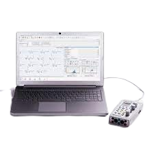
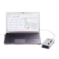
In coloproctology centers, EMG of external sphincter and pelvic floor muscles is often required. Portable EMG and NCS system Neuro-MEP-Micro packed with the necessary techniques is the right solution for these needs.
Typically 10-21 working days – excluding furniture and heavy/bulky equipment. Please contact us for further information.
Description
Features
Specific techniques for neurophysiological assessment of external sphincter and other pelvic floor neuromuscular structures
The system is used to perform various neurophysiological examinations of pelvic floor for diagnostic and scientific purposes, i.e.:
- bulbocavernosus and other sacral reflex tests with electrical stimulation of pudendal nerve to assess the conduction of reflex arcs (S2-S4);
- surface EMG of external sphincter and pelvic floor muscles to assess the level of tonic contraction;
- needle EMG with quantitative motor unit potential (MUP) analysis coupled with sacral reflex test to detect sacral denervation;
- stimulation EMG used with a disposable St. Mark’s electrode to stimulate the distal part of pudendal nerve;
- pudendal nerve somatosensory evoked potentials (pSEPs) especially in patients with preserved sacral reflexes and hypoesthesia of perineum;
- sympathetic skin responses (SSR) recorded from perineal area to assess the conduction velocity of sympathetic unmyelinated fibers and myelinated sensory nerve fibers;
- analysis of motor evoked potentials (MEP) in perineal muscles during the cortical and sacral magnetic stimulation (the assessment of corticospinal tract conduction with recording of pelvic floor motor evoked potentials (if the magnetic stimulator is available)).
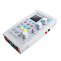
Portable design
Neuro-MEP-Micro requires very little space and is connected to a computer via the USB cable which ensures data uploading and power supply of the device. If not powered from mains, the device can operate from the notebook battery.
Electrical stimulator with two outputs
Two software switchable outputs to plug in the electrical stimulator allow a specialist to place two pairs of stimulating electrodes on a patient and connect them to the device. Thus, there is no need to switch the electrodes as the stimulating electrode is software-defined.
Quick Comparison
| Settings | Neuro MEP Micro-Portable EMG and NCS System remove | Sonoscape P15 Ultrasound Machine With Four Probes remove | Littman Stethoscope remove | Timesco Jumper Fingertip Pulse Oximeter remove | Sonoscape P50 Ultrasound Machine remove | Bettermed BT666EPZ Multi-function Electric Delivery Bed remove |
|---|---|---|---|---|---|---|
| Name | Neuro MEP Micro-Portable EMG and NCS System remove | Sonoscape P15 Ultrasound Machine With Four Probes remove | Littman Stethoscope remove | Timesco Jumper Fingertip Pulse Oximeter remove | Sonoscape P50 Ultrasound Machine remove | Bettermed BT666EPZ Multi-function Electric Delivery Bed remove |
| Image | 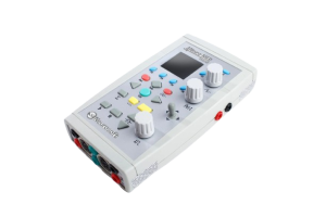 |  |  | 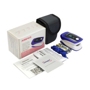 |  |  |
| SKU | SF1033560012-8 | SF1033560084-5 | SF1033560084-2 | SF1033560012-11 | SF1033560084-51 | |
| Rating | ||||||
| Price |
| $13,900.00 | $13.20 | $17.00 |
| $2,024.00 |
| Stock | ||||||
| Availability | ||||||
| Add to cart | ||||||
| Description | Shipped From Abroad
In coloproctology centers, EMG of external sphincter and pelvic floor muscles is often required. Portable EMG and NCS system Neuro-MEP-Micro packed with the necessary techniques is the right solution for these needs.
Delivery & Availability:
Typically 10-21 working days – excluding furniture and heavy/bulky equipment. Please contact us for further information.
| In Stock A feature-rich system inheriting the Wi-Sono high-end platform, the P15 uses an array of advanced tools to help enhance the image quality. It's a cost-effective, simplified console with an intuitive user interface and multiple intelligent functions. Delivery & Availability: Typically 2 working days – excluding furniture and heavy/bulky equipment. Please contact us for further information. | In Stock
Delivery & Availability: Typically 2 working days – excluding furniture and heavy/bulky equipment. Please contact us for further information. | In Stock Integrated with SPO2 probe and processing display module. Small in volume、light in weight and convenient in carrying. Operation of the product is simple ,low power consumption. SPO2 , PR and Ridisplay,. Pulse rate value display, bar graph display. Low-voltage indication: low-voltage indicator appears before working abnormally which is due to low-voltage. Automatically power off function: when the device is under the state of measuring interface . it will automatically power off within 5 seconds if the finger falls out of probe. Delivery & Availability: Typically 2 working days – excluding furniture and heavy/bulky equipment. Please contact us for further information. | Shipped from Abroad Easily accomplish more with SonoScape’s new P50 ultrasound system. Incorporating single crystal clarity, automatic corrections and calculation, and user defined flexibility promises a confident diagnostic experience as well as opening new doors of opportunity for ultrasound use. Delivery & Availability: Typically 7-14 working days – excluding furniture and heavy/bulky equipment. Please contact us for further information. | In Stock
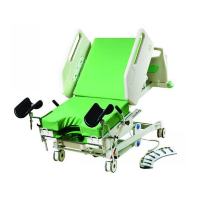
Function:Backrest tilting From 0°to 75° ±5°
Hi-lo function
Reverse:14°±1°
Trendelenburg:12°±1°
Delivery & Availability: Typically 2 working days – excluding furniture and heavy/bulky equipment. Please contact us for further information. |
| Content | FeaturesSpecific techniques for neurophysiological assessment of external sphincter and other pelvic floor neuromuscular structures The system is used to perform various neurophysiological examinations of pelvic floor for diagnostic and scientific purposes, i.e.:
| DETAILS
Super Wide-bandwidth Platform
Inheriting Wi-sono's ultra-wide system platform and with the advanced probe technology, high-resolution and deep penetration images are provided for precision medicine.
Spatial Compound Imaging
Spatial Compound Imaging utilizes several lines of sight for optimal contrast resolution, speckle reduction and border detection, with which P15 is ideal for superficial and abdominal imaging with better clarity and improved continuity of structures.
μ-Scan+
The new generation μ-Scan imaging technology gives you better image quality by reducing noise, improving signal strength and improving visualization.
Dynamic Color
Dynamic color improves upon already existing color Doppler technologies for a clearer capture of color flow and detailed visualization of even tiny veins with lower velocities.
Real-time Panoramic
With real-time panoramic, you can acquire an extended field of view for large organs or long vessels for easy measurement and diagnostic efficiency. Accomplished in real-time for the convenience of the sonographers, any mistake can also be easily back tracked and corrected without interrupting the scan.
3D/4D
Outstanding volume performance with speed and convenience makes P15 outshine others on volume imaging.
Tissue Doppler Imaging
Tissue Doppler Imaging allows clinical doctors to quantitatively evaluate local myocardial movements and functions, facilitating them with the ability to analyze and compare the motions of the different parts of the patient's heart.
Auto IMT
Quick measurement of intra-media vessel thickness ensures good reproducibility and high diagnostic efficiency.
Click Here To Download Catalogue | Littman Stethoscope Features:
| Integrated with SPO2 probe and processing display module • Small in volume, light in weight, convenient in carrying • Simple operation, lower power consumption • OLED screen display • SPO2, PR, bar graph of pulse, plethysmogram, blood perfusion index, battery indication display • 4 directions display, 6 display models, large fonts display • Brightness control, Demo mode, Setup menu • Movement interference • Alarm function and adjustable alarm parameters • Can be connected to mobile phones through Bluetooth; can support Android and iOS client • Auto power-off when no signal after 10s • Powered by 2*AAA alkaline batteries Specification: • Size: 62 x 37 x 32mm • Peak wavelength range of the light emitted from the probe: red light 660nm±3; infrared light 905nm±5 • Maximum optical output power of the probe: 1.2mW for infrared light (905nm) • Bluetooth module: 4.0 (single-mode) • Working temperature: 5℃ to 40℃ (41°F to 104°F) • Relative humidity: 15% to 80%, non-condensing • Atmospheric pressure: 70kPa to 106kPa • Rated voltage: DC 3V Package included: • 1 x JUMPER JPD-500F Bluetooth Fingertip Pulse Oximeter • 2 x AAA Batteries • 1 x Carry Case • 1 x Lanyard • 1 x English User Manual | DETAILS
Powerful Compact Precision
Taking into consideration the evolving expectations and needs for ultrasound, the P50 is a slim and unobtrusive trolley system that is comfortable in tight, congested spaces with little room to work in. Providing everything you need for a comfortable examination in a small space for both you and your patient.
Single Crystal Transducer
Wideband single crystal probes greatly improve the signal ratio, acquire stunning images and provide superior sensitivity and resolution for both the near and far-fields.
μ-Scan+
The new generation μ-Scan imaging technologies give you better image quality by reducing noise, improving signal strength and improving visualization.
Dynamic Color
Dynamic colour improves upon already existing colour Doppler technologies for clear capture of colour flow and detail visualization of even tiny veins with lower velocities.
Solution for Radiology
P50, is a leading-edge ultrasound system that can meet the demands of any clinical setting. You can experience a superior performance in multi-dimensional imaging for a full range of clinical applications – abdominal, breast and cardiovascular.
C-xlasto Imaging
By understanding that tissue stiffness varies depending on the type of tissue, we can use C-xlasto Imaging to easily find abnormalities and tumours within soft tissue. The differences in tissue responses are detected and visualized in real-time by the elastography algorithms through different representations, which can be particularly helpful in analyzing breast, thyroid and musculoskeletal structures. Predominately used only in linear probes, SonoScape’s new transvaginal and bi-plane probe for gynaecology and urology are breaking the mould and expanding elastography applications.
Real-time Color Panoramic
With the combination of colour flow and real-time panoramic, visualizing the blood flow of an entire vein or artery is now an easy task. Accomplished in real-time for the convenience of the sonographers, any mistakes can also be easily backtracked and corrected without interrupting the scan.
Contrast Imaging
Contrast Imaging on P50 makes full use of the infra harmonic signal and second harmonic signal to improve the image resolution and deep penetration. What’s more, the Dynamic Acoustic Control technology effectively controls the acoustic pressure for the contrast agent, decreasing the required agent dose and assures uniform image quality, guaranteeing longer contrast agent duration and better lesion perfusion of delayed phase observation.
Solution for OB/GYN
P50 has superior image quality, automated measurement tools, and a variety of volume technologies to provide ideal solutions for clinical examinations such as pregnancy examinations, and gynecologic disease diagnosis. With a new 4D transvaginal probe, P50 helps you to see and detect fetal abnormalities and significantly improves your diagnostic confidence during your examinations.
S-Live Silhouette
A unique transparent 3D anatomical image of the fetus for improved initial anatomical review. By using this new application, the system can create completely different fetal images from conventional ultrasound images, which can depict the fetal's intracorporeal anatomical structure.
Pelvic Floor 4D
Working in conjunction with SonoScape’s latest transvaginal probes, trans-perineal 4D pelvic floor ultrasound provides a useful clinical assessment of the impact of vaginal delivery on the female anterior compartment. Allowing doctors to judge whether the pelvic organs prolapsed or not, the extent of prolapse, and determining whether the pelvic muscles tore correctly.
S-Guide
S-Guide gives the user an extensive list of example obstetric ultrasound images as reference guides and a convenient checklist system to keep track of their progress during their obstetrics examination.
Auto Face
Automatically removes masking layers in front of the fetus’s face for a clearer vision of the fetus’s face.
AVC Follicle
AVC Follicle automatically identifies how many follicles are present and calculates their individual volumes.
Solution for Cardiology
P50 provides clear 2D clinical images and Doppler sensitivity to assess critical cardiac performance. Compatible with SonoScape’s single crystal probes, the P50 can provide images with better resolution and penetration in Cardiac diagnosis.
Tissue Doppler Imaging
Tissue Doppler Imaging allows clinical doctors to quantitatively evaluate local myocardial movements and functions, facilitating them with the ability to analyze and compare the motions of the different parts of the patient’s heart.
Stress Echo
Stress echocardiography is the combination of 2D echocardiography with physical, pharmacological or electrical stress of the patient. It also then provides users with report management tools such as configurable template editor, multiple loops to select one for storage, wall motion scoring, stress echo report, etc
Auto IMT
Auto IMT is used when determining the level of vascular sclerosis present in the patient by automatically tracing and calculating the thickness of the carotid vessels. What distinguishes the P50 is that it provides an instant and accurate Mean and Max index at the touch of a single button.
Auto EF
Automated 2D Cardiac Quantification is a fully intelligent trace function for endocardium with 19 easily-adjustable points providing rapid access to proven 2D EF and volumes.
Click Here To Download Catalogue |
|
| Weight | N/A | N/A | N/A | N/A | N/A | N/A |
| Dimensions | N/A | N/A | N/A | N/A | N/A | N/A |
| Additional information |


Reviews
There are no reviews yet.