
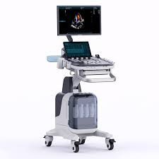
Premium Diagnostic Ultrasound System PU-MT241A
$0.00
Shipped From Abroad
Delivery & Availability:
Typically 10-21 working days – excluding furniture and heavy/bulky equipment. Please contact us for further information.
Typically 10-21 working days – excluding furniture and heavy/bulky equipment. Please contact us for further information.
Description
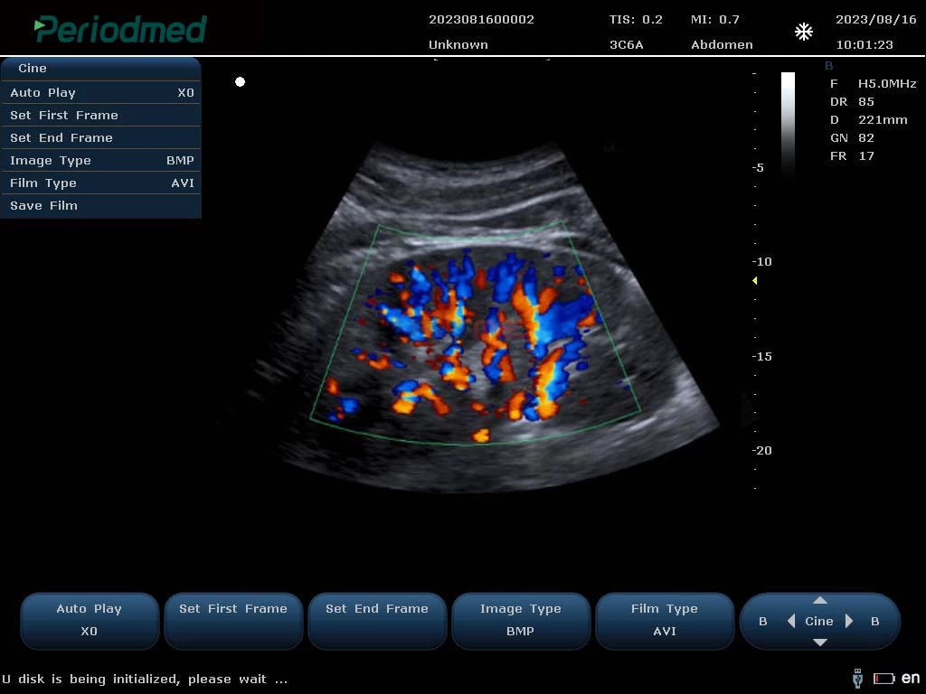
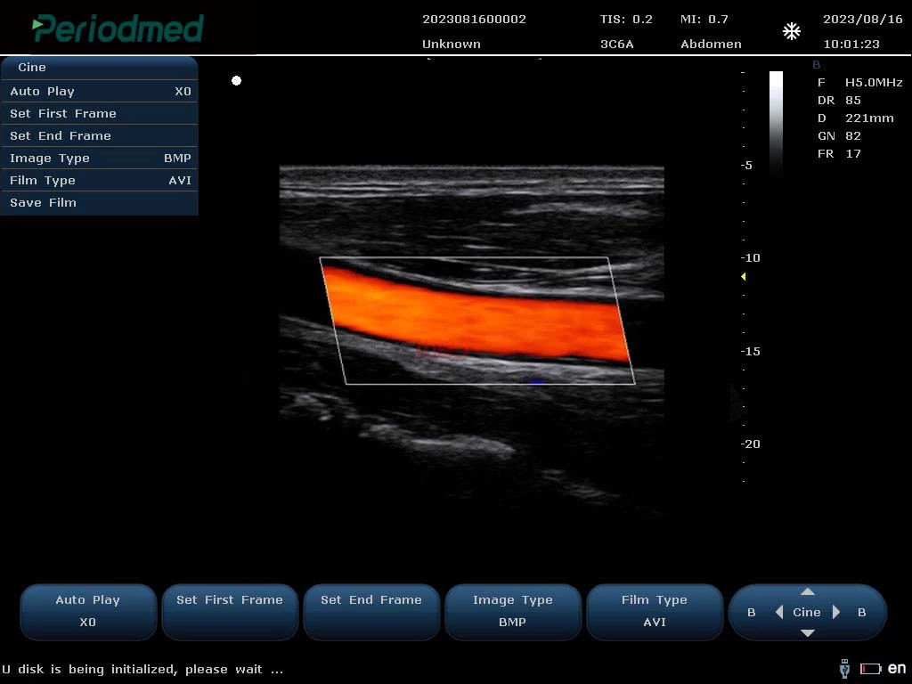
Ultrasound General imaging Images
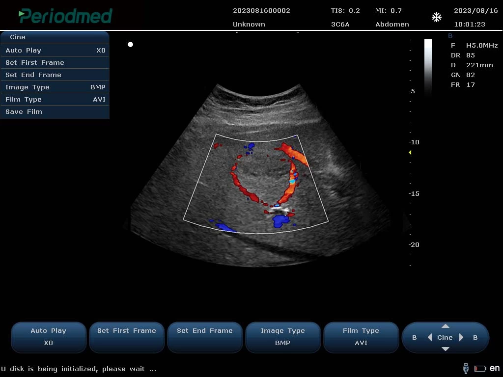
Convex Probe-Color Mode-Liver6
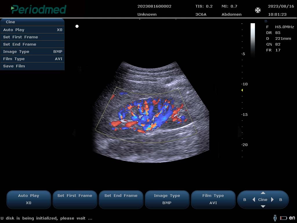
Convex Probe-Color Mode-Kidney 1
Convex Probe-Color Mode-Kidney 2
Ultrasound Carotid Images
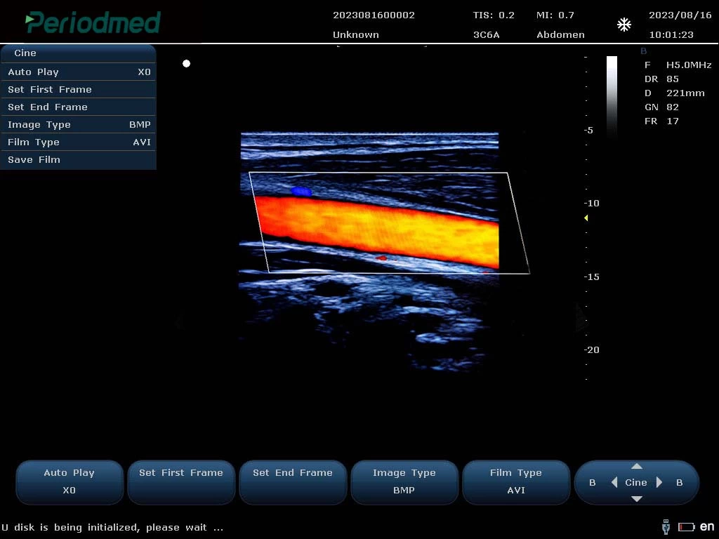
Linear Probe-Color Mode-Carotid1
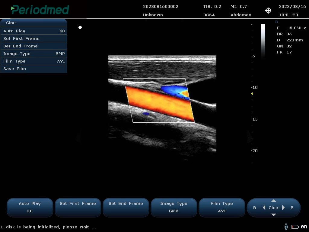
Linear Probe-Color Mode-Carotid2
Linear Probe-Color Mode-Carotid3
Ultrasound Cardiovascular Images
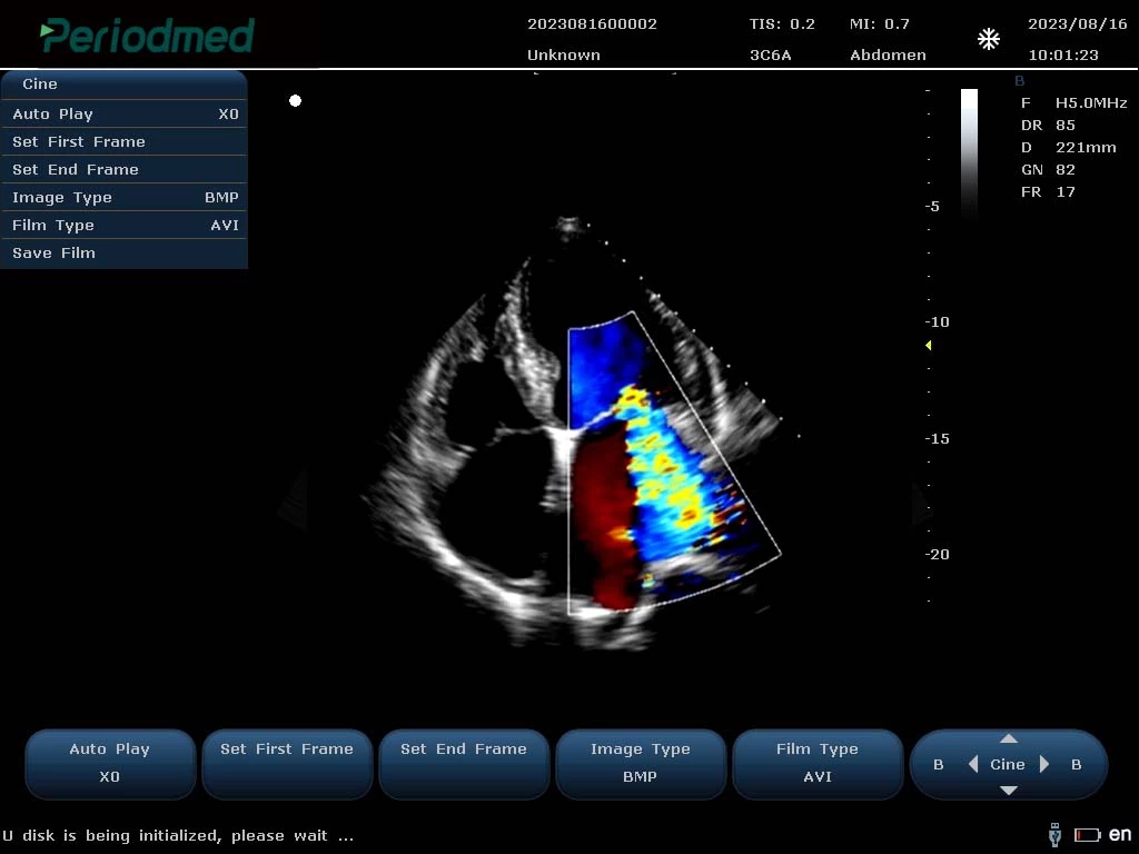
Phased Array Probe-Color Mode-Cardiac1
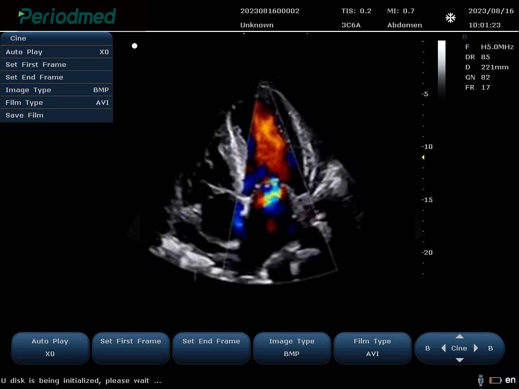
Phased Array Probe-Color Mode-Cardiac2
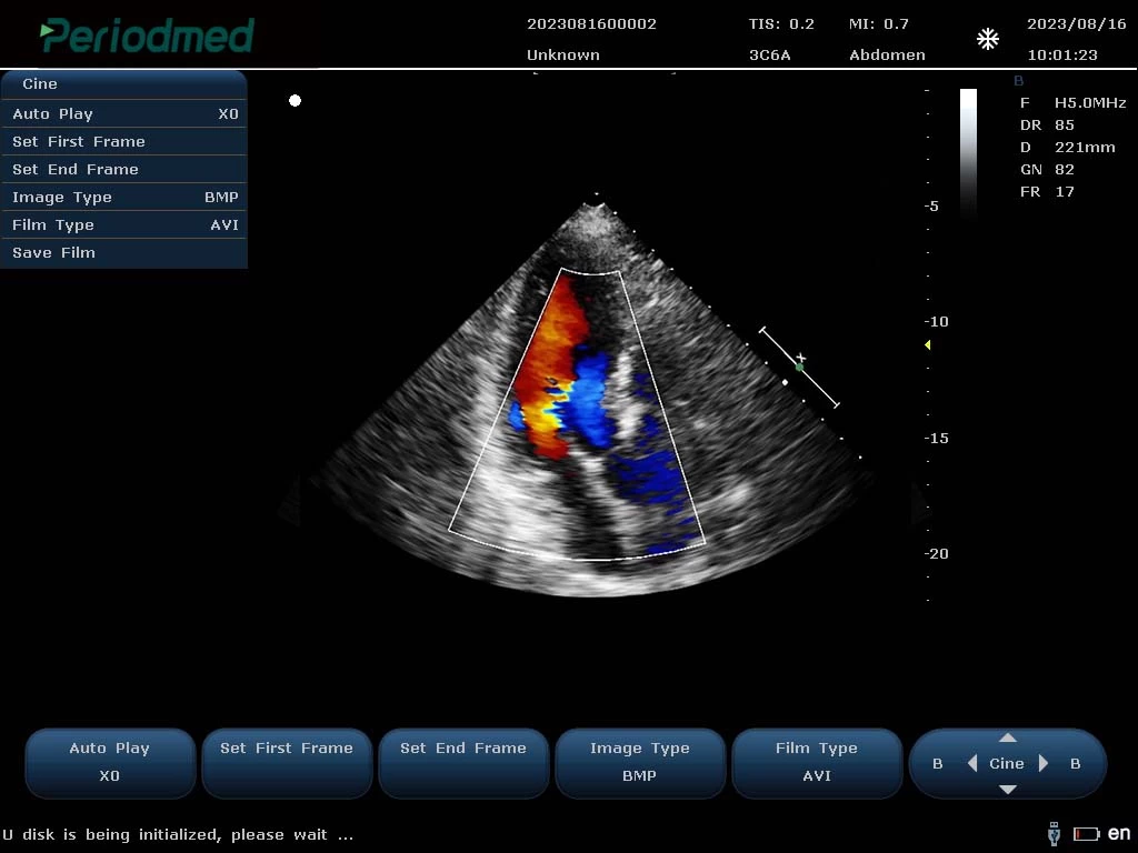
Phased Array Probe-Color Mode-Cardiac3
PU-MT241A Specification
| Multiple Display Modes: | B/2B/4B/BMM/PDI/DPDICF/CW/B+CF/DPDI+PW/B+CF+PW |
| Product Function: | Color Flow(CF) Power Doppler(PDl) Pulsed Doppler(PW) Directional Power Doppler (DPDl) Continuous Doppler (CW) Tissue Doppler (TDI) Color M Type,Anatomical M Type |
| Imaging Technology: | High Pulsed Repetition Frequency(Hprf) Tissue Harmonic Imaging(Thi) Pulsed Reverse Harmonic Imaging(Ithi) Tissue Specific Imaging(Tsi) Spatial Composite Imaging Picture In Picture Imaging Mode |
| Advanced Technology: | 1.Point-By-Point Focusing Application Technology Replaces The Traditional 1-4 Point Multi-Point Transmission Focusing Mode,Making The Transmission Focusing More Accurate 2.The Soft Beamformer Technology Of Gpu Architecture Provides More Powerful Computing Power For The Implementation Of The New Algorithm,And Fundamentally Improves The Quality Of Ultrasound Image 3.192 Elements 128 Channels,With Sound Speed Calibration Technology,16 Beam Compound Algonithm To Provide Dynamic And Accaurate Calculation Parameters,Effectively Improve The Focusing Accuracy 4.The Fast Sound Velocity Calibration Technology Can Automatically Correct The Actual Sound Velocity Of Different Human Bodies And Different Tissues In Real Time,And Provide Dynamic And Accurate Calculation Parameters For Beam Synthesis Algorithm |
| Optional: | Wide Field Imaging Puncture Enhancement Contrast Imaging Elastography Imaging 3D/4D Imaging |
Quick Comparison
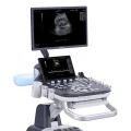
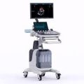
| Premium Diagnostic Ultrasound System PU-MT241A remove | MIR Spirolab Spirometer remove | FlowMir Disposable Turbine with Cardboard Mouthpiece remove | Bistos BT- 720 Patient Monitor remove | ASPEL AsCARD Green B/W ECG Machine remove | Sonoscape P20 Ultrasound Machine remove | |||||||||||||||||||||||||||||||||||||||||||||||||||||||||||||||||||||||||||||||||||||||||||
|---|---|---|---|---|---|---|---|---|---|---|---|---|---|---|---|---|---|---|---|---|---|---|---|---|---|---|---|---|---|---|---|---|---|---|---|---|---|---|---|---|---|---|---|---|---|---|---|---|---|---|---|---|---|---|---|---|---|---|---|---|---|---|---|---|---|---|---|---|---|---|---|---|---|---|---|---|---|---|---|---|---|---|---|---|---|---|---|---|---|---|---|---|---|---|---|---|
| Name | Premium Diagnostic Ultrasound System PU-MT241A remove | MIR Spirolab Spirometer remove | FlowMir Disposable Turbine with Cardboard Mouthpiece remove | Bistos BT- 720 Patient Monitor remove | ASPEL AsCARD Green B/W ECG Machine remove | Sonoscape P20 Ultrasound Machine remove | ||||||||||||||||||||||||||||||||||||||||||||||||||||||||||||||||||||||||||||||||||||||||||
| Image | 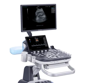 | 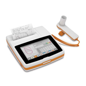 | 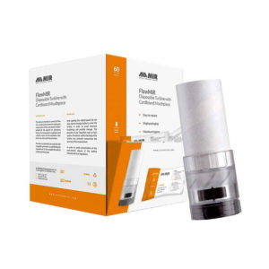 | 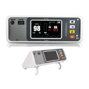 |  |  | ||||||||||||||||||||||||||||||||||||||||||||||||||||||||||||||||||||||||||||||||||||||||||
| SKU | SF1033560130124-1 | SF1033560084-13 | SF1033560084-22 | SF1033560059-8 | SF1033560075-8 | SF1033560012-9 | ||||||||||||||||||||||||||||||||||||||||||||||||||||||||||||||||||||||||||||||||||||||||||
| Rating | ||||||||||||||||||||||||||||||||||||||||||||||||||||||||||||||||||||||||||||||||||||||||||||||||
| Price |
| $2,101.00 | $145.00 | $330.00 |
|
| ||||||||||||||||||||||||||||||||||||||||||||||||||||||||||||||||||||||||||||||||||||||||||
| Stock | ||||||||||||||||||||||||||||||||||||||||||||||||||||||||||||||||||||||||||||||||||||||||||||||||
| Availability | ||||||||||||||||||||||||||||||||||||||||||||||||||||||||||||||||||||||||||||||||||||||||||||||||
| Add to cart | ||||||||||||||||||||||||||||||||||||||||||||||||||||||||||||||||||||||||||||||||||||||||||||||||
| Description | Shipped From Abroad
Delivery & Availability:
Typically 10-21 working days – excluding furniture and heavy/bulky equipment. Please contact us for further information.
| In Stock
Spirometry test:
| In stock Spirometry testing requires maximum accuracy and hygiene. Each Flowmir disposable turbine, which includes a cardboard mouthpiece, has been individually factory calibrated with a computerized system and it is packaged individually. FlowMIR® is an inexpensive alternative to a costly reusable flowmeter and replaces the need for an antibacterial filter Typically 2 working days – excluding furniture and heavy/bulky equipment. Please contact us for further information. | Shipped from abroad Bistos BT - 720 Patient Monitor: Bistos Portable Patient Vital Signs Monitor, SpO2, Pulse, *NIBP, BT-720. The Bistos BT-720 is a compact size vital signs monitor with the standard parameters of SpO2 and Pulse. You can also add the NIBP and/or Masimo SpO2 at an additional cost. The BT-720 has a 4.3" color touch screen The PC software allows the vital signs information to be transferred and analyzed on a computer. Delivery & Availability: Typically 7 working days – excluding furniture and heavy/bulky equipment. Please contact us for further information. | Shipped from Abroad AsCARD Green electrocardiograph is a 1- and 3-channel ECG unit which enables to make electrocardiogram in full 12 leads. Intended for ECG examinations of adult and paediatric patients aimed at identification of cardiological abnormalities, myocardial ischaemia or infarction. The device is intended for use in healthcare facilities by duly trained personnel. ECG examination may be recorded in manual or automatic mode with the ability to perform the analysis and interpretation. Delivery & Availability: Typically 10 working days – excluding furniture and heavy/bulky equipment. Please contact us for further information. | Shipped from Abroad Incorporating innovative technologies, P20’s user-friendly design with a simple operation panel, intuitive user interface and a variety of intelligent auxiliary scanning tools, will significantly improve your daily examination experience. Besides general imaging applications, P20 has entitled with diagnostic 4D technology which has an extraordinary performance in obstetrics and gynecology applications. Delivery & Availability: Typically 5-7 working days – excluding furniture and heavy/bulky equipment. Please contact us for further information. | ||||||||||||||||||||||||||||||||||||||||||||||||||||||||||||||||||||||||||||||||||||||||||
| Content | https://youtu.be/K2dzsICPG_s
Ultrasound General imaging Images
Convex Probe-Color Mode-Liver6
Convex Probe-Color Mode-Kidney 1
Convex Probe-Color Mode-Kidney 2 Ultrasound Carotid Images
Linear Probe-Color Mode-Carotid1
Linear Probe-Color Mode-Carotid2
Linear Probe-Color Mode-Carotid3 Ultrasound Cardiovascular Images
Phased Array Probe-Color Mode-Cardiac1
Phased Array Probe-Color Mode-Cardiac2
Phased Array Probe-Color Mode-Cardiac3 PU-MT241A Specification
| Spirometry test:
Spirometry parameters:
FVC, FEV1, FEV1/FVC, FEV1/VC, PEF, FEF25, FEF50, FEF75, FEF25–75, FEF75–85, Lung Age, Extrapolated Volume, FET, Time to PEF, FEV0.5, FEV0.5/FVC, FEV0.75, FEV0.75/FVC, FEV2, FEV2/FVC, FEV3, FEV3/FVC, FEV6, FEV1/ FEV6, FEV1/PEF, FEV1/ FEV0.5, FIVC, FIV1, FIV1/FIVC, PIF, FIF25, FIF50, FIF75, FEF50/FIF50, VC, IVC, IC, ERV, IRV, Rf, VE, VT, tI, tE, VT/tI, tE/tTOT, MVV (measured), MVV (calculated).
Click Here To Download Catalogue |
| Bistos BT - 720 Patient Monitor: Bistos Portable Patient Vital Signs Monitor, SpO2, Pulse, *NIBP, BT-720. The Bistos BT-720 is a compact size vital signs monitor with the standard parameters of SpO2 and Pulse. You can also add the NIBP and/or Masimo SpO2 at an additional cost. The BT-720 has a 4.3" color touch screen The PC software allows the vital signs information to be transferred and analyzed on a computer.
Features:
Click Here To Download Catalogue | AsCARD Green electrocardiograph is a 1- and 3-channel ECG unit which enables to make electrocardiogram in full 12 leads. Intended for ECG examinations of adult and paediatric patients aimed at identification of cardiological abnormalities, myocardial ischaemia or infarction. The device is intended for use in healthcare facilities by duly trained personnel. ECG examination may be recorded in manual or automatic mode with the ability to perform the analysis and interpretation.
Electrocardiograph is based on advanced microprocessor technology. It is equipped with a thermal printer with high-resolution head and graphical LCD display. A hightech membrane keyboard makes the AsCARD Green device operation intuitive, and its menu navigation exceptionally easy. This light-weight, small-footprint and battery powered cause that device can be easily transported to any location. With plastic casing and foil covered keyboard, the device is neat and easy to clean.
Technical Specifications:
Click Here To Download Catalogue | DETAILS
Upgraded Images with More Clarity
SonoScape never stops making progress in improving the image quality of its ultrasound products to enhance the confidence of diagnosis for doctors. With extraordinary images provided by P20, the anatomy structures are clearer than ever.
C-Xlasto Imaging
With C-xlasto Imaging, P20 enables comprehensive quantitative elastic analysis. Meanwhile, C-xlasto on P20 is supported by linear, convex and transvaginal probes, to ensure good reproducibility and highly consistent quantitative elastic results.
S-Live
S-Live allows for detailed visualization of subtle anatomical features, thereby enabling intuitive diagnosis with real-time 3D images and enriching patient communication.
Pelvic Floor 4D
Transperineal 4D pelvic floor ultrasound can provide useful clinical values in assessing the vaginal delivery impact on the female anterior compartment, judging whether the pelvic organs are prolapsed or not and the extent, determining if the pelvic muscles were torn accurately.
Anatomic M Mode
Anatomic M Mode helps you observe the myocardial motion at different phases by freely placing sample lines. It accurately measures the myocardial thickness and the heart size of even difficult patients and supports the myocardial function and LV wall-motion assessment.
Tissue Doppler Imaging
P20 is endowed with Tissue Doppler Imaging which provides velocities and other clinical information on myocardial functions, facilitating clinical doctors with the ability to analyze and compare the motions of different parts of the patient's heart.
Click Here To Download Catalogue | ||||||||||||||||||||||||||||||||||||||||||||||||||||||||||||||||||||||||||||||||||||||||||
| Weight | N/A | N/A | N/A | N/A | N/A | N/A | ||||||||||||||||||||||||||||||||||||||||||||||||||||||||||||||||||||||||||||||||||||||||||
| Dimensions | N/A | N/A | N/A | N/A | N/A | N/A | ||||||||||||||||||||||||||||||||||||||||||||||||||||||||||||||||||||||||||||||||||||||||||
| Additional information | ||||||||||||||||||||||||||||||||||||||||||||||||||||||||||||||||||||||||||||||||||||||||||||||||


Reviews
There are no reviews yet.