- Sorry, this product cannot be purchased.
Progetti Manual Defibrillator
$4,695.00
Ship from abroad
This is designed to be the ideal device to deal with cardiovascular emergencies and to monitor the patient in hospital and non-hospital environments. A helping Hand in hard times.
Delivery & Availability:
Typically 5-7 working days – excluding furniture and heavy/bulky equipment. Please contact us for further information.
Description
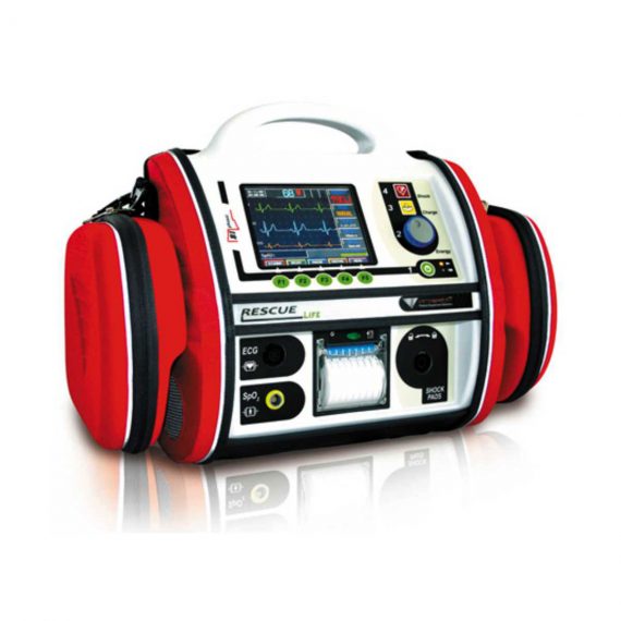
- The combined defibrillator monitor Rescue Life is designed to be used by out-of-hospital and hospital users. Having an innovative and fully featured design, it provides for defibrillation and monitoring in a single portable device.
- Brilliant, high contrast, wide-angle TFT LCD colour display allows the simultaneous visualization of up to 3 ECG channels. Information such as heart rate, SpO2 value, alarms and operational functions are clearly visible on the display even in low visibility conditions. In AED or Advisory mode, text messages and voice prompts guide the operator during the CPR operations.
- Rescue Life is impact proof, compact and lightweight. The dedicated comfortable and washable carrying bag keeps all the accessories at hand and protects Rescue Life during the emergency procedures. The case is built with washable materials and has a comfortable handle for safe transportation. The paddles have dedicated seat on the back of Rescue Life with a fast hook-unhook procedure. The IPX4 standard permits the operation under adverse climatic situations.
- Rescue Life includes manual, AED and Advisory operation modes allowing the best use in different situations. It is designed to be used by doctors and trained individuals. The manual mode provides very easy three steps operation: energy selection from 1-230J, energy charge and shock. In AED mode, the voice prompts and display messages are assisting the operator during CPR operations, charging automatically to 150J when a shockable ECG rhythm is detected. In Advisory mode, the system alerts when the shock is necessary (using voice prompts and display messages) leaving the energy level and charging choice to the operator.
- The Rescue Life defibrillator uses a biphasic truncated exponentia(BTE) waveform with the HiCap technology which brings the benefits of current controlled systems but with higher reliability. Controlling the waveform duration based on the body impedance measurement, the Rescue Life is able to maintain the ideal duration, peak current and voltage of the debrillation shock. The optimal biphasic waveform adopted by Rescue Life provides the confidence of the first shock success without the need for escalating energy.
Quick Comparison
| Settings | Progetti Manual Defibrillator remove | Topaz Digital X-ray Machine remove | ASPEL AsCARD Green B/W ECG Machine remove | ASPEL Ambulatory BP Machine remove | Sonoscape P20 Ultrasound Machine remove | DrGem Ceiling Mounted Digital X-ray remove |
|---|---|---|---|---|---|---|
| Name | Progetti Manual Defibrillator remove | Topaz Digital X-ray Machine remove | ASPEL AsCARD Green B/W ECG Machine remove | ASPEL Ambulatory BP Machine remove | Sonoscape P20 Ultrasound Machine remove | DrGem Ceiling Mounted Digital X-ray remove |
| Image |  |  |  | 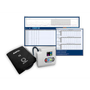 |  |  |
| SKU | SF1033560095-1 | SF1033560074-1 | SF1033560075-8 | SF1033560075-13 | SF1033560012-9 | SF1033560074-4 |
| Rating | ||||||
| Price | $4,695.00 |
|
| $920.00 |
|
|
| Stock | ||||||
| Availability | ||||||
| Add to cart | ||||||
| Description | Ship from abroad
This is designed to be the ideal device to deal with cardiovascular emergencies and to monitor the patient in hospital and non-hospital environments. A helping Hand in hard times.
| In Stock DRGEM’s TOPAZ X-ray machine is a state-of-the-art mobile digital radiography system, designed with maximum comfort for patients and users in mind. From its user-friendly software to smooth movements, TOPAZ is made to improve your workflow and provide you with high-quality images. Delivery & Availability: Typically 21 working days – excluding furniture and heavy/bulky equipment. Please contact us for further information. | Shipped from Abroad AsCARD Green electrocardiograph is a 1- and 3-channel ECG unit which enables to make electrocardiogram in full 12 leads. Intended for ECG examinations of adult and paediatric patients aimed at identification of cardiological abnormalities, myocardial ischaemia or infarction. The device is intended for use in healthcare facilities by duly trained personnel. ECG examination may be recorded in manual or automatic mode with the ability to perform the analysis and interpretation. Delivery & Availability: Typically 10 working days – excluding furniture and heavy/bulky equipment. Please contact us for further information. | Shipped from Abroad ASPEL Ambulatory BP Machine - is a recorder of long-term records of non-invasive measurement of blood pressure intended for use in clinics, hospitals, outpatient centers and specialist surgeries. The recorder enables the assessment of blood pressure by the oscillometric method in adult patients, pregnant women, including preeclampsia and pediatric patients (from 3 years of age). Blood pressure is assessed by using an inflatable cuff, an accurate pressure transducer, and a deflation valve. Delivery & Availability: Typically 10 working days – excluding furniture and heavy/bulky equipment. Please contact us for further information. | Shipped from Abroad Incorporating innovative technologies, P20’s user-friendly design with a simple operation panel, intuitive user interface and a variety of intelligent auxiliary scanning tools, will significantly improve your daily examination experience. Besides general imaging applications, P20 has entitled with diagnostic 4D technology which has an extraordinary performance in obstetrics and gynecology applications. Delivery & Availability: Typically 5-7 working days – excluding furniture and heavy/bulky equipment. Please contact us for further information. | In Stock The GXR-SD is a diagnostic digital radiography system that provides reliable high quality digital radiographic images with a reduced dose. The GXR-SD DR systems offer comprehensive digital solutions to all radiography needs, featuring ACQUIDR digital imaging system with stationary or portable digital flat-panel detectors as well as reliable high-frequency x-ray generators that are known worldwide for their excellent performance, lifetime and stability. Patient tables and wall stands are also offered. Delivery & Availability: Typically 21 working days – excluding furniture and heavy/bulky equipment. Please contact us for further information. |
| Content |
| TOPAZ X-ray machine is among the high end X-ray machine manufactured by DRGEM, a digital X-ray system that provides quality images with little or no effort.
It begins with Advanced Technology
Integrating high technology and over a decade of experience in conventional and digital radiography systems, DRGEM’s TOPAZ X-ray machine is a state-of-the-art mobile digital radiography system, designed with maximum comfort for patients and users. From its user-friendly software to smooth movements, TOPAZ X-ray machine is made to improve your workflow and provide you with high-quality images.
Full Featured Imaging Software & Excellent Digital Image Processing
With a high-performance, built-in touchscreen, TOPAZ X-ray machine offers a user-friendly interface and powerful software for easy operation and increased workflow. The anatomical view-based digital image processing, automatically optimizes and enhances the quality of the image. it also comes with automatic image storage and print with DICOM 3.0 networking capability. additionally, the system offers increasing exam throughput while decreasing examination time.
Click Here To Download Catalogue | AsCARD Green electrocardiograph is a 1- and 3-channel ECG unit which enables to make electrocardiogram in full 12 leads. Intended for ECG examinations of adult and paediatric patients aimed at identification of cardiological abnormalities, myocardial ischaemia or infarction. The device is intended for use in healthcare facilities by duly trained personnel. ECG examination may be recorded in manual or automatic mode with the ability to perform the analysis and interpretation.
Electrocardiograph is based on advanced microprocessor technology. It is equipped with a thermal printer with high-resolution head and graphical LCD display. A hightech membrane keyboard makes the AsCARD Green device operation intuitive, and its menu navigation exceptionally easy. This light-weight, small-footprint and battery powered cause that device can be easily transported to any location. With plastic casing and foil covered keyboard, the device is neat and easy to clean.
Technical Specifications:
Click Here To Download Catalogue | ASPEL Ambulatory BP Machine - is a recorder of long-term records of non-invasive measurement of blood pressure intended for use in clinics, hospitals, outpatient centers and specialist surgeries. The recorder enables the assessment of blood pressure by the oscillometric method in adult patients, pregnant women, including preeclampsia and pediatric patients (from 3 years of age). Blood pressure is assessed by using an inflatable cuff, an accurate pressure transducer, and a deflation valve.
Features:
Save-2-Safe: Double security system
Thanks to the use of two independent measuring systems with an additional valve, it meets the highest standards and takes care of patient safety even better.
Start-Easy: Quick start in two moves
The quick launch function allows you to use the device instantly, easily allows you to start recording in holter mode.
Memo-Care: Cuff pressure memory
Recorder remembers the pressure in the cuff. Thanks to the use of Intelligent Solutions, it adapts individually to the patient.
Power-Usb: USB connection
The device can work without batteries: by connecting to a computer via a USB cable.
Technical Specification:
Click Here To Download Catalogue | DETAILS
Upgraded Images with More Clarity
SonoScape never stops making progress in improving the image quality of its ultrasound products to enhance the confidence of diagnosis for doctors. With extraordinary images provided by P20, the anatomy structures are clearer than ever.
C-Xlasto Imaging
With C-xlasto Imaging, P20 enables comprehensive quantitative elastic analysis. Meanwhile, C-xlasto on P20 is supported by linear, convex and transvaginal probes, to ensure good reproducibility and highly consistent quantitative elastic results.
S-Live
S-Live allows for detailed visualization of subtle anatomical features, thereby enabling intuitive diagnosis with real-time 3D images and enriching patient communication.
Pelvic Floor 4D
Transperineal 4D pelvic floor ultrasound can provide useful clinical values in assessing the vaginal delivery impact on the female anterior compartment, judging whether the pelvic organs are prolapsed or not and the extent, determining if the pelvic muscles were torn accurately.
Anatomic M Mode
Anatomic M Mode helps you observe the myocardial motion at different phases by freely placing sample lines. It accurately measures the myocardial thickness and the heart size of even difficult patients and supports the myocardial function and LV wall-motion assessment.
Tissue Doppler Imaging
P20 is endowed with Tissue Doppler Imaging which provides velocities and other clinical information on myocardial functions, facilitating clinical doctors with the ability to analyze and compare the motions of different parts of the patient's heart.
Click Here To Download Catalogue | DrGem Ceiling Mounted Digital X-ray is a diagnostic digital radiography system that provides reliable high quality digital radiographic images with a reduced dose. The GXR-SD DR systems offer comprehensive digital solutions to all radiography needs, featuring ACQUIDR digital imaging system with stationary or portable digital flat-panel detectors as well as reliable high-frequency x-ray generators that are known worldwide for their excellent performance, lifetime and stability. Patient tables and wall stands are also offered.
Features:
Click Here To Download Catalogue |
| Weight | N/A | N/A | N/A | N/A | N/A | N/A |
| Dimensions | N/A | N/A | N/A | N/A | N/A | N/A |
| Additional information |


Reviews
There are no reviews yet.