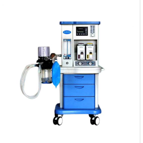
Render SD-M2000B Anesthetic Machine
$4,840.00
Shipped from Abroad
- Contains Output interface for Anesthesia information management system.
- Complete with backup battery for working more than 30 minutes.
Delivery & Availability:
Typically 21 working days – excluding furniture and heavy/bulky equipment. Please contact us for further information.
Description
Features:
- Contains Output interface for Anesthesia information management system.
- Complete with backup battery for working more than 30 minutes.
Technical Specifications:
| Model: | SD-M2000B |
|---|---|
| Brand: | RENDER |
| Origin: | Made In China |
| Category: | Electronics & Electricity / Electronic Instrument / Electricity Meter & Instrument |
| Label: | anesthesia machine , anesthesia ventilator , anesthesia machine |
Click Here To Download Catalogue
Quick Comparison
| Render SD-M2000B Anesthetic Machine remove | Sonoscape S22 Ultrasound Machine remove | DrGem GXR-SD 400mA Floor Mounted Digital X-ray remove | Sonoscape E2 Ultrasound Machine remove | DrGem Ceiling Analogue X-ray Machine remove | ASPEL AsCARD Coral PC Based ECG Machine remove | |||||||||||
|---|---|---|---|---|---|---|---|---|---|---|---|---|---|---|---|---|
| Name | Render SD-M2000B Anesthetic Machine remove | Sonoscape S22 Ultrasound Machine remove | DrGem GXR-SD 400mA Floor Mounted Digital X-ray remove | Sonoscape E2 Ultrasound Machine remove | DrGem Ceiling Analogue X-ray Machine remove | ASPEL AsCARD Coral PC Based ECG Machine remove | ||||||||||
| Image |  |  |  |  |  | 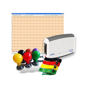 | ||||||||||
| SKU | SF1033560011-10 | SF1033560012-3 | SF1033560074-5 | SF1033560012-17 | SF1033560074-7 | SF1033560075-11 | ||||||||||
| Rating | ||||||||||||||||
| Price | $4,840.00 | $9,350.00 |
| $5,500.00 |
| $486.00 | ||||||||||
| Stock | ||||||||||||||||
| Availability | ||||||||||||||||
| Add to cart | ||||||||||||||||
| Description | Shipped from Abroad
| Shipped from Abroad As SonoScape steps forward to add value and efficiency to ultrasound, the latest S22 was designed in a user-friendly platform to address current and future demanding needs. It represents an excellent mix in performance and price. Delivery & Availability: Typically 5-7 working days – excluding furniture and heavy/bulky equipment. Please contact us for further information. | In Stock The GXR-SD Digital X-ray is a diagnostic digital radiography system that provides reliable high quality digital radiographic images with a reduced dose. The GXR-SD DR systems offer comprehensive digital solutions to all radiography needs, featuring ACQUIDR digital imaging system with stationary or portable digital flat-panel detectors as well as reliable high-frequency x-ray generators that are known worldwide for their excellent performance, lifetime and stability. Patient tables and wall stands are also offered. Delivery & Availability: Typically 21 working days – excluding furniture and heavy/bulky equipment. Please contact us for further information. | Shipped from Abroad Sonoscape E2 portable ultrasound machine is a color Doppler ultrasound system that reaches beyond your expectations due to its compact and fashionable appearance. It fulfills GI, OB/GYN, Cardiac and POC applications to fit your routine scanning needs while its color mode will help you for more accurate and efficient diagnosis of lesions. E2 provides a wide range of applications to assist users with routine scanning. E2 provides automatic calculations to enhance your diagnostic confidence and save you time for patient communication. Delivery & Availability: Typically 14 working days – excluding furniture and heavy/bulky equipment. Please contact us for further information. | Shipped from abroad The DrGem Ceiling Analogue X-ray Machine is a diagnostic radiography system that provides reliable high quality radiographic images with a reduced dose. The reliable high-frequency x-ray generators that are known worldwide for their excellent performance, lifetime and stability. Patient tables and wall stands are also offered. Delivery & Availability: Typically 21 working days – excluding furniture and heavy/bulky equipment. Please contact us for further information. | Shipped from Abroad AsCARD Coral electrocardiograph is a 3-, 6-, 12-channel ECG equipped with CardioTEKA software allows transmission of full 12 ECG leads to the user PC through USB interface. It is intended for carrying out ECG examinations in adults and pediatric patients in all types of health care centres. ECG procedures can be performed by qualified personnel only. AsCARD Coral can cooperate also with CardioTEST system as 12-channel ECG device allows transmission of full 12 ECG leads to the user PC through USB interface. Delivery & Availability: Typically 10 working days – excluding furniture and heavy/bulky equipment. Please contact us for further information. | ||||||||||
| Content |
Click Here To Download Catalogue | DETAILS
As SonoScape steps forward to add value and efficiency to ultrasound, the latest S22 was designed in a user-friendly platform to address current and future demanding needs. It represents an excellent mix in performance and price.
S22, is a shared service ultrasound system with a slim and elegant package that has combined mobility with utility to fit in specific clinical situations including emergency department, ICU, operating room and so on. Furthermore, its ergonomic design, easy operating and flexible data management will give you a memorable experience.
SPECIFICATION
• Large high-resolution widescreen LED
• Sensitive touch screen
• Four transducer sockets plus one socket for pencil probe
• A comprehensive selection of probes: linear, Convex, Micro-convex, Volumetric, Endocavity, Bi-plane, Phased Array, TEE, Intraoperative, Pencil
• Premium application technology: 4D, μ-scan speckle reduction, compound imaging, Pulse Inversion Harmonic Imaging, Color M-Mode, Steer M-Mode, PDI, TDI, Real-time Panoramic Imaging, Trapezoid Imaging, Auto-IMT…
• Full patient database and image management solutions: DICOM 3.0, AVI/JPG, USB 2.0, HDD, DVD, PDF report
• Multi-Language Input Keyboard
• Built-in battery
Click Here To Download Catalogue | DrGem GXR-SD 400mA Floor Mounted Digital X-ray system matches with a radiographic room which perfectly fits your workow and can be easily upgraded to DR system with the help of DR interface and PC interface in GXR generator as well as Bucky suitable to Flat Panel Detector. GXR X-ray system is equipped with a high frequency X-ray generator which consistently produces high quality radiograph in favor of high quality X-ray output with a very small kV ripple and accurate mA and mAs. GXR X-ray system is designed to provide convenience to operator and comfort to patient
Features of DrGem GXR-SD 400mA Floor Mounted Digital X-ray:
Click Here To Download Catalogue | SONOSCAPE E2 DETAILS
Auto Image Optimization
A portable ultrasound machine with the press of a button, the image is automatically adjusted and optimized, saving you time with parameter adjustments. Additionally, with Auto Focus on, the focus area follows the depth of the ROI box as it is moved in the scanning field, providing users with excellent image quality in the desired area of interest.
Automated Calculation
Auto IMT is used when determining the level of vascular sclerosis present in the patient by automatically tracing the thickness of the carotid vessels.
Auto trace provides users sensitive and accurate wave tracing, avoiding the error of manual trace and giving out calculation result in no time
In-Build Battery pack
This portable ultrasound machine was equipped with an in-build battery pack which enable the user to perform image scanning when AC power is not available.
Click Here To Download Catalogue | DrGem Ceiling Analogue X-ray Machine is a diagnostic radiography system X-ray Machine that provides reliable high quality radiographic images with a reduced dose. The reliable high-frequency x-ray generators that are known worldwide for their excellent performance, lifetime and stability. Patient tables and wall stands are also offered.
Features of DrGem Ceiling Analogue X-ray Machine
Click Here To Download Catalogue |
AsCARD Coral electrocardiograph is a 3-, 6-, 12-channel ECG equipped with CardioTEKA software allows transmission of full 12 ECG leads to the user PC through USB interface. It is intended for carrying out ECG examinations in adults and pediatric patients in all types of health care centres. ECG procedures can be performed by qualified personnel only. AsCARD Coral can cooperate also with CardioTEST system as 12-channel ECG device allows transmission of full 12 ECG leads to the user PC through USB interface.
Technical Specification:
Click Here To Download Catalogue | ||||||||||
| Weight | N/A | N/A | N/A | N/A | N/A | N/A | ||||||||||
| Dimensions | N/A | N/A | N/A | N/A | N/A | N/A | ||||||||||
| Additional information |


Reviews
There are no reviews yet.