

Tensiomed ArterioGraph
$0.00
Shipped From Abroad
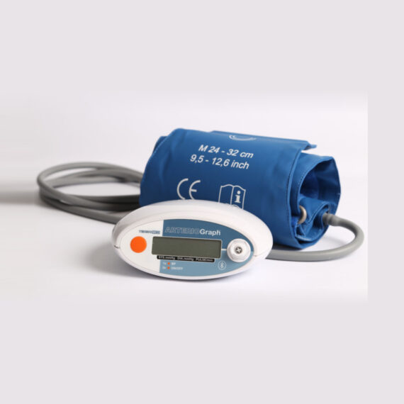
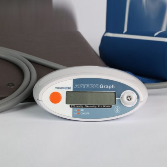
The accuracy of Arteriograph is validated invasively & it is the first patented method (US Pat. No. 20070106162) for oscillometric measurement and determination of fundamental central hemodynamic parameters (SBPao, AIXao, PWV), based on a simple upper arm cuff measurement. What is unique to the Arteriograph are the real-time transmission and visualisation of the detected pressure curves by the Arteriograph software.
Delivery & Availability:
Typically 14-21 Working Days – excluding furniture and heavy/bulky equipment. Please contact us for further information.
Description
The accuracy of Arteriograph is validated invasively & it is the first patented method (US Pat. No. 20070106162) for oscillometric measurement and determination of fundamental central hemodynamic parameters (SBPao, AIXao, PWV), based on a simple upper arm cuff measurement. What is unique to the Arteriograph are the real-time transmission and visualisation of the detected pressure curves by the Arteriograph software.
The novelty of the Arteriograph device in detecting the mentioned parameters is that a single upper arm cuff is used as a sensor, but in a very special condition; the cuff is pressurised suprasystolically, ensuring to obtain pure pressure signals (waves). Arteriograph is an office device, but portable and ideal tool for screening purposes.
The Arteriograph device measures the next parameters, within 2 minutes! absolutely user independently (only a push bottom), and provides the following parameters:
Click Here To Download Catalogue
Quick Comparison
| Tensiomed ArterioGraph remove | Sonoscape P20 Ultrasound Machine remove | ASPEL AsCARD Coral PC Based ECG Machine remove | Sonoscape P10 Ultrasound Machine remove | Sonoscape S11 Ultrasound Machine remove | ASPEL Stress ECG with Ergometer and Software remove | |
|---|---|---|---|---|---|---|
| Name | Tensiomed ArterioGraph remove | Sonoscape P20 Ultrasound Machine remove | ASPEL AsCARD Coral PC Based ECG Machine remove | Sonoscape P10 Ultrasound Machine remove | Sonoscape S11 Ultrasound Machine remove | ASPEL Stress ECG with Ergometer and Software remove |
| Image |  |  |  |  |  |  |
| SKU | SF10335601276 | SF1033560012-9 | SF1033560075-11 | SF1033560012-7 | SF1033560012-1 | SF1033560075-1 |
| Rating | ||||||
| Price |
|
| $486.00 | $9,350.00 | $6,380.00 | $4,202.00 |
| Stock | ||||||
| Availability | ||||||
| Add to cart | ||||||
| Description | Shipped From Abroad
The accuracy of Arteriograph is validated invasively & it is the first patented method (US Pat. No. 20070106162) for oscillometric measurement and determination of fundamental central hemodynamic parameters (SBPao, AIXao, PWV), based on a simple upper arm cuff measurement. What is unique to the Arteriograph are the real-time transmission and visualisation of the detected pressure curves by the Arteriograph software.
Delivery & Availability:
Typically 14-21 Working Days – excluding furniture and heavy/bulky equipment. Please contact us for further information.
| Shipped from Abroad Incorporating innovative technologies, P20’s user-friendly design with a simple operation panel, intuitive user interface and a variety of intelligent auxiliary scanning tools, will significantly improve your daily examination experience. Besides general imaging applications, P20 has entitled with diagnostic 4D technology which has an extraordinary performance in obstetrics and gynecology applications. Delivery & Availability: Typically 5-7 working days – excluding furniture and heavy/bulky equipment. Please contact us for further information. | Shipped from Abroad AsCARD Coral electrocardiograph is a 3-, 6-, 12-channel ECG equipped with CardioTEKA software allows transmission of full 12 ECG leads to the user PC through USB interface. It is intended for carrying out ECG examinations in adults and pediatric patients in all types of health care centres. ECG procedures can be performed by qualified personnel only. AsCARD Coral can cooperate also with CardioTEST system as 12-channel ECG device allows transmission of full 12 ECG leads to the user PC through USB interface. Delivery & Availability: Typically 10 working days – excluding furniture and heavy/bulky equipment. Please contact us for further information. | Shipped from Abroad The P10 color Doppler ultrasound system is a new generation product from SonoScape. It is designed to give high quality images, rich probe configurations, various clinical tools and automatic analysis software to provide you with comprehensive solutions for your growing demand for clinical applications. Delivery & Availability: Typically 5-7 working days – excluding furniture and heavy/bulky equipment. Please contact us for further information. | In Stock A Value Choice beyond Your Expectation. SonoScape’s trolley color Doppler system S11 redefines price and performance with practical design. The S11 will go beyond your expectations but not your budget. Delivery & Availability: Typically 2 working days – excluding furniture and heavy/bulky equipment. Please contact us for further information. | Shipped from Abroad Ergometer CRG 200 is dedicated for Exercise Stress Tests System CardioTEST. The Ergometer has been designed according to modern technologies. It is controlled from PC equipped in CardioTEST software. Load level is controlled by a microprocessor, therefore it does not depend on speed, which in turn can be adjusted according to patient’s individual needs. Ergometer is equipped with ECG mode recording 12 standard leads. Delivery & Availability: Typically 21 working days – excluding furniture and heavy/bulky equipment. Please contact us for further information. |
| Content | The accuracy of Arteriograph is validated invasively & it is the first patented method (US Pat. No. 20070106162) for oscillometric measurement and determination of fundamental central hemodynamic parameters (SBPao, AIXao, PWV), based on a simple upper arm cuff measurement. What is unique to the Arteriograph are the real-time transmission and visualisation of the detected pressure curves by the Arteriograph software. The novelty of the Arteriograph device in detecting the mentioned parameters is that a single upper arm cuff is used as a sensor, but in a very special condition; the cuff is pressurised suprasystolically, ensuring to obtain pure pressure signals (waves). Arteriograph is an office device, but portable and ideal tool for screening purposes. The Arteriograph device measures the next parameters, within 2 minutes! absolutely user independently (only a push bottom), and provides the following parameters:Click Here To Download Catalogue | DETAILS
Upgraded Images with More Clarity
SonoScape never stops making progress in improving the image quality of its ultrasound products to enhance the confidence of diagnosis for doctors. With extraordinary images provided by P20, the anatomy structures are clearer than ever.
C-Xlasto Imaging
With C-xlasto Imaging, P20 enables comprehensive quantitative elastic analysis. Meanwhile, C-xlasto on P20 is supported by linear, convex and transvaginal probes, to ensure good reproducibility and highly consistent quantitative elastic results.
S-Live
S-Live allows for detailed visualization of subtle anatomical features, thereby enabling intuitive diagnosis with real-time 3D images and enriching patient communication.
Pelvic Floor 4D
Transperineal 4D pelvic floor ultrasound can provide useful clinical values in assessing the vaginal delivery impact on the female anterior compartment, judging whether the pelvic organs are prolapsed or not and the extent, determining if the pelvic muscles were torn accurately.
Anatomic M Mode
Anatomic M Mode helps you observe the myocardial motion at different phases by freely placing sample lines. It accurately measures the myocardial thickness and the heart size of even difficult patients and supports the myocardial function and LV wall-motion assessment.
Tissue Doppler Imaging
P20 is endowed with Tissue Doppler Imaging which provides velocities and other clinical information on myocardial functions, facilitating clinical doctors with the ability to analyze and compare the motions of different parts of the patient's heart.
Click Here To Download Catalogue |
AsCARD Coral electrocardiograph is a 3-, 6-, 12-channel ECG equipped with CardioTEKA software allows transmission of full 12 ECG leads to the user PC through USB interface. It is intended for carrying out ECG examinations in adults and pediatric patients in all types of health care centres. ECG procedures can be performed by qualified personnel only. AsCARD Coral can cooperate also with CardioTEST system as 12-channel ECG device allows transmission of full 12 ECG leads to the user PC through USB interface.
Technical Specification:
Click Here To Download Catalogue | DETAILS
B + Compound
B + Compound utilizes several lines of sight for optimal contrast resolution, speckle reduction and border detection, with which P10 is ideal for superficial and abdominal imaging with better clarity and improved continuity of structures.
μ-Scan
The new generation μ-Scan imaging technology gives you better image quality by reducing noise, improving signal strength and improving visualization.
P10 offers a comprehensive selection of electronic probes to maximize its capabilities to meet a wide range of applications including abdomen, pediatric, OB/GYN, cardiovascular, musculoskeletal, etc. The advanced probe technologies also effectively enhance the image quality and confidence in reaching clinical diagnoses, even in difficult patients.
Convex Probe 3C-A
Ideal for an abundant of application such as abdomen, gynecology, obstetrics, urology and even abdomen biopsy.
Linear Probe L741
This linear probe is designed to satisfy vascular, breast, thyroid, and other small parts diagnosis, and its adjustable parameters could also present users a clear view of MSK and deep vessels.
Phase Array Probe 3P-A
For the purpose of adult and pediatric cardiology and emergency, the phase array probe provides elaborate presets for different exam modes, even for difficult patients.
Intracavitary Probe 6V1
Intracavitary probe could face application of gynecology, urology, prostate, and its temperature detection technology not only protects the patient but also extends the service life.
Click Here To Download Catalogue | DETAILS
SonoScape’s trolley colour Doppler system S11 redefines price and performance with practical design. The S11 will go beyond your expectations but not your budget. As an easy-to-use ultrasound system, the S11 is integrated with a new software platform, especially optimized for a smooth workflow and convenient operation. The system speeds up the exam process and makes file management easier.
SPECIFICATION
- 15-inch high definition LCD monitor with articulating arm
- Compact and agile trolley design
- 3 active transducer sockets available for a wide range of applications
- Duplex, Color Doppler, DPI, PW Doppler, tissue harmonic imaging, μ-scan speckle reduction imaging, compound imaging, trapezoidal imaging
- Customized settings based on your own working style
- Full patient database and image management solutions
Click Here To Download Catalogue | Ergometer CRG 200 is dedicated for Exercise Stress Tests System CardioTEST. The Ergometer has been designed according to modern technologies. It is controlled from PC equipped in CardioTEST software. Load level is controlled by a microprocessor, therefore it does not depend on speed, which in turn can be adjusted according to patient’s individual needs. Ergometer is equipped with ECG mode recording 12 standard leads.
Technical Specifications:
Click Here To Download Catalogue |
| Weight | N/A | N/A | N/A | N/A | N/A | N/A |
| Dimensions | N/A | N/A | N/A | N/A | N/A | N/A |
| Additional information |


Reviews
There are no reviews yet.