| Description | In Stock
- Light weight lead core for protection; Velcro enclosure
- 0.5mm Lead (pb) Equivalency Protection
- Stain resistant and washable material
- Easy to wear ; One size fits all
| Shipped from Abroad
Sonoscape E2 portable ultrasound machine is a color Doppler ultrasound system that reaches beyond your expectations due to its compact and fashionable appearance. It fulfills GI, OB/GYN, Cardiac and POC applications to fit your routine scanning needs while its color mode will help you for more accurate and efficient diagnosis of lesions. E2 provides a wide range of applications to assist users with routine scanning. E2 provides automatic calculations to enhance your diagnostic confidence and save you time for patient communication.
Delivery & Availability:
Typically 14 working days – excluding furniture and heavy/bulky equipment. Please contact us for further information. | In stock
- Length: 355 mm
- Height: 560 mm
- Diameter: 150 mm
- Power: 30 w
- Scattering diffusion: Greater than 0.9
Delivery & Availability:
Typically 5-7 working days – excluding furniture and heavy/bulky equipment. Please contact us for further information. |
Shipped from Abroad
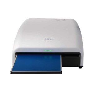
SUPiA made by Signers offers such a better clinic environment with no chemicals, ideal space, high-resolution image quality, and affordability.
Delivery & Availability:
Typically 14 working days – excluding furniture and heavy/bulky equipment. Please contact us for further information. | Shipped from Abroad
This Machine gives a possibility to perform computed tomography without any problems and on high quality level. This device is used to conduct exams of internal organs and their functioning. With its help, a physician has a possibility to assess the condition of the human body as a whole.
Delivery & Availability:
Typically 90 working days – excluding furniture and heavy/bulky equipment. Please contact us for further information. | In Stock
DRGEM’s TOPAZ X-ray machine is a state-of-the-art mobile digital radiography system, designed with maximum comfort for patients and users in mind. From its user-friendly software to smooth movements, TOPAZ is made to improve your workflow and provide you with high-quality images.
Delivery & Availability:
Typically 21 working days – excluding furniture and heavy/bulky equipment. Please contact us for further information. |
| Content |
- Light weight lead core for protection; Velcro enclosure
- 0.5mm Lead (pb) Equivalency Protection
- Stain resistant and washable material
- Easy to wear ; One size fits all
| SONOSCAPE E2 DETAILS
Auto Image Optimization
A portable ultrasound machine with the press of a button, the image is automatically adjusted and optimized, saving you time with parameter adjustments. Additionally, with Auto Focus on, the focus area follows the depth of the ROI box as it is moved in the scanning field, providing users with excellent image quality in the desired area of interest.
Automated Calculation
Auto IMT is used when determining the level of vascular sclerosis present in the patient by automatically tracing the thickness of the carotid vessels.
Auto trace provides users sensitive and accurate wave tracing, avoiding the error of manual trace and giving out calculation result in no time
In-Build Battery pack
This portable ultrasound machine was equipped with an in-build battery pack which enable the user to perform image scanning when AC power is not available.
|
- Length: 355 mm
- Height: 560 mm
- Diameter: 150 mm
- Power: 30 w
- Scattering diffusion: Greater than 0.9
| SUPiA X-ray Digitizer made by Signers offers such a better clinic environment with no chemicals, ideal space, high-resolution image quality, and affordability
FEATURE
Rigid Type
• No damage or scratch on image plates during scanning & erasing
• Scanning & Erasing without a roller
• No cut-off image during winter and cold period
Durability
• Extremely simple structure design
• Strong aluminum base plate
• Flip covers preventing dust from inside scanner
Barcode System
• Automatically recognising cassette sizes(14x17", 10x12", 18x24cm) by barcode reader
Compact & lightweight design
- Very small and compatible CR on desktop (only 63.5cm)
- Only 21.5kg (47.4lbs)
Cassette
Strong structure
• Strong enough against external impact
• Totally metal frame
• Enduring under 150kg on cassette
Featherlight
• 14x17" : 2.05kg 10x12" : 0.99kg 18x24cm : 0.75kg
Dust free & Easy cleaning
• Easy to clean up dust on IPs
• Prevent dust from outside
User friendly design
• Various colors 14x17"(Green),
10x12"(Blue), 18x24cm(Pink))
• Barcode label
TECHNICAL SPECIFICATION
|
SUPiA Specifications |
|
| Cassette |
SUPiA CR Cassette 14x17 (inch)
SUPiA CR Cassette 10x12 (inch)
SUPiA CR Cassette 18x24 (cm) |
| Throughput |
Up to 94 IPs/hour (14x17"/160μm) |
| Slots |
Single Cassette feed |
| Dimensions (W x D x H) |
436 x 636 x 196mm |
| Weight |
21.5kg (47.4lbs) |
| Grayscale Resolution |
Acquisition : 16 bits per pixel
Display : 12 bits per pixel |
| Power Supply Conditions |
Single Phase 50 ~ 60Hz
AC 90 ~ 264V |
| Network |
100 MBit |
| PC Connection |
USB 2.0 |
| Computer Min.
requirements |
OS : Win 7 or 10
CPU : Intel i5
RAM : 4GB
Graphic Card : Intel HD 2500
HDD : 500GB
Monitor : FHD 1920 x 1080
|
| Operating conditions |
Temperature : 10 ~ 30˚C
Humidity : 15 ~ 85% RH |
| This Machine gives a possibility to perform computed tomography without any problems and on high quality level. This device is used to conduct exams of internal organs and their functioning. With its help, a physician has a possibility to assess the condition of the human body as a whole.
Features:
- It is easy to use;
- Convenience;
- Multi functionality;
- Obtained images are of high definition;
- High-definition 3D images of the area under study;
- The procedure is pain-free;
- The data is processed fast;
- The image can be stored in the computer memory;
- The diagnostics does not take a lot of time;
- Acceptable radiation dose.
Technical Specifications:
| No. |
Technical Features |
Descriptions |
| 1 |
Gantry |
|
| 1.01 |
Gantry type |
Low voltage slip-ring |
| 1.02 |
Gantry driven type |
Strap-driven |
| 1.03 |
Patient opening |
70cm |
| 1.04 |
Gantry tilt mode |
Digital gantry tilt |
| 1.05 |
Digital tilt capability |
±50° |
| 1.06 |
Detector type |
OptiWave rare-earth ceramic detector |
| 1.07 |
Numbers of detector rows |
16 |
| 1.08 |
Width of Z-axle detector |
20mm |
| 1.09 |
Detector columns of channels per row |
848 |
| 1.10 |
Numbers of detector columns |
13568 |
| 1.11 |
Data-transfer type |
RF, optical fiber communication |
| 1.12 |
Distance of focus-ISO-center |
53cm |
| 1.13 |
Distance of focus-detector |
94cm |
| 1.14 |
3D laser orientation |
Provided |
| 1.15 |
13" integrated display panel |
Provided |
| 1.16 |
Adose automatic exposure control (mA
Modulation) |
Provided |
| 1.17 |
Auto-voice manager |
Breath Graphical Display
Hold Message (Record/Playback)
Breath Message (Record/Playback) |
| 1.18 |
AccuSaving energy conservation management |
Provided |
| 2 |
HVPS and X-ray tube |
|
| 2.01 |
Maximum continuous output of HVgenerator |
42kW |
| 2.02 |
Tube kV selections |
70kV, 80kV, 100 kV, 120 kV, 140 kV |
| 2.03 |
Tube mA range |
10~350mA |
| 2.04 |
Tube anode heat capacity |
3.5MHU |
| 2.05 |
Max. anode cooling rate |
735kHU/min |
| 2.06 |
Type of cooling |
Oil cooling + Air cooling |
| 2.07 |
Tube focus |
Large: 1.2mm×1.4mm
Small: 0.7mm×0.8mm |
| 2.08 |
Collimator width selection |
4-level election |
| 2.09 |
Focus spot tracking technology |
Provided |
| 3 |
Patient table |
|
| 3.01 |
Maximum horizontal-movable range |
1850mm |
| 3.02 |
Table horizontal-scannablerange |
1800mm |
| 3.03 |
Table horizontal-position repeatability |
±0.25mm |
| 3.04 |
Minimum height above floor |
430mm |
| 3.05 |
Maximum vertical-movable range |
500mm |
| 3.06 |
Maximum speed of vertical movement |
35mm |
| 3.07 |
Maximum speed of horizontal movement |
150mm/s |
| 3.08 |
Maximum patient weight |
205kg |
| 3.09 |
Foot pedal of patient table control |
Provided |
| 4 |
Computer |
|
| 4.01 |
CPU |
3.5GHz |
| 4.02 |
Memory |
32GB |
| 4.03 |
Storage of hard-disk |
1TB×2 |
| 4.04 |
Monitor |
24’’ LCD Monitor |
| 4.05 |
Resolution of monitor |
1920×1200 |
| 4.06 |
Image-data external storage type |
CD/DVD/USB |
| 4.07 |
Time of image reconstruction (512×512) |
33.3ms/image |
| 4.08 |
Speed of image reconstruction (512×12) |
30fps |
| 4.09 |
DICOM 3.0 interface |
Provided |
| 4.10 |
Printer DICOM 3.0 interface |
Provided |
| 4.11 |
Auto filming |
Provided |
| 4.12 |
Worklist function |
Provided |
| 5 |
Scan parameters |
|
| 5.01 |
Shortest 360 degree rotation time |
0.75s |
| 5.02 |
Allowed rotation times |
0.75s, 1.0s, 1.5s, 2.0s, 3.0s, 4.0s |
| 5.03 |
Maximum slice numbers per rotation |
32 |
| 5.04 |
Minimum slice thickness of scan |
1.25mm |
| 5.05 |
Minimum slice thickness of reconstruction |
0.625mm |
| 5.06 |
Maximum slice thickness of scan |
20mm |
| 5.07 |
Nominal reconstruction slice thickness |
0.625mm, 1.25mm, 2.5mm, 5.0mm, 7.5mm,
10mm, 20mm |
| 5.08 |
Speed of image reconstruction (512×512) |
30 frames/s |
| 5.09 |
Scan FOV |
50cm |
| 5.10 |
Image reconstruction matrix |
512×512, 1024×1024 (Optional) |
| 5.11 |
Image reconstruction matrix |
512×512, 1024×1024 (Optional) |
| 5.12 |
Image display matrix |
512×512, 1024×1024 (Optional) |
| 5.13 |
Maximum continuous scan duration |
120s |
| 5.14 |
Maximum continuous scan length |
180cm |
| 5.15 |
Direction of TOPO |
Front-back, Left-right |
| 5.16 |
Max. length of TOPO |
180cm |
| 5.17 |
Range of pitch |
0.5~1.5 |
| 5.18 |
Scan mode |
Scout scan
Axial scan
Helical scan
Cine scan |
| 6 |
Image Quality |
|
| 6.01 |
High contrast resolution |
21lp/cm@0%MTF |
| 6.02 |
Low contrast resolution |
2.0mm@0.30% |
| 6.03 |
Isotropic imaging resolution |
0.24mm |
| 6.04 |
Range of CT numbers |
-32767~32768 |
| 6.05 |
Image noise |
≤0.29@28mGy |
| 7 |
Advanced application |
|
| 7.01 |
Multi-Planar Reconstruction (MPR) |
Provided |
| 7.02 |
Curve Multi-Planar Reconstruction (CPR) |
Provided |
| 7.03 |
Surface Shaded Display (SSD) |
Provided |
| 7.04 |
Volume Rendering (VR) |
Provided |
| 7.05 |
Maximum Intensity Projection (MIP) |
Provided |
| 7.06 |
Minimum Intensity Projection (MinIP) |
Provided |
| 7.07 |
Virtual Endoscopy (VE) |
Provided |
| 7.08 |
CT angiography (CTA) |
Provided |
| 7.09 |
Tissue segmentation |
Provided |
| 7.10 |
One click bone remove |
Provided |
| 7.11 |
One click patient table remove |
Provided |
| 7.12 |
Bolus-tracking Technology |
Provided |
| 7.13 |
Spiral auto start |
Provided |
| 7.14 |
Cine display |
Provided |
| 7.15 |
AbastTM bone artifact suppression technology |
Provided |
| 7.16 |
AmastTM metal artifact suppression technology |
Provided |
| 7.17 |
Admir3D all-domain iterative reconstruction |
Provided |
| 7.18 |
Low-dose pediatric scan technology |
Provided |
| 7.19 |
Low-dose lung scan technology |
Provided |
| 7.20 |
AccuHead grey-white matter enhanced
technology |
Provided |
| 7.21 |
AccuOrgan lung high resolution scan technology |
Provided |
| 7.22 |
AccuOrgan inner-ear high resolution scan
technology |
Provided |
| 7.23 |
AccuOrgan body high resolution scan technology |
Provided |
| 7.24 |
AccuOrgan bone high resolution scan technology |
Provided |
| 7.25 |
AccuMatter dual-energy with Admir3D for new
application |
Provided |
| TOPAZ X-ray machine is among the high end X-ray machine manufactured by DRGEM, a digital X-ray system that provides quality images with little or no effort.
It begins with Advanced Technology
Integrating high technology and over a decade of experience in conventional and digital radiography systems, DRGEM’s TOPAZ X-ray machine is a state-of-the-art mobile digital radiography system, designed with maximum comfort for patients and users. From its user-friendly software to smooth movements, TOPAZ X-ray machine is made to improve your workflow and provide you with high-quality images.
Full Featured Imaging Software & Excellent Digital Image Processing
With a high-performance, built-in touchscreen, TOPAZ X-ray machine offers a user-friendly interface and powerful software for easy operation and increased workflow. The anatomical view-based digital image processing, automatically optimizes and enhances the quality of the image. it also comes with automatic image storage and print with DICOM 3.0 networking capability. additionally, the system offers increasing exam throughput while decreasing examination time.
- Provides convenient user interface and easy operation
- Anatomical view-based digital image processing
- Radiographic stand and automatic collimator control function
- DICOM 3.0 networking interface features include: work-list, print, store, and query for
- integration with any PACS or RIS.
Features of Topaz X-ray Machine:
- Outstanding image quality by optimized digital image processing
- Easy driving and maneuverable with ergonomic and compact design
- Convenient and enough space for detector, battery and other necessary stuff
- Swift mobility with 5km/h speed allows you to save time, cost and satisfy your patient with quick processing
- Accurate positioning and precise movement provided with 4 direction buttons on this control panel
- Longer arm stroke and high column provide wider coverage and patient-friendly operation service
- A safety function with front safety bumper & brake, spring loaded front wheel and status LED indicator
- Provide best satisfaction and convenience for your patient and operator. It will prevent any unexpected and secondary accident
Technical Specification:
Topaz X-ray Machine
- Output Rating - 32kW/40kW
- kVRange - 40 to 125V/150kV
- mA Range - 10 to 400mA/500mA
- mAs Range - 0.1 to 400mA/500mAs
Digital Flat-panel Detector
- Size – 14X17inch
- Scintillator - Csl/GOS
- Type - Wireless
|


Reviews
There are no reviews yet.