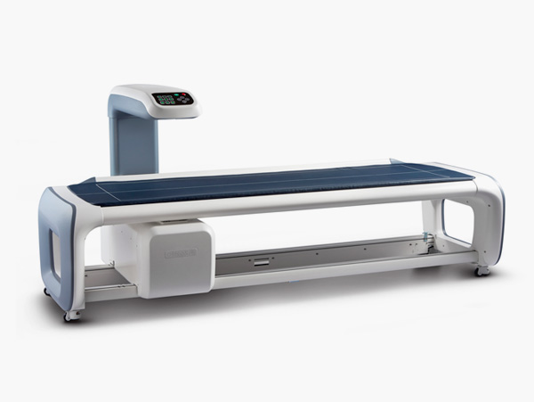
BONE DENSITOMETER (PRIMUS)
$0.00
Shipped From Abroad
PRIMUS is a whole body analyzer for the BMD(Bone Mineral Density), body composition, skeletal morphology, and Sarcopenia by scanning the whole body or a specific area of the body with its cutting edge Dual X-ray Absorptiometry(DXA) technology.
Typically 10-21 working days – excluding furniture and heavy/bulky equipment. Please contact us for further information.
Description
Description
16 Channel Whole Body DXA for BMD and Body Analysis
State-of-the-art DXA Whole Body Scanning System
PRIMUS quantitatively analyzes the BMD, lean mass and fat mass. You can help your patients to keep their whole body balance and to maintain a healthier life by following the process of continuing diagnosis and management.
Features
- Optimized Narrow Fan Beam DXA Technology
Based on the optimized fan beam DXA technology, PRIMUS is leading a new design trend of medical devices with its combination of sophisticated feature and a cutting-edge touch-type console panel
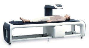
- Patient-Oriented Scan Table
A specially devised height of the bed helps the senior patients and those with relatively low height to lie down comfortably, so that they can complete their examination safely.
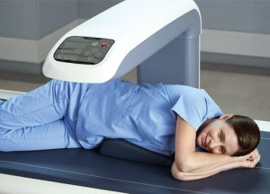
- Multi-channel Detectors for Wide and Fast Scan
Because the multi-channel detectors enable to cover a wide area with higher speed, it can measure the whole body of a patient efficiently.
- Convenient Examination
Along with its widened scan area, PRIMUS shortens the examination time with a higher scan speed resulting to a more convenient examination process.
- High Resolution for Skeletal Morphological Analysis
Besides the BMD analysis, you can perform the various skeletal morphology analysis by utilizing OsteoSys’s exclusive high-resolution image analysis functions.

- TBS(Trabecular Bone Score) on Primus
TBS iNsight™ is a software tool that installs on Primus. This simple, rapid and reproducible method estimates fracture risk based on a determination of bone texture (an index correlated to bone microarchitecture). The result is expressed as a Trabecular Bone Score (TBS). It requires no additional scan time or additional radiation exposure nor extra work for the technician. Once the spine scan is completed on Primus, TBS results are displayed automatically within seconds.
- From Osteoporosis to Sarcopenia and Obesity
The body composition function can quantitatively measure the bone, fat, and lean mass respectively and calculate the weight of a patient by adding up all the data of these elements. Moreover, it helps the diagnosis of Osteoporosis, Sarcopenia, obesity and Lipodystrophy.
Specifications
| Measurement Type | Whole body DXA (Total body Composition and assessment) |
| Measurement Method | Narrow fan beam |
| Scan site | Whole body, AP spine, Femur(Dual femur), Forearm, Lateral spine, LVA(VFA) |
| Scan area | 2020 × 580mm / 2020 × 620mm(Optional) |
| Scan time | AP spine – 30Sec. (± 2Sec.) Femur – 25Sec. (± 2Sec.) Forearm – 23Sec. (± 2Sec.) Whole body : 7Min.(Ergonomic) / 11min.(Standard mode) * Depends on height |
| Reproducibility | ≤ 1.0% C.V. |
| Measured parameter | BMD, BMC, BMI, T-score, Z-score, Area, Total body BMD, Total body Composition(Fat/Lean/BMC), HA(Hip Analysis), Dual femur Total body composition and various whole body assessment Orthopedics / Pediatrics / FRAX / B-Scope(body-Scope) / Color mapping / Ergonomic scan / Trend report / DICOM & PACS |
| Dimension | (W)2784mm × (D)1045mm × (H)1258mm (Standard) (W)2284mm × (D)1045mm × (H)1258mm (SB) |
| Weight | 210kg |
| Power consumption | 110VAC / 220VAC(+/- 10%) |
Quick Comparison
| BONE DENSITOMETER (PRIMUS) remove | DrGem Floor Mounted Analogue X-ray remove | Sonoscape S11 Ultrasound Machine remove | Sonoscape E2 Ultrasound Machine remove | DrGem Ceiling Analogue X-ray Machine remove | Sonoscape S8 Exp Portable Ultrasound remove | |||||||||||||||||||||
|---|---|---|---|---|---|---|---|---|---|---|---|---|---|---|---|---|---|---|---|---|---|---|---|---|---|---|
| Name | BONE DENSITOMETER (PRIMUS) remove | DrGem Floor Mounted Analogue X-ray remove | Sonoscape S11 Ultrasound Machine remove | Sonoscape E2 Ultrasound Machine remove | DrGem Ceiling Analogue X-ray Machine remove | Sonoscape S8 Exp Portable Ultrasound remove | ||||||||||||||||||||
| Image |  |  |  |  |  |  | ||||||||||||||||||||
| SKU | SF1033560130103-2 | SF1033560074-6 | SF1033560012-1 | SF1033560012-17 | SF1033560074-7 | SF1033560012-15 | ||||||||||||||||||||
| Rating | ||||||||||||||||||||||||||
| Price |
|
| $6,380.00 | $5,500.00 |
| $9,350.00 | ||||||||||||||||||||
| Stock | ||||||||||||||||||||||||||
| Availability | ||||||||||||||||||||||||||
| Add to cart | ||||||||||||||||||||||||||
| Description | Shipped From Abroad
PRIMUS is a whole body analyzer for the BMD(Bone Mineral Density), body composition, skeletal morphology, and Sarcopenia by scanning the whole body or a specific area of the body with its cutting edge Dual X-ray Absorptiometry(DXA) technology.
Delivery & Availability:
Typically 10-21 working days – excluding furniture and heavy/bulky equipment. Please contact us for further information.
| In Stock GXR Analogue X-ray system matches with a radiographic room which perfectly fits your workow and can be easily upgraded to DR system with the help of DR interface and PC interface in GXR generator as well as Bucky suitable to Flat Panel Detector. GXR X-ray system is equipped with a high frequency X-ray generator which consistently produces high quality radiograph in favor of high quality X-ray output with a very small kV ripple and accurate mA and mAs. GXR X-ray system is designed to provide convenience to operator and comfort to patient. Delivery & Availability: Typically 21 working days – excluding furniture and heavy/bulky equipment. Please contact us for further information. | In Stock A Value Choice beyond Your Expectation. SonoScape’s trolley color Doppler system S11 redefines price and performance with practical design. The S11 will go beyond your expectations but not your budget. Delivery & Availability: Typically 2 working days – excluding furniture and heavy/bulky equipment. Please contact us for further information. | Shipped from Abroad Sonoscape E2 portable ultrasound machine is a color Doppler ultrasound system that reaches beyond your expectations due to its compact and fashionable appearance. It fulfills GI, OB/GYN, Cardiac and POC applications to fit your routine scanning needs while its color mode will help you for more accurate and efficient diagnosis of lesions. E2 provides a wide range of applications to assist users with routine scanning. E2 provides automatic calculations to enhance your diagnostic confidence and save you time for patient communication. Delivery & Availability: Typically 14 working days – excluding furniture and heavy/bulky equipment. Please contact us for further information. | Shipped from abroad The DrGem Ceiling Analogue X-ray Machine is a diagnostic radiography system that provides reliable high quality radiographic images with a reduced dose. The reliable high-frequency x-ray generators that are known worldwide for their excellent performance, lifetime and stability. Patient tables and wall stands are also offered. Delivery & Availability: Typically 21 working days – excluding furniture and heavy/bulky equipment. Please contact us for further information. | Shipped from Abroad With ultra-modern innovative design and the clinically-proven technologies, S8 Exp is portable ultrasound scanner well equipped as a low-physical-effort and enhanced-image-quality ultrasound scanner, which not only provides optimized solutions for versatile applications, but does help to improve the user-experience for both routine and non-traditional challenges. Delivery & Availability: Typically 5-7 working days – excluding furniture and heavy/bulky equipment. Please contact us for further information. | ||||||||||||||||||||
| Content | Description
https://youtu.be/At-pJ5Ed7MM?si=hHrMXb81amiKZn5h
16 Channel Whole Body DXA for BMD and Body AnalysisState-of-the-art DXA Whole Body Scanning System
PRIMUS quantitatively analyzes the BMD, lean mass and fat mass. You can help your patients to keep their whole body balance and to maintain a healthier life by following the process of continuing diagnosis and management.
Features
Specifications
| DrGem GXR Floor Mounted Analogue X-ray system matches with a radiographic room which perfectly fits your workflow and can be easily upgraded to DR system with the help of DR interface and PC interface in GXR generator as well as Bucky suitable to Flat Panel Detector. GXR (Analogue X-ray)system is equipped with a high frequency X-ray generator which consistently produces high quality radiograph in favor of high quality X-ray output with a very small kV ripple and accurate mA and mAs. GXR (Analogue X-ray) system is designed to provide convenience to operator and comfort to patient.
Features of DrGem GXR Floor Mounted Analogue X-ray:
Click Here To Download Catalogue | DETAILS
SonoScape’s trolley colour Doppler system S11 redefines price and performance with practical design. The S11 will go beyond your expectations but not your budget. As an easy-to-use ultrasound system, the S11 is integrated with a new software platform, especially optimized for a smooth workflow and convenient operation. The system speeds up the exam process and makes file management easier.
SPECIFICATION
- 15-inch high definition LCD monitor with articulating arm
- Compact and agile trolley design
- 3 active transducer sockets available for a wide range of applications
- Duplex, Color Doppler, DPI, PW Doppler, tissue harmonic imaging, μ-scan speckle reduction imaging, compound imaging, trapezoidal imaging
- Customized settings based on your own working style
- Full patient database and image management solutions
Click Here To Download Catalogue | SONOSCAPE E2 DETAILS
Auto Image Optimization
A portable ultrasound machine with the press of a button, the image is automatically adjusted and optimized, saving you time with parameter adjustments. Additionally, with Auto Focus on, the focus area follows the depth of the ROI box as it is moved in the scanning field, providing users with excellent image quality in the desired area of interest.
Automated Calculation
Auto IMT is used when determining the level of vascular sclerosis present in the patient by automatically tracing the thickness of the carotid vessels.
Auto trace provides users sensitive and accurate wave tracing, avoiding the error of manual trace and giving out calculation result in no time
In-Build Battery pack
This portable ultrasound machine was equipped with an in-build battery pack which enable the user to perform image scanning when AC power is not available.
Click Here To Download Catalogue | DrGem Ceiling Analogue X-ray Machine is a diagnostic radiography system X-ray Machine that provides reliable high quality radiographic images with a reduced dose. The reliable high-frequency x-ray generators that are known worldwide for their excellent performance, lifetime and stability. Patient tables and wall stands are also offered.
Features of DrGem Ceiling Analogue X-ray Machine
Click Here To Download Catalogue | Sonoscape S8 Exp Portable Ultrasound scannerDETAILS Agile and Versatile With ultra-modern innovative design and the clinically-proven technologies, S8 Exp Portable Ultrasound scanner is well equipped as a low-physical-effort and enhanced-image-quality ultrasound scanner, which not only provides optimized solutions for versatile applications but does help to improve the user experience for both routine and non-traditional challenges. Working with S8 Exp, it will trigger your unlimited reverie and endow you with endless charm. Carrying forward the classical design of SonoScape's portable ultrasound products, S8 Exp successfully combines the best ergonomics, attractive design and ease of use. This charismatic identity is also enhanced by a sophisticated color palette—with sedate grey as its interior paint color and pearl white as exterior cover, S8 Exp reveals a style of aristocrat and strong character among SonoScape's ultrasound systems. Workflow The S8 Exp is a portable ultrasound scanner that adapts to your workflow, whether you are in the consulting room, at the bedside, or at a remote location. With easy-to-use new platform designed for sonographers' needs and full connection interfaces for easy connectivity and data sharing, S8 Exp leads to improved user comfort and clinical outcome as well as patient throughput and working efficiency. Powerful Platform Embedded with SonoScape's core imaging technologies such as μ-scan, PHI and Spatial Compound, S8 Exp boasts exceptional 2D image, sensitive spectral, Color and Power Doppler, displaying well-defined anatomy and pathology and facilitating a highly optimized diagnostic user environment for conclusive diagnoses. Besides, S8 Exp offers a comprehensive selection of electronic probes to maximally extend its capabilities to meet a wide range of applications including the abdomen, pediatric, OB/GYN, cardiovascular, musculoskeletal, etc. The advanced probe technologies also effectively enhance the image quality and confidence in reaching clinical diagnoses even in difficult patients.Click Here To Download Catalogue | ||||||||||||||||||||
| Weight | N/A | N/A | N/A | N/A | N/A | N/A | ||||||||||||||||||||
| Dimensions | N/A | N/A | N/A | N/A | N/A | N/A | ||||||||||||||||||||
| Additional information |


Reviews
There are no reviews yet.