BONE DENSITOMETER (PRIMUS)
$0.00
Shipped From Abroad
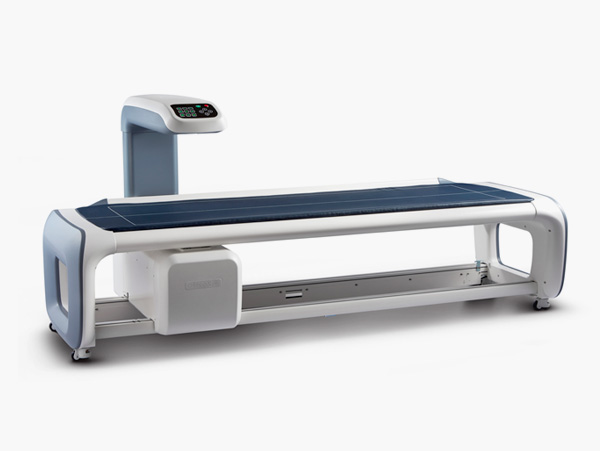
PRIMUS is a whole body analyzer for the BMD(Bone Mineral Density), body composition, skeletal morphology, and Sarcopenia by scanning the whole body or a specific area of the body with its cutting edge Dual X-ray Absorptiometry(DXA) technology.
Typically 10-21 working days – excluding furniture and heavy/bulky equipment. Please contact us for further information.
Description
Description
16 Channel Whole Body DXA for BMD and Body Analysis
State-of-the-art DXA Whole Body Scanning System
PRIMUS quantitatively analyzes the BMD, lean mass and fat mass. You can help your patients to keep their whole body balance and to maintain a healthier life by following the process of continuing diagnosis and management.
Features
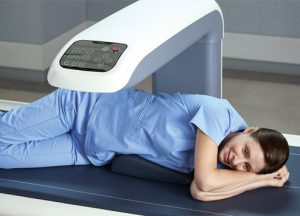
- Optimized Narrow Fan Beam DXA Technology
Based on the optimized fan beam DXA technology, PRIMUS is leading a new design trend of medical devices with its combination of sophisticated feature and a cutting-edge touch-type console panel
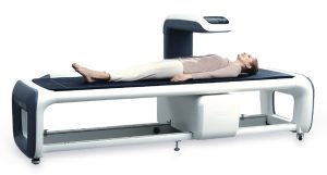
- Patient-Oriented Scan Table
A specially devised height of the bed helps the senior patients and those with relatively low height to lie down comfortably, so that they can complete their examination safely.
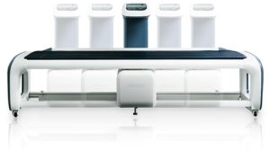
- Multi-channel Detectors for Wide and Fast Scan
Because the multi-channel detectors enable to cover a wide area with higher speed, it can measure the whole body of a patient efficiently.
- Convenient Examination
Along with its widened scan area, PRIMUS shortens the examination time with a higher scan speed resulting to a more convenient examination process.
- High Resolution for Skeletal Morphological Analysis
Besides the BMD analysis, you can perform the various skeletal morphology analysis by utilizing OsteoSys’s exclusive high-resolution image analysis functions.

- TBS(Trabecular Bone Score) on Primus
TBS iNsight™ is a software tool that installs on Primus. This simple, rapid and reproducible method estimates fracture risk based on a determination of bone texture (an index correlated to bone microarchitecture). The result is expressed as a Trabecular Bone Score (TBS). It requires no additional scan time or additional radiation exposure nor extra work for the technician. Once the spine scan is completed on Primus, TBS results are displayed automatically within seconds.
- From Osteoporosis to Sarcopenia and Obesity
The body composition function can quantitatively measure the bone, fat, and lean mass respectively and calculate the weight of a patient by adding up all the data of these elements. Moreover, it helps the diagnosis of Osteoporosis, Sarcopenia, obesity and Lipodystrophy.
Specifications
| Measurement Type | Whole body DXA (Total body Composition and assessment) |
| Measurement Method | Narrow fan beam |
| Scan site | Whole body, AP spine, Femur(Dual femur), Forearm, Lateral spine, LVA(VFA) |
| Scan area | 2020 × 580mm / 2020 × 620mm(Optional) |
| Scan time | AP spine – 30Sec. (± 2Sec.) Femur – 25Sec. (± 2Sec.) Forearm – 23Sec. (± 2Sec.) Whole body : 7Min.(Ergonomic) / 11min.(Standard mode) * Depends on height |
| Reproducibility | ≤ 1.0% C.V. |
| Measured parameter | BMD, BMC, BMI, T-score, Z-score, Area, Total body BMD, Total body Composition(Fat/Lean/BMC), HA(Hip Analysis), Dual femur Total body composition and various whole body assessment Orthopedics / Pediatrics / FRAX / B-Scope(body-Scope) / Color mapping / Ergonomic scan / Trend report / DICOM & PACS |
| Dimension | (W)2784mm × (D)1045mm × (H)1258mm (Standard) (W)2284mm × (D)1045mm × (H)1258mm (SB) |
| Weight | 210kg |
| Power consumption | 110VAC / 220VAC(+/- 10%) |
Quick Comparison
| Settings | BONE DENSITOMETER (PRIMUS) remove | LED Double X-ray Viewing Box remove | DrGem Floor Mounted Analogue X-ray remove | DrGem Ceiling Analogue X-ray Machine remove | Sonoscape S11 Ultrasound Machine remove | Sonoscape P20 Ultrasound Machine remove | ||||||||||||||||||||
|---|---|---|---|---|---|---|---|---|---|---|---|---|---|---|---|---|---|---|---|---|---|---|---|---|---|---|
| Name | BONE DENSITOMETER (PRIMUS) remove | LED Double X-ray Viewing Box remove | DrGem Floor Mounted Analogue X-ray remove | DrGem Ceiling Analogue X-ray Machine remove | Sonoscape S11 Ultrasound Machine remove | Sonoscape P20 Ultrasound Machine remove | ||||||||||||||||||||
| Image |  |  |  |  |  |  | ||||||||||||||||||||
| SKU | SF1033560130103-2 | SF1033560084-193 | SF1033560074-6 | SF1033560074-7 | SF1033560012-1 | SF1033560012-9 | ||||||||||||||||||||
| Rating | ||||||||||||||||||||||||||
| Price |
| $151.00 |
|
| $6,380.00 |
| ||||||||||||||||||||
| Stock | ||||||||||||||||||||||||||
| Availability | ||||||||||||||||||||||||||
| Add to cart | ||||||||||||||||||||||||||
| Description | Shipped From Abroad
PRIMUS is a whole body analyzer for the BMD(Bone Mineral Density), body composition, skeletal morphology, and Sarcopenia by scanning the whole body or a specific area of the body with its cutting edge Dual X-ray Absorptiometry(DXA) technology.
Delivery & Availability:
Typically 10-21 working days – excluding furniture and heavy/bulky equipment. Please contact us for further information.
| In stock
Double x-ray film viewer, Compact, Solid with Backlight of LED’s based panel, Long Life Approximate LED’s life 50, 000 Hrs., Uniform Light at the total surface area, No Heat Emission, Wall Mounted, Can be used for tracing on X-Ray, Auto-sensor, Screen. Size: 430 mm x 710 mm.
| In Stock GXR Analogue X-ray system matches with a radiographic room which perfectly fits your workow and can be easily upgraded to DR system with the help of DR interface and PC interface in GXR generator as well as Bucky suitable to Flat Panel Detector. GXR X-ray system is equipped with a high frequency X-ray generator which consistently produces high quality radiograph in favor of high quality X-ray output with a very small kV ripple and accurate mA and mAs. GXR X-ray system is designed to provide convenience to operator and comfort to patient. Delivery & Availability: Typically 21 working days – excluding furniture and heavy/bulky equipment. Please contact us for further information. | Shipped from abroad The DrGem Ceiling Analogue X-ray Machine is a diagnostic radiography system that provides reliable high quality radiographic images with a reduced dose. The reliable high-frequency x-ray generators that are known worldwide for their excellent performance, lifetime and stability. Patient tables and wall stands are also offered. Delivery & Availability: Typically 21 working days – excluding furniture and heavy/bulky equipment. Please contact us for further information. | In Stock A Value Choice beyond Your Expectation. SonoScape’s trolley color Doppler system S11 redefines price and performance with practical design. The S11 will go beyond your expectations but not your budget. Delivery & Availability: Typically 2 working days – excluding furniture and heavy/bulky equipment. Please contact us for further information. | Shipped from Abroad Incorporating innovative technologies, P20’s user-friendly design with a simple operation panel, intuitive user interface and a variety of intelligent auxiliary scanning tools, will significantly improve your daily examination experience. Besides general imaging applications, P20 has entitled with diagnostic 4D technology which has an extraordinary performance in obstetrics and gynecology applications. Delivery & Availability: Typically 5-7 working days – excluding furniture and heavy/bulky equipment. Please contact us for further information. | ||||||||||||||||||||
| Content | Description
https://youtu.be/At-pJ5Ed7MM?si=hHrMXb81amiKZn5h
16 Channel Whole Body DXA for BMD and Body AnalysisState-of-the-art DXA Whole Body Scanning System
PRIMUS quantitatively analyzes the BMD, lean mass and fat mass. You can help your patients to keep their whole body balance and to maintain a healthier life by following the process of continuing diagnosis and management.
Features
Specifications
| Double x-ray film viewer, Compact, Solid with Backlight of LED’s based panel, Long Life Approximate LED’s life 50, 000 Hrs., Uniform Light at the total surface area, No Heat Emission, Wall Mounted, Can be used for tracing on X-Ray, Auto-sensor, Screen. Size: 430 mm x 710 mm. | DrGem GXR Floor Mounted Analogue X-ray system matches with a radiographic room which perfectly fits your workflow and can be easily upgraded to DR system with the help of DR interface and PC interface in GXR generator as well as Bucky suitable to Flat Panel Detector. GXR (Analogue X-ray)system is equipped with a high frequency X-ray generator which consistently produces high quality radiograph in favor of high quality X-ray output with a very small kV ripple and accurate mA and mAs. GXR (Analogue X-ray) system is designed to provide convenience to operator and comfort to patient.
Features of DrGem GXR Floor Mounted Analogue X-ray:
Click Here To Download Catalogue | DrGem Ceiling Analogue X-ray Machine is a diagnostic radiography system X-ray Machine that provides reliable high quality radiographic images with a reduced dose. The reliable high-frequency x-ray generators that are known worldwide for their excellent performance, lifetime and stability. Patient tables and wall stands are also offered.
Features of DrGem Ceiling Analogue X-ray Machine
Click Here To Download Catalogue | DETAILS
SonoScape’s trolley colour Doppler system S11 redefines price and performance with practical design. The S11 will go beyond your expectations but not your budget. As an easy-to-use ultrasound system, the S11 is integrated with a new software platform, especially optimized for a smooth workflow and convenient operation. The system speeds up the exam process and makes file management easier.
SPECIFICATION
- 15-inch high definition LCD monitor with articulating arm
- Compact and agile trolley design
- 3 active transducer sockets available for a wide range of applications
- Duplex, Color Doppler, DPI, PW Doppler, tissue harmonic imaging, μ-scan speckle reduction imaging, compound imaging, trapezoidal imaging
- Customized settings based on your own working style
- Full patient database and image management solutions
Click Here To Download Catalogue | DETAILS
Upgraded Images with More Clarity
SonoScape never stops making progress in improving the image quality of its ultrasound products to enhance the confidence of diagnosis for doctors. With extraordinary images provided by P20, the anatomy structures are clearer than ever.
C-Xlasto Imaging
With C-xlasto Imaging, P20 enables comprehensive quantitative elastic analysis. Meanwhile, C-xlasto on P20 is supported by linear, convex and transvaginal probes, to ensure good reproducibility and highly consistent quantitative elastic results.
S-Live
S-Live allows for detailed visualization of subtle anatomical features, thereby enabling intuitive diagnosis with real-time 3D images and enriching patient communication.
Pelvic Floor 4D
Transperineal 4D pelvic floor ultrasound can provide useful clinical values in assessing the vaginal delivery impact on the female anterior compartment, judging whether the pelvic organs are prolapsed or not and the extent, determining if the pelvic muscles were torn accurately.
Anatomic M Mode
Anatomic M Mode helps you observe the myocardial motion at different phases by freely placing sample lines. It accurately measures the myocardial thickness and the heart size of even difficult patients and supports the myocardial function and LV wall-motion assessment.
Tissue Doppler Imaging
P20 is endowed with Tissue Doppler Imaging which provides velocities and other clinical information on myocardial functions, facilitating clinical doctors with the ability to analyze and compare the motions of different parts of the patient's heart.
Click Here To Download Catalogue | ||||||||||||||||||||
| Weight | N/A | N/A | N/A | N/A | N/A | N/A | ||||||||||||||||||||
| Dimensions | N/A | N/A | N/A | N/A | N/A | N/A | ||||||||||||||||||||
| Additional information |


Reviews
There are no reviews yet.